http://www.uem.br/acta ISSN printed: 1679-9275 ISSN on-line: 1807-8621
Doi: 10.4025/actasciagron.v36i1.16972
Evaluation of the mycoflora and aflatoxins from the pre-harvest to
storage of peanuts: a case study
Gisele Ferreira de Souza1, Simone Aparecida Galerani Mossini1, Carla Cristina Arrotéia2, Carlos Kemmelmeier2 and Miguel Machinski Junior1*
1
Laboratório de Toxicologia, Departamento de Ciências Básicas da Saúde, Universidade Estadual de Maringá, Av. Colombo, 5790, 87020-900, Maringá, Paraná, Brazil. 2
Departamento de Bioquímica, Universidade Estadual de Maringá, Maringá, Paraná, Brazil. *Author for correspondence. E-mail: [email protected]
ABSTRACT. Aflatoxins are carcinogens produced by Aspergillus flavus, A.parasiticus and A. nomius. In the present study, peanut samples were collected at different phenological stages of the plant during the 2007/2008 and 2008/2009 seasons and from stored peanuts harvested in 2007/2008. The mycoflora and aflatoxins in the peanuts were evaluated. The results showed the presence of Fusarium spp., Macrophomina spp., Trichoderma spp., Aspergillus spp. and Cladosporium spp. during the period of peanut maturation (39.8, 17.9, 8.2, 2.7 and 1.7%, respectively) and storage (49.8, 27.8, 12.5, 8.8 and 1.0%, respectively). However, aflatoxins were not detected in the samples. Of the 25 Aspergillus spp. isolates, 24 (96%) were producers of aflatoxin B1 (96%), 10 (40%) of aflatoxin B2, 17 (68%) of aflatoxin G1, and 10 (40%) of aflatoxin G2. The
isolation of Aspergillus spp. during storage was not influenced by the temperature, relative humidity or water activity (p > 0.05). The detection of aflatoxin-producing Aspergillus spp. in the samples analysed at different phenological stages, aerial gynophore, pod filling (seeds), mature fruits (pod), and dry fruits (harvest), indicates the importance of good agricultural practices from the cultivation to storage of peanuts in southern Brazil.
Keywords: mycotoxins, fungi, Aspergillus spp., toxigenic potential, Arachis hypogaea L.
Avaliação da micoflora e aflatoxinas da produção ao armazenamento de amendoim:
estudo de caso
RESUMO. Aflatoxinas são metabólitos carcinogênicos produzidos pelo Aspergillus flavus, A. parasiticus e A. nomius. No presente estudo, amostras de amendoim foram coletadas em diferentes estágios fenológicos da planta durante as safras de 2007/2008 e 2008/2009, bem como no período de armazenamento do amendoim referente à safra de 2007/2008. A micoflora e os níveis de aflatoxinas foram determinados. Os resultados demonstraram a presença de Fusarium spp., Macrophomina spp., Trichoderma spp., Aspergillus spp. e Cladosporium spp. no período de maturação (39,8; 17,9; 8,2; 2,7 e 1,7%, respectivamente) e no armazenamento (49,8; 27,8; 12,5; 8,8 e 1,0%). As aflatoxinas não foram detectadas nas amostras coletadas. Dos 25 isolados de Aspergillus spp., 24 foram produtores de aflatoxina B1 (96%), 10 de B2 (40%), 17 de G1
(68%) e 10 de G2 (40%). O isolamento de Aspergillus spp. durante o armazenamento não foi influenciado
pela temperatura, umidade relativa e atividade de água (p > 0,05). A detecção de Aspergillus spp. produtores de aflatoxinas nos diferentes estágios fenológicos: ginóforo aéreo, granação (sementes), frutos maduros (vagem) e frutos secos (colheita) demonstra a importância de boas práticas agrícolas da produção ao armazenamento do amendoim no sul do Brasil.
Palavras-chave: micotoxinas, fungos, Aspergillus spp., potencial toxigênico, Arachis hypogaea L.
Introduction
Peanuts (Arachis hypogaea L.) are rich in protein and
a high-energy food. Peanut plants are currently grown on a large scale in China, India, Africa and the United States, with a world production of approximately 37,6 million tons (FAO, 2010). In Brazil, peanut production increased by 14.4% from 226,500 tons in 2010/2011 to 259,100 tons in 2011/2012, with this rise being a consequence of the expansion of the cultivated area (15.8%) (CONAB, 2012).
Peanut crops are highly susceptible to fungal contamination, including toxin-producing fungi. Aflatoxins are secondary metabolites produced by
different species of the genus Aspergillus, particularly
by the producers. The climatic conditions of high humidity and temperature found in Brazil also increase
the probability of the growth of Aspergillus and
production of aflatoxins, a situation that is aggravated during the rainy season and under conditions of stress, such as drought or high insect proliferation (SCUSSEL, 2005).
Aflatoxin B1 is a potent carcinogen classified by
the International Agency for Research on Cancer as a group I carcinogen, i.e., a potent hepatocarcinogen for animals and humans (IARC, 2002). It is estimated that 35% of human cancer cases are directly related to diet, and the presence of aflatoxins in foods is considered to be an important risk factor for the genesis of hepatocellular carcinoma, particularly in tropical countries (LIU; WU, 2010).
In Brazil, maximum levels of aflatoxins in foods
were established by legislation and is 20 μg kg-1 for
the sum of aflatoxins B1, B2, G1 and G2 in peanuts
(BRASIL, 2011). However, despite the current legislation, high levels of aflatoxins are frequently detected in peanut-based foods destined for human consumption in Brazil (MAGRINE et al., 2011; GONÇALEZ et al., 2008a; NAKAI et al., 2008; SCUSSEL, 2005).
The elaboration of food safety programs for the control of aflatoxins in peanuts is not an easy task. Prevention through pre-harvest management is the best approach to control contamination with mycotoxins. However, the associated risks also depend on the post-harvest procedures, including the period of harvest, temperature, humidity, water activity, selection of the product for processing, decontamination conditions, use of chemical agents, storage, and transport (TOREGEANI-MENDES et al., 2011).
Peanuts can be colonised by a variety of fungi. The intimate contact with the soil during the stages of peanut growth and the storage period provides an ideal situation for fungal colonisation. The objectives of the present study were to identify the mycoflora in field samples of peanuts collected at different stages of maturity and during the period of storage to determine the presence of aflatoxins and to evaluate the aflatoxigenic potential of the Aspergillus isolates.
Material and methods
Sampling site
The study was conducted on a farm located in Maringá, Paraná State, Brazil, at a latitude of 23°19’38.14”, longitude of 51°57’47.24” and elevation of 560 m. The size of the cultivated area
was 73,200 m2, with the plantings in rows. Hill
cultivation, also known as weeding, was performed
and consists of the accumulation of soil at the foot of the plants during the first cleaning or hoeing.
Virginia peanut seeds (Arachis hypogaea hypogaea)
were used because, even though they present a longer phenological cycle than the Valencia-type variety, the productivity of the former variety increases compared to the previous harvest.
The botanical characteristics of A. hypogaea hypogaea
include its creeping habit, extra-long seed size
(approx. 90 g 100 seeds-1), and large pods containing
two seeds of beige colour. The side branches of the plant can reach 1.30 m, but the main stem does not exceed 15 cm. The samples corresponding to the different stages of maturity of the plant were collected during the 2007/2008 and 2008/2009 harvest, i.e., from a planting in October 2007 to the harvest in May 2008 and from a planting in September 2008 to the harvest in April 2009, respectively. Stored samples harvested in 2007/2008 were collected between May 2008 and January 2009.
Sample collection
Field samples of the plants at different stages of development (phenological stages) were collected for identification of mycoflora during the two harvest periods, as follows: stage I, aerial gynophore; stage II, gynophore penetrated into soil; stage III, pod filling (seeds); stage IV, mature fruits (pod); and stage V, dry fruits (harvest). A random block design was used. For this purpose, the plantation area was divided into 10 equal plots, with 10 samples containing approximately 50 g, corresponding to the stages I-IIII, and 500 g for stages IV and V. The 50 samples corresponding to the five peanut maturity stages were homogenised, and 50 g was used for the identification of mycoflora and the evaluation of the aflatoxigenic potential of the Aspergillus isolates A 450 g sample (stages IV and V) was used for the determination of aflatoxins using thin-layer chromatography (TLC). For the aflatoxin determination, the samples were ground in a blender model 51BL31 (Waring Co., Torrington, CT, USA), passed through a 20-mesh sieve, and homogenised. The samples were stored in plastic bags at -20°C.
2009). During each sampling, 10 samples of 100 g each were randomly collected from various sacks in the storage shed to obtain a sample of 1,000 g. The samples were divided into parts of 50 g for the analysis of the water activity, 50 g for the identification of the mycobiota and toxigenic potential, and 900 g for the aflatoxin analysis.
Isolation and identification of the mycoflora
The samples of the peanut fruits at the different stages of maturity were disinfected with 0.4% sodium hypochlorite solution for 3 min. and then washed with sterile water. For each sample, three gynophores and three peanuts grains each were directly seeded onto 10 Petri dishes containing potato-dextrose agar (PDA), as described by Pitt and Hocking (1997), 10 dishes containing Dichloran Rose Bengal Chloramphenicol agar (DRBC) according to Pitt et al. (1979), and 10
dishes containing Aspergillus flavus and Aspergillus
parasiticus agar (AFPA), as described by Pitt et al. (1983). The plates were incubated for 5 days at 20°C for PDA, 5 days at 25°C for DRBC, and 2 days at 30°C for AFPA. The same procedure was adopted for the samples obtained during the period of storage.
The results were expressed as the percentage of the sample contaminated with fungi in relation to all of the samples. Colonies with different morphologies were isolated and identified according to Nelson et al. (1983) and Pitt and Hocking (1997).
Aflatoxin standards
The aflatoxin standards were purchased from Sigma Chemical Co. (St. Louis, USA). Standard stock and working solutions were prepared according to the Manual of the Official Methods of Analysis of the Association of Official Agricultural Chemists International (AOAC, 2005). Individual stock solutions were prepared for each toxin, and the concentration was determined by ultraviolet spectrophotometry (Shimadzu UV-1601P, Tokyo, Japan) at 350 nm. The working solution was
prepared in benzene-acetonitrile (98:2, v v-1)
containing a mixture of the following toxins:
5 μg mL-1 AFB
1 and AFG1 and 1.5 μg mL-1 AFB2 and
AFG2.
Determination of aflatoxins by thin-layer chromatography (TLC)
The presence of aflatoxins was evaluated as described by Soares and Rodriguez-Amaya (1989). Each sample was homogenised, and two portions of 50 g each were removed for analysis in duplicate. One portion of 50 g of the sample was mixed with
270 mL methanol and 30 mL 4% potassium chloride and homogenised in a blender (Warning Co., Torrington, CT) at a low rotation for 5 min. The mixture was filtered through standard filter paper, and 150 mL of the filtrate was transferred to a beaker. The filtrate was clarified with 150 mL 10% copper sulphate and 20 g Celite and left to stand at room temperature for 5 min. The mixture was then filtered through standard filter paper, and 150 mL of the filtrate was transferred to a separation funnel. After adding 150 mL water, this mixture was partitioned twice with 10 mL chloroform. Five mL of the first and second chloroform partitions were combined and evaporated to dryness in a water bath at 60ºC.
The residue obtained was dissolved in 200 μL
benzene-acetonitrile (98:2). For screening, 5 μL of
the extract was applied to a TLC plate (Alugram® SIL G Silica gel 60, Macherey-Nagel, Duren, Germany), 2 cm from the baseline. The standards were applied separately. The plate was placed in an unsaturated chamber containing toluene-ethyl
acetate-chloroform-formic acid (70:50:50:20, v v-1
v v-1). The aflatoxins were visualised under UV
light (350 nm) and quantified by comparing the fluorescence of the samples with the aflatoxin standards. The quantification limit of the method
was 2 μg kg-1 for each aflatoxin. All of the reagents
used were of p.a. grade (Merck, Darmstadt, Germany).
In this experiment, five spiked samples of
20 g kg-1 of AFB
1 and AFG1 and 6 g kg-1 of AFB2
and AFG2 were used for the recovery test. The
recovery average for aflatoxins B1, B2, G1 and G2
were 97.4, 97.5, 97.4 and 111.2%, respectively.
Evaluation of the toxigenic potential of A. flavus and
A. parasiticus
For the evaluation of the aflatoxin-producing
potential, the fungal isolates identified as A. flavus
and A.parasiticus were inoculated into the centre of a coconut agar plate and incubated in a BOD oven at 25°C for 7 days. The growing colonies were monitored under ultraviolet light for the presence of the characteristic fluorescence of the genera. Next, the entire contents of the plate was transferred to a blender, and 30 mL chloroform was added. The extracts obtained were filtered through filter paper, concentrated in a water bath at 60°C, and
resuspended in 200 μL chloroform. TLC was
performed as described above.
Determination of the water activity
The water activity (Aw) was determined in
(Decagon Devices, Inc., Pullman, WA, USA) at a temperature of 25ºC.
Climatic data
The temperature, relative air humidity and rainfall data for the period from September 2007 to April 2009 were obtained from the Maringá Climatological Station at State University of Maringá, Brazil.
Statistical analyses
The results were analysed using the SAS 9.1 statistical software. An analysis of variance (ANOVA) and linear regression were used to determine the influence of the variables of the 2007/2008 and 2008/2009 harvest and of storage
on the frequency of the isolation of Aspergillus spp.
Regression analysis was also used to select the dependent variables: frequency of isolation of Aspergillus spp. and aflatoxin production. The independent variables were the culture medium (PDA, AFPA, and DRBC), temperature, relative air humidity, rainfall, and water activity. The water activity was measured during the period of storage of the 2007/2008 harvest. The level of significance was set at 5% (p < 0.05) for all of the tests.
Results and discussion
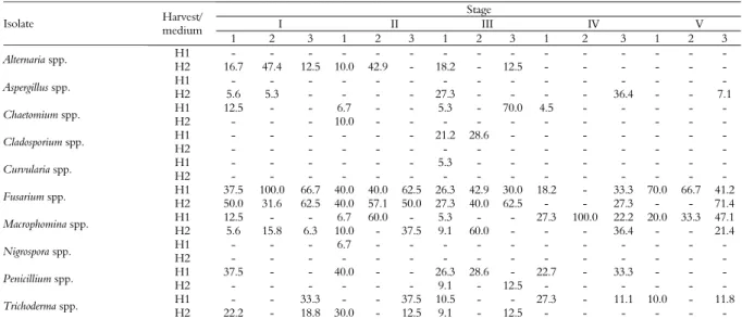
The fungal population isolated during the different peanut maturity stages of the 2007/2008
harvest consisted of Fusarium spp. (38.4 to 62.4%
depending on the culture medium, i.e., PDA,
DRBC and AFPA), Penicillium spp. (28.6 to
33.3%), Macrophomina spp. (14.3 to 34.6%),
Chaetomium spp. (0 to 70.0%), Cladosporium spp.
(0 to 28.6%), Trichoderma spp. (0 to 23.4%),
Nigrospora spp. (0 to 6.7%), and Curvularia spp. (0 to 5.3%). Similar results were obtained for the 2008/2009 production (p = 0.1457), with the
fungal population consisting of Fusarium spp.
(23.5 to 54.7% depending on the culture
medium), Penicillium spp. (9.1 to 37.9%),
Alternaria spp. (8.3 to 45.1%), Macrophomina spp.
(8.2 to 31.3%), Chaetomium spp. (0 to 10.0%), and
Trichoderma spp. (0 to 20.4%). An exception was
that the Aspergillus spp. were detected in stages I,
III, IV and V at a frequency of 5.3 to 36.4% (Table 1). The similar frequencies of isolation obtained for the two production periods can be explained by the presence of the same experimental and climatic conditions (Table 2). Similar results have been reported by Mphane et al. (2004), Gonçalez et al. (2008a) and Nakai et al. (2008) who isolated Aspergillus spp., Fusarium spp., Penicillium spp., Cladosporium spp., and Trichoderma spp. as the main contaminants of peanuts. These studies were conducted in Botswana and the Junqueiropolis and Tupã regions, São Paulo State, Brazil, respectively.
Three culture media described in the literature
were tested for the isolation of Aspergillus spp.: PDA,
as described by Pitt and Hocking (1997); DRBC, as described by Pitt et al. (1979); and AFPA, as described by Pitt et al. (1983). Our statistical analysis revealed no significant differences among the media (p = 0.7287).
Table 1. Relative frequency (%) of fungal species isolated from peanuts at different stages of maturity by culture on different media.
Stage
I II III IV V
Isolate Harvest/ medium
1 2 3 1 2 3 1 2 3 1 2 3 1 2 3 H1 - - - -
Alternaria spp.
H2 16.7 47.4 12.5 10.0 42.9 - 18.2 - 12.5 - - - - H1 - - - -
Aspergillus spp.
H2 5.6 5.3 - - - - 27.3 - - - - 36.4 - - 7.1 H1 12.5 - - 6.7 - - 5.3 - 70.0 4.5 - - - - -
Chaetomium spp.
H2 - - - 10.0 - - - - H1 - - - 21.2 28.6 - - - -
Cladosporium spp.
H2 - - - - H1 - - - 5.3 - - - -
Curvularia spp.
H2 - - - - H1 37.5 100.0 66.7 40.0 40.0 62.5 26.3 42.9 30.0 18.2 - 33.3 70.0 66.7 41.2
Fusarium spp.
H2 50.0 31.6 62.5 40.0 57.1 50.0 27.3 40.0 62.5 - - 27.3 - - 71.4 H1 12.5 - - 6.7 60.0 - 5.3 - - 27.3 100.0 22.2 20.0 33.3 47.1
Macrophomina spp.
H2 5.6 15.8 6.3 10.0 - 37.5 9.1 60.0 - - - 36.4 - - 21.4 H1 - - - 6.7 - - - -
Nigrospora spp.
H2 - - - - H1 37.5 - - 40.0 - - 26.3 28.6 - 22.7 - 33.3 - - -
Penicillium spp.
H2 - - - 9.1 - 12.5 - - - - H1 - - 33.3 - - 37.5 10.5 - - 27.3 - 11.1 10.0 - 11.8
Trichoderma spp.
H2 22.2 - 18.8 30.0 - 12.5 9.1 - 12.5 - - - -
Table 2. Meteorological data referring to the period of collection of peanut samples at different stages of maturity during the 2007/2008 and 2008/2009 harvest in Maringá, Brasila.
Harvest
2007/2008 2008/2009
Stage
TMin (°C) TMean (°C) TMax (°C) RU (%) RI (mm) TMin (°C) TMean (°C) TMax (°C) RU (%) RI (mm)
I 20.6 24.0 27.5 69 737.6 20.6 24.7 28.9 66 754 II 22.1 25.0 27.9 73 165.9 22.0 26.1 30.3 70 147 III 20.5 23.4 26.3 74 35.5 24.2 27.5 30.8 68 54 IV 22.4 24.7 27.0 64 59.1 22.3 25.1 27.9 68 102 V 16.9 22.2 27.6 74 162.4 22.8 24.4 26.1 58 0
aLatitude = 23°19’38.14”; longitude = 51°57’47.24”; altitude = 560 m. T
Min, TMean, TMax = minimum, mean and maximum temperature; RU = relative humidity; RI = rainfall index.
Stages 2007/2008 = I – 10/16/07 to 02/18/08; II – 02/18/08 to 03/11/08; III – 03/11/08 to 03/25/08; IV – 03/25/08 to 04/08/08; V - 04/08/08 to 05/13/08. Stages 2008/2009 = I – 09/23/08 to 02/09/09; II - 02/09/09 to 03/05/09; III - 03/05/09 to 03/20/09; IV – 03/20/09 to 04/16/09; V – 04/16/09 to 04/27/09.
During the peanut pod formation (stages of development) and the beginning of pod filling, the fruits contain a high amount of water, and these stages were characterised by a high incidence of Fusarium spp., the fungus best adapted to substrates with a high water content (HORN, 2005). Similar results have been reported by Pitt and Hocking (1997) and Gonçalez et al. (2008a). In stage IV, the
most frequently isolated species was Macrophomina
spp. (PDA, 27.3%; AFPA, 100.0%; and DRBC, 22.2%); this stage coincided with a decline in the
isolation of Fusarium spp. (PDA, 18.2%; AFPA,
0.0%; and DRBC, 33.3%) (Table 1). The genera Macrophomina and Fusarium share some hosts and tend to develop mixed infections under certain conditions of humidity. These two fungi are soil
contaminants, but no toxigenic Macrophomina species
have been described (JANA et al., 2003).
The samples collected at the different stages of peanut maturity were not contaminated with aflatoxins, a result similar to that reported by
Gonçalez et al. (2008b). In 2007/2008, Aspergillus spp.
were not isolated at any of the peanut maturity stages analysed, a finding that explains the lack of detection of aflatoxins. Immature peanut fruits are more resistant to fungal invasion and contamination with aflatoxins because they produce higher amounts of phytoalexins than the mature fruits (DORNER et al., 1989). Another important factor was that the maximum temperature observed during the peanut growth was 30.8°C (Table 2), whereas temperatures
higher than 32°C are ideal for the growth of Aspergillus
spp. (PITT; HOCKING, 1997). With respect to the relative humidity, the values observed (64 to 74%) were also below the levels considered to be optimal
for the growth of Aspergillus (83 to 85%)
(CHRISTENSEN et al., 1977). The statistical analysis showed no correlation between the presence of Aspergillus spp. and the temperature (p = 0.7416,
r² = 0.0089), relative humidity (p = 0.7391, r² = 0.010),
or rainfall (p = 0.7416, r² = 0.0075). In contrast, Dorner et al. (1989) and Gonçalez et al. (2008a) demonstrated a good correlation between drought and high temperatures.
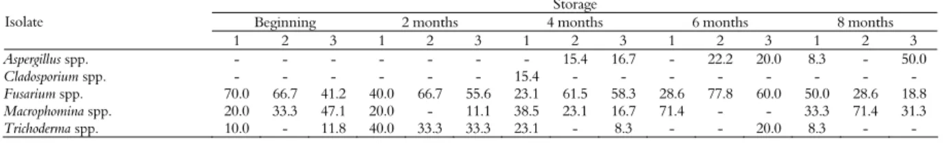
The fungal population isolated from the peanut samples collected during storage is shown in Table 3. The following species were isolated during
our monitoring: Fusarium spp., Macrophomina spp.,
Trichoderma spp., Cladosporium spp., and Aspergillus
spp. Fusarium spp. was the most frequent species due
to the contact of peanuts with the soil (means of 59.3% at the beginning of pod filling, 66.7% after 2 months of storage, 61.5% after 4 months, 77.8% after 6 months, and 50% after 8 months). This soil fungus can survive for several months during storage (PITT; HOCKING, 1997), and results similar to the present findings have been reported by Nakai et al. (2008) for peanut varieties stored for
12 months. Aspergillus spp. was only isolated after 4,
6 and 8 months of storages, with frequencies of isolation of 15.4% (AFPA) and 16.7% (DRBC), 22.2% (AFPA) and 20% (DRBC), and 8.3% (PDA) and 50% (DRBC), respectively.
A clear decline in the isolation of Fusarium spp.
was observed after 8 months of storage (18.8% on AFPA). Because this fungus is best adapted to substrates with a high water content (HORN, 2005), the low water content of the samples after 8 months of storage favoured the growth of Aspergillus spp. (50% on AFPA).
Table 3. Relative frequency (%) of fungal species isolated from samples of peanuts stored between May 2008 and January 2009.
Storage
Beginning 2 months 4 months 6 months 8 months Isolate
1 2 3 1 2 3 1 2 3 1 2 3 1 2 3
Aspergillus spp. - - - 15.4 16.7 - 22.2 20.0 8.3 - 50.0
Cladosporium spp. - - - 15.4 - - - -
Fusarium spp. 70.0 66.7 41.2 40.0 66.7 55.6 23.1 61.5 58.3 28.6 77.8 60.0 50.0 28.6 18.8
Macrophomina spp. 20.0 33.3 47.1 20.0 - 11.1 38.5 23.1 16.7 71.4 - - 33.3 71.4 31.3
Trichoderma spp. 10.0 - 11.8 40.0 33.3 33.3 23.1 - 8.3 - - 20.0 8.3 - -
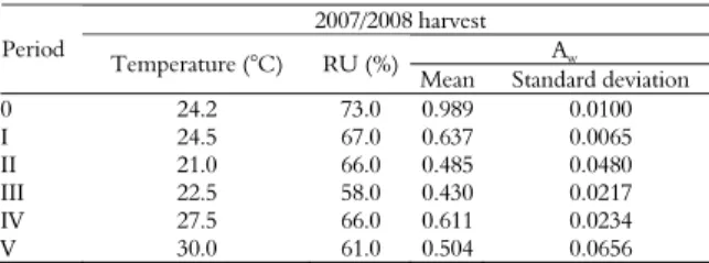
The mean water activity before drying was 0.989; similar values ranging from 0.90 to 0.99 were reported by Gonçalez et al. (2008b). Studies have shown that peanut fruits with a high water activity
do not favour the growth of A. flavus or A. parasiticus
(DORNER et al., 1989; HORN, 2005), as observed
in the present study in which no Aspergillus spp. were
detected. After drying, the Aw ranged from 0.430 to
0.637, values below the range of 0.78-0.80
established as optimal for the growth of Aspergillus
spp. by Pitt and Hocking (1997). However, despite
the low Aw, the growth of Aspergillus spp. was
observed after 4 months of storage. This finding agrees with other studies reporting the isolation of Aspergillus from samples stored at a low Aw
(DORNER et al., 1989; HORN, 2005; NAKAI et al., 2008).
In the present study, the mean temperature ranged from 21 to 30°C, and the relative humidity ranged from 58 to 73% during the 8-month storage period (Table 4). According to Christensen et al. (1977), and Pitt and Hocking (1997), these temperature (< 32-33°C) and humidity (< 83-85%) conditions, respectively, are optimal for the growth of Aspergillus spp.
The ANOVA showed that the mycoflora
detected during storage, particularly Aspergillus spp.,
were not correlated with the duration of storage (p = 0.12, r² = 0.0102) temperature (p = 0.999, r² = 0.0137), relative air humidity (p = 0.999, r² = 0.1532), or water activity (p = 0.999, r² = 0.1268). These results show no differences in the mean frequency of isolation between the different periods of storage of peanuts, in contrast to the studies of Gonçalez et al. (2008a and b) and Nakai et al. (2008).
Table 4. Mean temperature, relative humidity and water activity recorded during the period of experimental storage.
2007/2008 harvest Aw
Period
Temperature (°C) RU (%)
Mean Standard deviation 0 24.2 73.0 0.989 0.0100 I 24.5 67.0 0.637 0.0065 II 21.0 66.0 0.485 0.0480 III 22.5 58.0 0.430 0.0217 IV 27.5 66.0 0.611 0.0234 V 30.0 61.0 0.504 0.0656
Period = 0 – freshly harvested (05/10/08); I – 05/10/08 to 05/13/08; II – 05/13/08 to 07/14/08; III – 07/14/08 to 09/12/08; IV – 09/12/08 to 11/11/08; V - 11/11/08 to 01/12/09. RU = relative humidity; Aw = water activity.
Aflatoxins B1, B2, G1 and G2 were not detected
in the samples collected at different maturity stages in 2007/2008 or 2008/2009 or during the storage of the 2007/2008 harvest. These results disagree with studies conducted in Brazil reporting a high prevalence of contamination with aflatoxins in
peanuts and peanut products (MAGRINE et al., 2011; CALDAS et al., 2002). More recently, Nakai et al. (2008) found that 33.3 and 28.3% of seeds
were contaminated with aflatoxins B1 and B2,
respectively.
The evaluation of the toxigenic potential of the
isolates showed that 96% of the Aspergillus spp.
isolated from peanut samples (n = 25) of the 2008/2009 harvest and from stored samples presented
a toxigenic potential. Among the A. flavus (32%) and
A.parasiticus (68%) isolates, 24 (96%) were producers
of aflatoxin B1, 10 (40%) of aflatoxin B2, 17 (68%) of
aflatoxin G1, and 10 (25%) of aflatoxin G2. In previous
studies on peanuts, Nakai et al. (2008) showed that 93.8% of the isolates presented toxigenic potential, whereas Pildain et al. (2004) observed that 75% of the isolates of peanut samples from Argentina presented toxigenic potential.
Conclusion
This study demonstrated that the production conditions applied in southern Brazil contribute to the susceptibility of peanuts to the colonisation with aflatoxigenic fungi during different stages of maturity in the field and storage. The results showed that the storage period of peanut is a critical phase, as indicated by the high percentage of aflatoxigenic isolates. However, no correlation was observed between the presence of the isolates and temperature, relative humidity or water activity. The results demonstrate the importance of good agricultural practices during the production, harvest and storage of peanuts in Brazil.
Acknowledgements
This study was supported by the Brazilian government funding agency Conselho Nacional de Desenvolvimento Científico e Tecnológico (CNPq) (grant No. 401817/2005-9).
References
AOAC-Association of Official Agricultural Chemists. Official Methods of Analysis of the Association of Analytical Chemists. 17th ed. Arlington: AOAC, 2005. BRASIL, Agência Nacional de Vigilância Sanitária. RDC Nº 7, Limites máximos tolerados (LMT) para micotoxinas. Brasília: ANVISA, 2011.
CONAB-Companhia Nacional de Abastecimento. Acompanhamento da Safra Brasileira – Grãos [Internet]. Brasília: Conab, 2012. Available from: <http://www.conab.gov.br/OlalaCMS/uploads/arquivos/1 2_02_16_08_47_47_boletim_portugues__fevereiro_2012. pdf>. Access on: Mar. 21, 2012.
DORNER, J. W.; COLE, R. J.; SANDERS, T. H.; BLANKENSHIP, P. D. Interrelationship of kernel water activity, soil temperature, maturity, and phytoalexin production in preharvest aflatoxin contamination of drought-stressed peanuts. Mycopathologia, v. 105, n. 2, p. 117-128, 1989.
FAO-Food and Agriculture Organization of the United Nations. FAOSTAT. Rome: FAO, 2010.
GONÇALEZ, E.; NOGUEIRA, J. H. C.; FONSECA, H.; FELICIO, J. D.; PINO, F. A.; CORRÊA, B. Mycobiota and mycotoxins in Brazilian peanut kernels from sowing to harvest. International Journal of Food Microbiology, v. 123, n. 3, p. 184-190, 2008a.
GONÇALEZ, E.; SOUZA, T. N.; ROSSI, M. H.; FELICIO, J. D.; CORRÊA, B. Avaliação da micoflora e ocorrência de micotoxinas em cascas de amendoim em diferentes estágios de maturação da vagem. Ciência e Agrotecnologia, v. 32, n. 5, p. 1380-1386, 2008b.
HORN, B. W. Colonization of wounded peanuts seeds by soil fungi: selectivity for species from Aspergillus section Flavi. Mycologia, v. 97, n. 1, p. 202-217, 2005.
IARC-International Agency of Research on Cancer. Some traditional herbal medicines, some mycotoxins, naphthalene and styrene. IARC Monographs, 82. Lyon: IARC, 2002.
JANA, T.; SHARMA, T. R.; PRASAD, R. D.; ARORA, D. K. Molecular characterization of Macrophomina phaseolina and Fusarium species by a single primer RAPD technique. Microbiology Research, v. 158, n. 3, p. 249-257, 2003.
LIU, Y.; WU, F. Global burden of aflatoxin-induced hepatocellular carcinoma: a risk assessment. Environmental Health Perspectives, v. 118, n. 6, p. 818-824, 2010.
MAGRINE, I. C. O.; FERRARI, S. S. C.; SOUZA, G. F.; MINAMIHARA, L.; KEMMELMEIER, C.; BANDO, E.; MACHINSKI JR., M. Intake of aflatoxins through the consumption of peanut products in Brazil. Food Additives and Contaminants: Part B, v. 4, n. 2, p. 99-105, 2011.
MPHANE, F. A.; SIAME, B. A.; TAYLOR, J. E. Fungi, aflatoxin and cyclopiazonic acid associated with peanut retailing in Botswana. Journal of Food Protection, v. 67, n. 1, p. 96-102, 2004.
NAKAI, V. K.; ROCHA, L. O.; GONÇALEZ, E.; FONSECA, H.; ORTEGA, M. M.; CORRÊA, B. Distribution of fungi and aflatoxins in a stored peanut variety. Food Chemistry, v. 106, n. 1, p. 285-290, 2008. NELSON, P. E.; TOUSON, T. A.; MARASAS, W. F. O.
Fusarium species. An illustrated manual foridentification. London: The Pennsylvania State University, 1983.
PILDAIN, M. B.; VAAMONDE, G.; CABRAL, D. Analysis of population structure of Aspergillus flavus from peanut based on vegetative compatibility geographic origin, mycotoxin and sclerotia production. International Journal of Food Microbiology, v. 93, n. 1, p. 31-40, 2004.
PITT, J. I.; HOCKING, A. D. Aspergillus and related teleomorphs. In: PITT, J. I.; HOCKING, A. D. (Ed.). Fungi and food spoilage. 2. ed. Baltimore: Aspen Publishers Inc., 1997. p. 339-416.
PITT, J. I.; GLENN, D. R.; HOCKING, A. D. An improved medium for the detection of Aspergillus flavus and A. parasiticus. Journal of Applied Bacteriology, v. 54, n. 1, p. 109-114, 1983.
PITT, J. I.; KING, A. D.; HOCKING, A. D. Dichloran-rose Bengal medium for enumeration and isolation of molds from foods. Applied and Environmental Microbiology, v. 37, n. 5, p. 959-970, 1979.
SCUSSEL, V. M. Aflatoxin and food safety: recent South American perspectives. In: ABBAS, H. K. Aflatoxin and food safety. London: CRC Press, 2005. p. 29-58.
SOARES, L. M. V.; RODRIGUEZ-AMAYA, D. B. Survey of aflatoxins, ochratoxin A, zearalenone, and sterigmatocystin in some Brazilian foods by using multi-toxin thin-layer chromatographic method. Journal of Association of Official Analytical Chemists, v. 72, n. 1, p. 22-26, 1989.
TOREGEANI-MENDES, K. A.; ARROTEIA, C. C.; KEMMELMEIER, C.; DALPASQUALE, V. A.; BANDO, E.; ALVES, A. F.; MARQUES, O. J.; NISHIYAMA, P.; MOSSINI, S. A. G.; MACHINSKI JR., M. Application of hazard analysis critical control points system for the control of aflatoxins in the Brazilian groundnut-based food industry. International Journal of Food Science and Technology, v. 46, n. 12, p. 2611-2618, 2011.
Received on April 27, 2012. Accepted on August 6, 2012.