CERNE
Historic:
Received 06/03/2018 Accepted 22/06/2018
Keywords: Yerba-mate UPGMA TEF-1α Pathogenicity
1 Federal University of Santa Maria, Santa Maria, Rio Grande do Sul, Brazil - ORCID: 0000-0002-7392-9588a, 0000-0003-2737-7599b, 0000-0002-6162-4445c, 0000-0002-6534-8070d, 0000-0002-4398-7998e, 0000-0002-3243-671Xf, 0000-0001-7436-9589g
+Correspondence: [email protected]
DOI: 10.1590/01047760201824032535
Ricardo Mezzomo1a+, Jéssica Mengue Rolim1b, Tales Poletto1c, Victoria Cozer Rosenthal1d,
Lucas Graciolli Savian1e, Lia Rejane Silveira Reiniger1f, Marlove Fátima Brião Muniz1g
MORPHOLOGICAL AND MOLECULAR CHARACTERIZATION OF Fusarium spp. PATHOGENIC TO Ilex Paraguariensis
MEZZOMO, R.; ROLIM, J. M.; POLETTO, T.; ROSENTHAL, V. C.; SAVIAN, L. G.; REINIGER, L. R. S.; MUNIZ , M. F. B.Morphological and molecular characterization of Fusarium spp. pathogenic to Ilex Paraguariensis. CERNE, v. 24, n. 3, p. 209-218, 2018.
HIGHLIGHTS
Due to the low initial costs and ease of employment of agrosilvopastoral systems, the cultivation of yerba-mate constitutes an alternative for small properties in the RS to the diversification of production.
The isolates of Fusarium spp. were submitted to the UPGMA analysis and grouped by the similarity between the characters averages.
Through the UPGMA clustering test, the thirty-nine isolates of Fusarium spp. were allocated to seven large groups.
The analysis of the TEF 1-α gene region was enough to discriminate species of Fusarium spp.
The reflex symptoms resulted from the interaction of Fusarium spp. isolates with yerba-mate roots.
ABSTRACT
The cultivation of yerba-mate (Ilex paraguariensis) has been intensified in the State of Rio Grande do Sul, as result of the valuation of the price paid to the producer. However, studies related to Fusarium pathosystem vs. yerba-mate are still insufficient, so that many producers have faced significant losses in herbal production due to the occurrence of the roots rotting. Considering the above, the objectives of this study were: a) morphologically characterize the Fusarium spp. isolates collected in sick plants of yerba-mate; b) select efficient morphological characters on separating the Fusarium spp. isolates in similarity groups; c) identify the Fusarium spp. isolates to species level through the sequencing of the genomic region of elongation factor 1-α (TEF-1α); and d) testing the pathogenicity of the Fusarium spp. isolates selected based on the morphological characterization. For such, collections for the pathogen isolates were made in five cities and the collection points were georeferenced. Thirty-nine isolates were morphologically characterized by variables as mycelial growth, sporulation, macroconidia length and width, microconid format, colonies pigmentation and formation of chlamydospore, and tested for pathogenicity by substrate inoculation. The variables used in the morphological characterization were efficient in discriminating the isolates in seven groups, especially length of macronidia associated to sporulation. Seven pathogenic isolates of Fusarium spp. were identified, also through molecular sequencing of the TEF-1α gene region. The sequencing of the TEF-1α region consolidated the identification of pathogenic isolates as F. solani and F. oxysporum.
INTRODUCTION
The yerba-mate is a native species of the Mixed Ombrophilous Forest, with wide distribution and of great environmental and socioeconomic importance especially in southern Brazil, northeastern Argentina and a great part of Paraguay and Uruguay (Fowler et al., 2007). In an area corresponding to 94,945 hectares, 602,899 tons of green yerba-mate were produced in Brazil in the year of 2015, resulting in an average productivity of 7,126 kg.ha-1. The State of Rio Grande do Sul (RS) concentrates
the largest part of the marketed and exported yerba-mate production, with 292,386 tons, which corresponds to 48.5% of the total produced. The State of Paraná follows with 217,851 tons or 36.1% of the production (Brazilian Institute of Geography and Statistics, 2015).
However, these yerba-mate plantations are subject to pathogen attacks such as the Fusarium fungi, known to cause diseases such as the roots rotting, which lead to a reduction in productivity and, consequently, to plant death.
Among the main symptoms of root rotting are the ascending yellowing of leaves, the growth stagnation and excessive drop in older leaves and in many cases, the death of the plant. As result of the pathogen attack, the roots darken and rot, compromising the development of the plantations. In the attacked plantations, the production losses may be greater than 30% (Poletto et al., 2006).
To ensure that control and prevention strategies can be deployed there is the necessity for studies that seek to identify the fungus causing the disease correctly.
Morphological characterization is among the
main mechanisms employed in the identification of
phytopathogenic fungi. It is used widely, and various analyses such as colonies pigmentation, texture, shape, marginal form and mycelial growth rate, process of formation, spore size and shape are involved (Burgess et al., 1995). Although, due to their great similarity, the differentiation of fungi through morphological characters is imprecise. Nevertheless, by directly accessing the DNA, the use of molecular techniques become more
efficient and accurate for the discrimination of fungal
species (Steenkamp et al., 2000).
The objectives of this study were: a) morphologically characterize the Fusarium spp. isolates
collected in sick plants of yerba-mate; b) select efficient
morphological characters on separating the Fusarium
spp. isolates in similarity groups; c) identify the Fusarium
spp. isolates to species level through the sequencing of the genomic region of the elongation factor 1-α
(TEF-1α); and d) test the pathogenicity of the Fusarium spp. isolates based on the morphological characterization.
MATERIAL AND METHODS
Obtaining and purification of isolates
The samples collections were performed in yerba-mate plantations in the cities of Anta Gorda, Arvorezinha, Putinga, Ilópolis, and Machadinho of the State of Rio Grande do Sul (RS), from January through March 2015. All the collection (Table 1) points were georeferenced using a Global Positioning System (GPS) equipment.
The plant material collected in the field (roots of
adult plants and seedlings with tipping symptoms) was
duly identified and placed in thermal boxes containing
ice and transported for further analysis in the Elocy Minussi Phytopathology Laboratory of the Department of Plant Protection of the Federal University of Santa Maria (UFSM). Then, the material was placed in a humid chamber. After that, the material was observed in stereoscopic and optical microscopes to check for the presence of pathogen structures (mycelium and spores). If positive, with the aid of a histological needle, structures of the pathogen were transferred to Petri dishes containing potato-dextrose-agar (PDA) culture
medium, as recommended by Alfenas and Mafia (2007).
After seven days of growth, the isolates grown on PDA
were purified according to the technique of monosporic
culture, described by Fernandez (1993).
Morphophysiological characterization of Fusarium spp. isolates
The isolates obtained followed the methodology described by Michereff et al. (2003), where each isolate was evaluated according to the parameters displayed below. The mycelial growth rate (M.G.R.) was determined from the transfer of PDA medium discs with a 6 mm diameter, obtained from colonies with seven
days of growth, for other plates with PDA. For this, five
replications were used, each one constituted by one plate. The mycelial growth of each isolate was evaluated after seven days of incubation by measuring the mean diameter of the colony into two diametrically opposite directions. The sporulation was assessed at 10 days of incubation by adding 20 mL of sterile distilled water in four Petri plates, after the scraping of the colonies and sieving in a double layer of gauze for the subsequent estimation of the conidial concentration (conidia.mL-1),
pigmentation of the colonies and the aerial mycelium were observed with the aid of the Munsell Soil Color Chart (2009). Then, with the purpose of verifying the macroconidia form, microconidia formation and chlamydospore presence or absence, the isolates were minced for clovers-leaf agar - CLA culture medium (Nelson, 1983). For each isolate, 30 conidia were measured. It was also counted the number of septa of the macronidia. The width and length were measured in an optical microscope, with a coupled micrometer, at 40x magnitude (Leslie; Summerell, 2006).
Molecular characterization of Fusarium spp. isolates
From the morphological characterization, the
Fusarium spp. isolates were submitted to a pathogenicity pilot test in tobacco seeds (Nicotiana tabacum) according to the methodology described by Rego et al. (2012). The
percentage of non-germinated seeds and symptomatic seedlings were evaluated. Based on the three analyzes (morphophysiological characterization, multivariate analysis of grouping and pathogenicity pilot test) seven
Fusarium spp. isolates with the greatest potential pathogenicity revealed by the pilot test (I1AR1, R6AR2, I8AR1, M3AR2, M4, M5AR2, M7C1) were selected for the molecular characterization and pathogenicity test in yerba-mate seedlings. The seven Fusarium spp.
isolates were duly identified, with their respective
codes, forwarded to DNA extraction, as CTAB method described by Doyle and Doyle (1991). The samples of genomic DNA extracted were submitted to Polymerase
Chain Reaction (PCR) for the amplification of the region
of elongation factor -1- alpha (TEF-1α), rDNA with the oligonucleotide primers for sequencing with the pair of T (ATGGGTAAGGARGACAAGAC) and EF1-1567R (ACHGTRCCRATACCACCRATCTT) primers (REHNER; BUCKLEY, 2005).
The reaction was carried out with 30 ng of DNA, 10x buffer, 2.5 μM of each dNTP, 20 nM of MgCl2, 25 ρmoles of each of the oligonucleotide primers, 5 units of the Taq polymerase enzyme and ultrapure water to complete the reaction volume. The reactions were carried out in the MJ Research, Inc. thermocycler. PTC - 100MT, under the following thermal conditions: 94ºC for 2 min, 30 cycles of 94ºC for 45 s, 55ºC for 30 s, 72ºC for 35 s and 72ºC for 10 min. At the end of the reaction, the pCR products were kept at 4ºC. A negative control
without DNA was included in the pCR amplifications. The amplified fragments and the control were separated by
electrophoresis in agarose gel at 1.2%, in TBE 1x buffer (10.8 g tris base, 5.5 g boric acid, 4mL EDTA at 0.5 M and 4 mL of distilled water), containing ethidium bromide and observed under ultraviolet light.
Sequencing was performed in the Mega BACE 500 sequencer (Amersham). The sequenced fragments were analyzed using the BioEdit software (HALL, 1999). The nucleotide sequences obtained were compared with those already existing in the GenBank to the isolated pathogens. The GenBank sequences which presented the highest scores were selected and aligned with the sequences obtained in the sequencing by the ClustalW algorithm. The phylogenetic analysis was conducted using the Neighbor-joining statistical method with 1.000 replicates, by MEGA software version 4 (TAMURA et al., 2007). The similarity of the nucleotides sequences between isolates was calculated through the Basic Local Alignment Search Tool procedure - BLAST (http://blast. ncbi.nlm.nih.gov ).
TABLE 1 Areas of collection of Fusarium spp. isolates from Ilex
paraguariensis plantations in RS..
Area code Collection date Region Coordinates (GMS)
AT January 2015 Anta Gorda -28°55’48.5” S,
-52°10’40.3” W
AV January 2015 Arvorezinha -28°52’13.5” S,
-52°10’52.3” W
I1 January 2015 Ilópolis -28°55'42.5" S,
-52°08'32.6" W
I2 January 2015 Ilópolis -28°55'42.7" S,
-52°08'33.6" W
I3 January 2015 Ilópolis -28°56'53.5" S,
-52°08'15.1" W
I4 January 2015 Ilópolis -28°57'03.6" S,
-52°04'56.0" W
I5 January 2015 Ilópolis -28°54'17.6" S,
-52°07'31.6" W
I6 January 2015 Ilópolis -28°53'58.1" S,
-52°04'48.0" W
I7 January 2015 Ilópolis -28°55'55.4" S,
-52°11'00.3" W
I8 January 2015 Ilópolis -28°55'48.6" S, -52°10'40.1" W
PF January 2015 Putinga -28°00'34.1" S, -52°06'00.2" W
M1 March 2015 Machadinho -27°33'41.9" S, -51°39'51.4" W M2 March 2015 Machadinho -27°32'23.1" S, -51°,39'40.1" W
M3 March 2015 Machadinho -27°32'46.6" S, -51°40'47.7" W
M4 March 2015 Machadinho -27°32'45.9" S, -51°40'48.1" W
M5 March 2015 Machadinho -27°32'16.1" S, -51°42'32.4" W
M6 March 2015 Machadinho -27°33’36.6” S, -51°41’34.7” W
Pathogenicity Test
The Fusarium spp. isolates selected in the pathogenicity pilot test and used in the molecular characterization were multiplied in maize grains, and incorporated into the substrate, following the procedures described below. The grains of maize were soaked in water at ambient temperature for approximately eight hours, then, the excess water was discarded.
Approximately 80 g of these grains were placed in 100 mL glass bottles and autoclaved for 40 min, twice,
in an interval of 24 hours. After cooling, five culture
medium discs of 7 mm in diameter, containing the
pathogen mycelium, were transferred to each flask. The flasks were incubated at 24°C, with 12h of photoperiod,
during 14 days, according to the methodology of
Klingelfuss et al. (2007) modified by Lazarotto et al. (2014). For the control, five discs of culture medium without the pathogen were placed in flasks, and the other
procedures were performed as described previously. Subsequently, plastic pots (approximate 3.6 L capacity),
with holes in the bottom, were filled with commercial
substrate. Then, 80 cm³ of inoculum produced in maize grains were mixed to the substrate and moistened with sterile distilled water (Lazarotto et al., 2014). After 15 days, the yerba-mate seedlings were transplanted and divided in four replicates of three seedlings, where they were kept in a greenhouse for 180 days or until the manifestation of symptoms such as seedlings damping off, foliar necrosis and roots rotting in each isolate with subsequent pathogen re-isolation in PDA for proof of the Koch’s Postulate.
Statistical designs and analyzes
The quantitative data of morphological characterization (sporulation, mycelial growth rate, macroconidia length and width) were submitted to multivariate analysis for grouping of Fusarium spp. isolates, executed Genes (Version 2009.7.0) software. The array of standardized Euclidean distance (D2) was calculated as a measure of dissimilarity and used for grouping the isolates by the “Unweighted Pair Group Method with Arithmetic Mean” - UPGMA method (Cruz, 2008).
RESULTS
Thirty-nine isolates were obtained from yerba-mate root necrosis (one isolate by collected symptomatic plant). Isolates were also obtained from seedlings with damping off symptoms, collected in producer nurseries (AV, PF, and M7). Differences in the mycelial growth rate
(M.G.R.) during the morphological characterization of
Fusarium spp. isolates were observed. The I2AR1 isolate showed the slowest growth (4.85 mm.day-1), on the
other hand, the I5AR1 isolated showed the fastest rate of 11.15 mm.day-1 (Table 2).
For the sporulation variable, the highlight was for the I1AR2 isolate, with an average of 69.66x10⁶ spores.mL-1,
while the M4AR2 isolate had the lowest average number of spores.mL-1 (2.19x10⁶).
The colony pigmentation on PDA medium was decisive for the isolates separation in groups. Thus, these were gathered into seven categories, in accordance with the varying shades of pink and light pink, purple, yellow, yellow-tan, greenish yellow and olive yellow, brown and pale brown, and dark brown. It can also be observed in Table 2 that in all isolates the aerial mycelium exhibited a white color.
In the macroconidia, large variations of length and width dimensions were observed. At length, the averages varied from 24.02 to 58.90 µm in the I1AR1 and I4 isolates, respectively. At width, averages ranged from 2.90 (I1AR2) to 6.35 µm (PF2). In their great majority, the macronidia were detailed containing three septa, reaching up to seven. Among the morphologically characterized isolates, only the I5AR1 and M7C3 isolates did not present the formation of chlamydospores in CLA medium (Table 2).
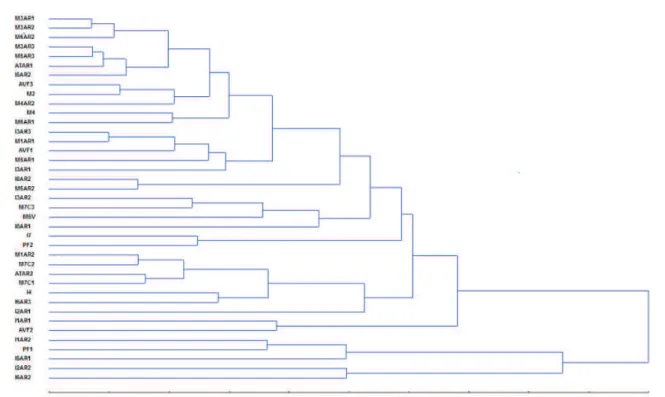
From the morphological characterization of the 39 isolates, the data related to the sporulation, mycelial growth, length and width of the macronidia were submitted to the UPGMA clustering method and, through the generated dendrogram, the isolates were grouped by the similarity between the average of the characters. By examining the dendrogram (Figure 1) the formation of the described seven major groups may be observed (Table 3).
Also in the dendrogram (Figure 1), note that the dissimilarity among the Group 7 isolates was greater
than 70%, which shows significant differences between
(26.77%); the remaining variables practically did not contribute to the divergence of the isolates.
The phylogenetic dendrogram was constructed by comparing the alignment of TEF-1α sequences of the isolates of this study with the sequences deposited at GenBank. The sequencing of the TEF-1α (Figure
2) identified the isolated I1AR1, I8AR1, M3AR2, M4,
M5AR2, M7C1 as F. solani and I6AR2 as F. oxysporum.
The high values of the bootstrap support, 99 and 100% respectively, increased the reliability of the alignment.
The sequencing of the TEF-1α region promoted
a more extensive intraspecific differentiation since the
F. solani isolates were distributed in different branches. The ones obtained in Machadinho (except M7C1) were assigned to the closest clades. Among the ones collected
in Ilópolis, the similarity was 88%.
TABLE 2 Morphological characteristics of isolates of Fusarium spp. obtained from symptomatic plants of Ilex paraguariensis.
Isolated Spo. (x106)1 M.G.R1
Macroconids1 Colony pigmentation1
Clamyd.2,3
Length. (µm)
Width. (µm)
Septa
No. Colony
Aerial mycelium I1AR1 4.02 6.69 24.02 4.95 3 or 4 Dark yellow White + I1AR2 21.03 9.29 36.03 2.90 3 or 4 Purple White + I2AR1 4.59 4.85 48.90 4.70 5 or 6 Brown White + I2AR2 69.66 9.95 37.10 4.65 3 Purple White + I3AR1 3.36 5.95 39.60 5.00 3-4-5 Purple White + I3AR2 7.54 9.62 36.10 4.82 3 or 4 Purple White + I3AR3 5.52 10.31 40.96 5.38 4 or 5 Olive green White + I4 16.30 6.57 58.90 5.20 5-6-7 Yellow White + I5AR1 14.35 11.15 31.00 4.05 3 Purple White -I5AR2 3.09 7.39 42.90 4.22 5 Yellow White + I6AR2 45.96 10.02 33.10 4.10 3 Purple White + I6AR3 19.85 7.23 55.70 4.60 5-6-7 Yellow tan White + I7 2.57 7.74 37.70 6.15 3-4-5 Yellow White + I8AR1 6.08 7.05 37.80 4.90 3 or 4 Yellow tan White + I8AR2 11.32 6.36 39.70 3.85 3 or 4 Olive yellow White + ATAR1 5.70 7.20 40.80 5.15 3 or 4 Pale brown White + ATAR2 5.32 7.47 51.00 4.65 4 or 5 Yellow tan White + PF1 17.05 9.39 28.40 3.55 3 Purple White + PF2 4.80 8.87 39.80 6.35 3-4-5 Yellow White + AVF1 11.32 7.45 38.20 4.40 3 or 5 Pale brown White + AVF2 4.68 6.96 30.90 5.75 3 Olive yellow White + AVF3 2.75 8.40 40.00 5.05 3 Olive yellow White + M1AR1 6.64 6.72 38.40 4.70 4 or 5 Dark brown White + M1AR2 6.39 7.30 47.90 4.95 5 Dark brown White + M2 2.73 7.92 38.40 4.93 3 or 4 Yellow White + M3AR1 8.00 7.90 45.50 5.03 3 or 4 Yellow White + M3AR2 6.39 7.30 46.30 5.10 3 or 4 Yellow White + M3AR3 8.72 7.48 41.50 5.12 3-4-5 Yellow tan White + M4 15.90 9.05 43.60 4.68 3 or 4 Yellow tan White + M4AR2 2.19 9.03 42.50 5.05 3-4-5 Light yellow White + M5AR1 2.60 6.95 31.80 4.75 3 or 4 Dark yellow White + M5AR2 14.84 6.48 42.40 4.10 3-4-5 Dark nut-brown White + M5AR3 9.16 7.32 40.90 4.95 3 or 4 Yellow White + M5V 6.45 8.38 32.50 4.28 3 or 4 Yellow White + M6AR1 13.85 8.49 40.60 5.05 3 or 4 Yellow White + M6AR2 11.88 7.66 44.20 5.10 3-4-5 Yellow White +
The main symptoms observed were the yellowing of the apex and leaves, yellowing followed by shrouding, yellowing accompanied of shrouding and subsequent leaf necrosis and leaf yellowing and necrosis. Within 30 days of the pathogenicity test, with the emergence of
reflex symptoms, a stagnation of seedlings growth was
perceived. In some cases, additionally to the growth stagnation, leaf fall and wither, it was possible to verify the death of plants. When removed from the substrate, the roots showed to be little evolved and dark, suggesting the
intimate relationship between the reflex symptoms and
the root rotting caused by the attack of F. oxysporum and
F. solani present in the substrate (Figure 3). All Fusarium
spp. isolates used in the seedlings of the yerba-mate
pathogenicity test were re-isolated, confirming their
pathogenicity and complying with the Koch’s Postulate.
DISCUSSION
In the taxonomy of fungi, it is of fundamental importance the stability of morphological characters in changing environmental conditions (TAYLOR et al.,
2001). Yet, the morphological identification of species of
Fusarium, in addition to being a task of difficult execution,
can generate ambiguity due to the variability of phenotypic
characters used in the taxonomic classification of this
genus (NELSON et al., 1983; LEWIS, 2000). However, Lima and Furtado (2007) emphasize that the situation
above does not prevent the species identification to
continue being made in a phenotypic approach, based on morphological and cultural characteristics. Poletto et al. (2006) succeeded in identifying species of Fusarium using morphological characters, such as macroconidia shape and size, microconidia and chlamydospores formation, following the methodology described by Lewis (2000).
Furthermore, in the present study, the physiological and morphological characters were relevant in the separation of the isolates into groups of similarity, mainly, sporulation and length of tuberculate. Lazarotto et al. (2014) considered valid the morphological characters of
FIGURE 1 Dendrogram showing the percentage of dissimilarity among 39 Fusarium spp. isolates. The dendrogram was obtained by UPGMA technique, from the analysis of the Euclidean distance matrix, with the four morphological characters (sporulation, mycelial growth, macronidia length and width).
TABLE 3 Groups of Fusarium spp. isolates based on the UPGMA clustering method.
Groups Isolates
1 M3AR1, M3AR2, M6AR2, M3AR3, M5AR3, ATAR1 and I5AR2
2 AVF3, M2, M4AR2, M4 and M6AR1
3 I8AR2, M5AR2, I8AR1, M1AR1, AVF1, M5AR1 and I3AR1
4 I3AR2, M7C3, M5V and R3AR3
5 I7, PF2, I1AR1 and AVF2
6 M1AR2, M7C2, ATAR2, M7C1, I4, I6AR3 and I2AR1
observed the formation of three groups among 12 isolates used in the study. The authors also reported that the macroconidia width was responsible for 99.98% of the divergence between the Fusarium spp. isolates. According to Walker et al. (2016), the multivariate analysis UPGMA
was efficient in the separation of 40 isolates of Cladosporium
spp. obtained from pecan nut into 5 groups with 30% of dissimilarity. According to the authors mentioned above, among the characters used in the analysis (mycelial growth, sporulation, conidia and ramoconides width and length) sporulation was responsible for the greatest relative contribution to the divergence (96.13%) followed by the mycelial growth (2.37%).
In the present study, the sequencing of the TEF-1α supported by high bootstrap values gave reliability
to the identification at the species level of pathogenic
isolates of Fusarium spp. The diffusion of the TEF-1α use may be associated to its high degree of information at the species level and to the fact that there have been
FIGURE 2 Phylogenetic dendrogram based on the neighbor-joining method from DNA sequences in the elongation factor 1-α (TEF-1α) region. The numbers on the branches indicate the bootstrap analysis percentage of repeats in which the repetitions were observed (1,000 replications). (*): Fusarium spp. isolates obtained in the present study. A sequence of Lasodiplodia subglobosa
was used as an outgroup.
FIGURE 3 Pathogenicity test in yerba-mate seedlings, 180 days after contact with substrate inoculated with Fusarium spp.: Reflex symptoms: (A) yellowing; (B) yellowing +
shrouding (C) yellowing + shrouding + necrosis; (D) yellowing + necrosis.
colony diameter, sporulation, macroconidial width and
length, for the grouping of pathogenic isolates of Fusarium
reports about the appearance of non-orthologue copies, as well as by the availability of ‘universal primers’ based on this gene (Geiser et al., 2004). For O’Donnell et al. (1998) and Seifert and Levesque (2004), studies involving genomic regions, performed with Fusarium
spp. demonstrated that the region of TEF-1α may be applied as an acceptable tool to distinguish fungus species, possessing a correlation of the Fusarium genus with mycotoxigenic capacity. Kristensen et al. (2005) emphasize the importance of the TEF-1α phylogenetic analysis because they have concluded that this is able to identify species of monophyletic Fusarium.
According to Du et al. (2012), the region TEF-1α was efficient in the classification, into four distinct
groups of species, when they sequenced 41 isolates of Fusarium since the similarity indices were higher than 95%. Hsuan et al. (2011) were successful in the
identification of isolates of Fusarium sp., belonging to the complex Gibberella fujikuroi obtained from maize (Zea mays), rice (Oryza sativa) and sugarcane (Saccharum
officinarum) in Malaysia. According to the authors cited, the sequencing of the TEF-1α consolidates itself as an important tool for the differentiation of the species of the Fusarium genus, supported by high bootstrap values. The polymorphism revealed by the analysis of the TEF 1-α was sufficient to discriminate even closely
related species of Fusarium. Furthermore, the resulting dendrogram was generated in a clear way, with good structure and resolution in branches distribution (Chandra et al., 2011; Nitschke et al., 2009).
Grigoletti Junior and Auer (2001) reported,
for the first time, the root rotting caused by Fusarium
spp. in mature plants of yerba-mate from plantations in the State of Paraná, Brazil. According to the authors, the trees had symptoms such as yellowing and loss of leaves, stagnation of growth, dark roots, and in more severe cases, death. Similar symptoms were mentioned by Poletto et al. (2006) and Poletto et al. (2007) where pathogenic isolates demonstrated, initially, yellowing and leaf fall, evolving to drought and death of plants. Still, according to the authors, the symptoms are typical in coppices distributed through the planting area. Poletto et al. (2015) observed that Fusarium spp. isolates pathogenic to yerba-mate produced signs of necrotic spots on the root system and darkening at the tips of the roots. Lazarotto et al. (2014) obtained similar results
to this study, with similar reflex symptoms in the plants
analyzed. According to the authors, isolates of Fusarium
spp. pathogenic to Carya illinoinensis triggered symptoms such as damping off, foliar necrosis, wilt of the aerial
part and plant death, consolidating the typical root rotting symptoms. Silva et al. (2011) exemplify that the colonization of the black pepper roots (Piper nigrum) by
Fusariumsolani f. sp. piperis reflects on the aerial part, the
leaves become yellowish and flaccid, causing premature
fall and as the disease progresses, the death of the plant.
CONCLUSIONS
The macroconid length and sporulation morphological characters are effective in differentiating Fusarium spp. isolates in yerba-mate, into groups. The isolates I1AR1, I8AR1, M3AR2, M4, M5AR2, M7C1, and I6AR2 are pathogenic to yerba-mate. The TEF-1α region is efficient for identification at the species
level. In this study, we identified Fusarium solani and
Fusariumoxysporum as the cause the root rotting in Ilex paraguariensis in Rio Grande do Sul state.
ACKNOWLEDGMENTS
The authors would like to thank the Coordenação de Aperfeiçoamento de Pessoal de Nível Superior
(CAPES) for granting a Ph.D. scholarship to the first
author and the CNPq for the Research Productivity Scholarship of the coauthors.
REFERENCES
ALFENAS, A. C.; MAFIA, R. G. Métodos em fitopatologia. Editora UFV, 2007. 382p.
BURGESS, T.; MALAJCZUK, N.; DELL, B. Variation in Pisolithus
and basidiospore morphology, culture characteristics and analysis of polypeptides using 1D SDS-PAGE. Mycological Research, v. 99, n. 1, p. 1-13, 1995.
CHANDRA, N. S.; WULFF, E. G.; UDAYASHANKAR, A. C.; NANDINI, B. P. NIRANJANA, S. R.; MORTENSEN, C. N.; PRAKASH, H.S. Prospects of molecular markers in
Fusarium species diversity. Applied Microbiology and
Biotechnology. v. 90, n. 5, p. 1625-1639, 2011.
CRUZ, C.D. Programa genes: diversidade genética. Universidade Federal de Viçosa, Viçosa, 2008.
DOYLE, J. J.; DOYLE, J. L. Isolation of Plant DNA from Fresh Tissue. Focus, v. 12, n. 1, p. 13-15, 1990.
DU, M.; REN, X.; SUN, Q.; WANG, I.; ZHANG, R. Characterization of Fusarium spp. causing potato dry rot in China and susceptibility evaluation of Chinese potato germplasm to the pathogen. Potato Research, v. 55, n. 2, p. 175–184, 2012.
FERREIRA, D.F. SISVAR: um programa para análises estatísticas e ensino de estatística. Revista Científica Symposium, v. 6, n. 2, p. 36-41, 2008.
FOWLER, J. A. P.; STURION, J. A.; ZUFFELLATO-RIBAS, K. C. Variação do desenvolvimento embrionário das sementes de erva-mate. Pesquisa Florestal Brasileira, n.54, p.105-108, 2007.
GEISER, D. M.; JIMÉNEZ-GUASCO, M. M.; KANG, S.; MAKALOWSKA, I.; VEERARAGHAVAN, N.; WARD, T. J.; KULDAU, G. A.; O’DONELL, K. FUSARIUM-ID v.1.0: A DNA sequence database for identifying Fusarium. European Journal of Plant Pathology, v. 110, n. 5-6, p. 473-479, 2004.
GRIGOLETTI JUNIOR, A.; AUER, C. Podridão de raízes em erva-mate (Ilex paraguariensis A. St.-Hill.) causada
por Fusarium sp. Fitopatologia Brasileira, v. 26:
p.572, 2001.
HALL, T. A. BioEdit: a user-friendly biological sequence alignment editor and analysis program for Windows 95/98. Nucleic Acids SymposiumSeries, v.41, p.95-98, 1999.
HSUAN, H. M.; SALLEH, B.; ZAKARIA, L. h B. Molecular
identification of Fusarium species in Gibberella fujikuroi
species complex from rice, sugarcane and maize from Peninsular Malaysia. International Journal of Molecular Sciences, v. 12, n. 10, p. 6722-6732, 2011.
IBGE. Instituto Brasileiro de Geografia e Estatística. Produção Agrícola e Municipal, v. 42, p.1-57, 2015.
JUNGES, E.; MUNIZ, M. F. B.; MEZZOMO, R.; BASTOS, B.; MACHADO, R. T. Trichoderma spp. na produção de mudas
de espécies florestais. Floresta e Ambiente v. 23, n. 2, p. 237-244, 2016.
KLINGELFUSS, L.H.; YORINORI, J.T.; DESTRO, D. Métodos de inoculação para quantificação de resistência em soja a Fusarium solani f. sp. glycines, em casa-de-vegetação. Fitopatologia Brasileira, v. 32, n.1, p. 50-55, 2007.
KRISTENSEN, R.; TORP, M.; KOSIAK, B.; HOLST-JENSEN, A. Phylogeny and toxigenic potential is correlated in Fusarium
species as revealed by partial translation elongation factor 1 alpha gene sequences. Mycological Research, v. 109, n. 2, p. 173-186, 2005.
LAZAROTTO, M; MILANESI, P. M.; MUNIZ. M. F. B.; REINIGER, L. R. S.; BELTRAME, R.; HARAKAVA, R.; BLUME, E. Morphological and molecular characterization
of Fusarium spp. pathogenic to pecan tree in Brazil.
Genetics and Molecular Research, v.13, n. 4, p. 9390-9402, 2014.
LESLIE, J.; SUMMERELL, B. A. The Fusarium Laboratory Manual. Blackwell Publishing, 2006. 388p.
MICHEREFF, S. J.; NORONHA, M. A.; ROCHA JUNIOR, O. M.; SILVA, J. A.; MIZUBUTI, E. S. G. Variabilidade de isolados de Alternaria brassicicola no estado de Pernambuco. Fitopatologia Brasileira, v. 28, n.6, p. 656-663, 2003.
MUNSELL SOIL COLOR CHARTS. Macbeth Division of Kollinorgen Instruments Corporation, 2009.
NELSON, P. E.; TOUSSOUN, T. A.; MARASAS, W. F.O. Fusarium Species: An Illustrated Manual for Identification. Pennsylvania State University Press, 1983. 193p.
NITSCHKE, E.; NIHLGARD, M.; VARRELMANN, M. Differentiation of eleven Fusarium spp. isolated from sugar beet, using restriction fragment analysis of a Polymerase Chain
Reaction–Amplified Translation Elongation Factor 1α Gene Fragment. Phytopathology, v. 99, n. 8, p. 921-929, 2009.
POLETTO, I.; MUNIZ, M. F. B.; CECONI, D. E.; SANTIN, D.;
WEBER, M. N. D.; BLUME, E. Zoneamento e identificação
de Fusarium spp. causador de podridão-de-raízes em
plantios de erva-mate (Ilex paraguariensis A. St.-Hil.) na região do Vale do Taquarí-RS. Ciência Florestal, v. 16, n. 1, p. 1-10, 2006.
POLETTO, I.; MUNIZ, M. F. B.; CECONI, D. E.; WEBER, M. N. D; BLUME, E. Primeira ocorrência de Pythium sp. e Rhizoctonia sp. causando podridão-de-raízes em ervais no Rio Grande do Sul. Ciência Florestal, v.17, n. 1, p. 65-69, 2007.
POLETTO, I.; MUNIZ, M. F. B.; CECONI, D. E.; POLETTO, T. Aspectos epidemiológicos da podridão-de-raízes da erva-mate (Ilex paraguariensis). Ciência Florestal, v. 25, n. 2, p. 281-291, 2015.
REGO, S. S.; SANTOS, A. F.; NOGUEIRA, A. C.; KUNIYOSHI, Y. S. Detection, transmission and pathogenicity of fungi
on Blepharocalyx salicifolius (H.B.K.) Berg. Seeds. Revista
Brasileira de Sementes, v. 34, n. 1, p. 9-13, 2012.
REHNER, S.A.; BUCKLEY, E. A Beauveria phylogeny inferred from nuclear ITS and EF1-alpha sequences: evidence for
cryptic diversification and links to Cordyceps teleomorphs. Mycologia, v. 97, n. 1, p. 84-98, 2005.
SEIFERT, K. A.; LEVESQUE, C. A. Phylogeny and molecular diagnosis of mycotoxigenic fungi. European Journal of Plant Pathology, v. 110, n. 5-6, p. 449–471, 2004.
SILVA, B. S. O.; DRUMOND NETO, A. P; SILVA, M. B.
Pimenta-do-reino: importância da defesa fitossanitária para
a sustentabilidade da atividade na região norte do Espírito Santo. Revista Brasileira de Agropecuária Sustentável, v. 1, n.1, p. 88-92, 2011.
TAMURA, K.; DUDLEY, J.; NEI, M.; KUMAR, S. MEGA4:
Molecular Evolutionary Genetics Analysis (MEGA) software
version 4.0. Molecular Biology and Evolution v. 24, n. 8,
p. 1596-1599, 2007.