_____________________________________________________________________________________________________________________________________________ (*)
Autor para correspondência/Corresponding author: Rua Dom Manoel de Medeiros, s/n, Dois Irmãos-CEP: 52171-900- Recife, Pernambuco-Brasil. e-mail:ninascanoni@yahoo.com.br
Histopathological assessment of the morphology and pigmentation of the skin of
dogs naturally infected by Leishmania infantum
[Avaliação histopatológica da morfologia e pigmentação da pele de cães com infecção
natural por Leishmania infantum]
“Artigo Científico/Scientific Article”
CS Maia1*, EM Sá Santos2, GGA Gonçalves3, FAB Santos3, RJR Padilha3 e LC Alves2 1 Departamento de Morfologia e Fisiologia Animal, Programa de Pós-Graduação em Biociência Animal/UFRPE, Recife, PE, Brasil. 2 Departamento de Medicina Veterinária, Laboratório de Doenças Parasitárias, /UFRPE, Recife, PE, Brasil.
3 Laboratório de Imunopatologia Keizo Azami/UFPE, Recife, PE, Brasil.
_______________________________________________________________________ Resumo
A pele representa o principal órgão envolvido na progressão da Leishmaniose Visceral Canina. O objetivo deste estudo foi para avaliar as alterações morfológicas e pigmentares da pele de cães com Leishmania infantum. Fragmentos de pele íntegra, lesionada e hiperpigmentada da região abdominal foram retirados com auxílio de um punch, de cada um dos 11 cães avaliados. Posteriormente, os tecidos coletados foram submetidos a técnicas de rotina histológica para análise em microscopia de luz e microscopia eletrônica de transmissão. Foi constatado infiltrado de células mononucleares na derme lesionada e formas amastigostas de Leishmania infantum no interior dos macrófagos de 81,8% (9/11) dos cães. Paraceratose e hiperqueratose foram observadas na pele lesionada de 54,5% (6/11) dos cães. Excesso de grânulos de melanina foi constatado tanto na epiderme como na derme da pele hiperpigmentada de 36,3% (4/11) dos cães. Na eletromicrografia, foi possível visualizar uma grande quantidade de grânulos de melanina ao redor do núcleo dos queratinócitos da camada espinhosa. Os resultados deste estudo sugerem uma possível relação entre mediadores inflamatórios produzidos na Leishmaniose Visceral Canina e o estímulo da melanogênese.
Palavras-chave: Leishmaniose Visceral Canina, pele, hiperpigmentação. Abstract
The skin is the main organ involved in the disease progression of Canine Visceral Leishmaniasis. The aim of this study was to assessment the morphological changes and pigmentary skin of dogs Leishmania infantum. Fragments of intact skin, and hyperpigmented lesions in the abdominal region were removed with the aid of a punch, each of the 11 dogs evaluated. Subsequently, the tissues collected were subjected to routine histological techniques for analysis in light microscopy and transmission electron microscopy. Was found mononuclear cell infiltrate in the dermis lesions and amastigostas forms of Leishmania infantum within macrophages of 81.8% (9/11) dogs. Parakeratosis and hyperkeratosis was observed in lesional skin of 54.5% (6/11) dogs. Excess melanin granules was noted both in the epidermis and in dermis of the skin hyperpigmented 36.3% (4/11) dogs. In the electron micrographs, it was possible to display a large amount of melanin granules around the nucleus of keratinocytes of the stratum spinosum. The results of this study suggest a possible relationship between inflammatory mediators produced in Canine Visceral Leishmaniasis and stimulation of melanogenesis.
Key words: Canine Visceral Leishmaniasis, skin, hyperpigmentation.
_____________________________________________________________________________ Introduction
Canine Visceral Leishmaniasis (CVL) is a severe systemic disease and in some
cases it can be fatal (GONTIJO and MELO 2004). Caused by the protozoan Leishmania. Infantum, it is transmitted by
the vector Lutzomya longipalpis (MISSAWA and LIMA, 2006) and is considered an immune-mediated disease due to alterations in the T and B cells, inducing large formation of circular immunocomplexes that are deposited on the walls of blood vessels causing vasculitis, uveitis, glomerulonephritis and arthritis (NOLI, 1999).
Dogs infected by L. infantum present a wide spectrum of clinical signs ranging of a health condition apparent to a severe form (FERRER, 1999). Despite de viscerotropic nature of the parasite, the skin represents the main organ involved in the progression of the disease (SILVA, 2007) and 90% of affected dogs, present skin lesions (SOLANO-GALLEGO et al., 2004. It is also possible to find high amount of parasites in infected dogs with normal skin. (SANTOS et al. 2010).
The most reported dermatological alterations are: exfoliative dermatitis, hyperkeratosis, ulcers and alopecia (SCOTT et al., 2001; TORRES-NETO et al., 2008). However, hyperpigmentation of the skin can also be present (SLAPPENDEL, 1988; AMUSATEGUI et al., 2003), although its histological description and association to CVL are little mentioned in the literature.
The aim of the study was to conduct a histopathological assessment to verify the morphological and skin pigmentation changes of dogs (Canis familiaris) naturally infected by Leishmania infantum.
Methods
Cylindrical fragments (punch 4 mm) of healthy, injured and hyperpigmentated skin were removed from the abdominal region, in each of the 11 evaluated dogs. The dogs were of different breeds and ages, of both sexes and they were from different municipalities in the state of Pernambuco, Brazil. All procedures were performed at the Hospital for Veterinary Medicine of the Universidade Federal Rural de Pernambuco (UFRPE) and after removed fragments, the dogs were euthanized according to current law in 2012.
All dogs used in the present study were
naturally infected by L. infantum, presented skin lesions, and were positive in the enzyme-linked immunosorbent assay (ELISA) and in the parasitological test of bone marrow.
To process the material that were observed by light microscopy, the skin fragments were initially fixed in 10% tamponade formalin. After fixation for 24 hours, the samples were dehydrated in increasing alcoholic solutions, classified with xilol, and saturated and included in paraffin. Later, the blocs obtained were cut in adjusted microtome to a thickness between 5μm and 3 μm, and mounted onto glass blades smeared with Meyer’s albumin. After this, the blades were stained with Hematoxylin-Eosin (HE) and mounted with coverslips and Entellan resin.
For the ultrastructural study of the skin fragments by transmission electron microscopy (TEM), the fragments were initially fixed in a glutaraldehyde solution of 4% in cacodylated buffer 0.1M, pH 7.4. Later, the pre-infiltration was carried out in a mixture of propylene oxide and Epon resin-812. The material was placed inside a heater (estufa) at 60C for 72 hours, for resin polymerization.
The blocs that were obtained were trimmed (aparados) and selected using an ultramicrotome and glass knives, obtaining semi-fine cuts of a thickness of 0.5 m. Later, these cuts were stained by an equal mixture of 1% AZUR II in distilled water and 1% methylene blue, and immediately after they were washed in water and after drying they were examined in a common optical microscope.
After selecting the area to be studied, the blocs were submitted to ultrafine cuts of a thickness of 40 to 80 mm, by using a diamond knife. The cuts obtained were mounted on copper mesh grids and contrasted with alcohol solution of 2% uranyle acetate, and underwent final treatment with lead citrate. After these procedures, the screens (telas) were taken to the TEM and electro micrographs obtained. The processing of skin fragments for light and electron microscopy were
performed at the Laboratory of Immunopathology Keizo Asami (LIKA) of the Universidade Federal de Pernambuco (UFPE).
To execute the experiment, the invasive procedures with the use of the punch were carried out after approval by the Comissão de Ética para Uso de Animais (CEUA) of the UFRPE, according to license 010/2011.
Results
In the derm of the lesioned abdominal skin of 81.8% (9/11) of the dogs, inflammatory infiltrate composed of macrophages, lymphocytes and plasmocytes (Figure 1) was found, as well as amastigote forms of L. infantum in the interior of the macrophages (Figure 2).
Figure 1. Photomicrograph of abdominal skin of dog with LV, dermis (De) presenting cells infiltrate monucleares (circular area). Epidermis (Ep). H-E.
Figure 2. Dermis (De) with macrophages with amastigotes of L. infantum.
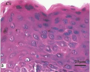
On the other hand, in the epidermis with lesion skin of 54.5% (6/11) of dogs, the alterations observed were parakeratosis, characterized by the presence of nuclei in
the stratum corneum (Figure 3) and hyperkeratosis with innumerous keratosis-hyaline presents in the cytoplasm of the keratinocytes of the granular layer in some skin fragments.
Figure 3. Abdominal skin dog with LV. Epidermal parakeratosis presenting with presence of nuclei in the stratum corneum (or Cc Ec).
When comparing fragments of healthy abdominal skin (Figure 4) with those of stained hyperpigmentation by H.E., a greater quantity of melanin granules in the keratinocytes of the basal and spinous
layers (Figure 5) was seen in the epiderm of 36.3% (4/11) of dogs with CVL. In the derm the presence of many melanophages was observed.
Figure 4. Photomicrograph of abdominal skin healthy dog with LV. We observe a small amount of melanin granules (brown) in the basal and spinous layers of the epidermis. H-E. F.
Figure 5. Photomicrograph of abdominal skin with hyperpigmentation dog with LV.
In the electro micrograph it was possible to observe the granules around the keratinocyte nuclei of the spinous layer of
dogs with hyperpigmentation (Figures 6 and 7).
Figures 6 and 7. Electron abdominal skin with hyperpigmentation dog. Keratinocytes of the spinous layer with melanin granules around the nucleus in keratinocytes.
Discussion
The infiltration of mononuclear cells observed in this study corroborate Costa et al. (2008) and Calabrese et al. (2010) which describe the same inflammatory pattern in lesional skin of dogs with VL. However, they differ from results found by Torres-Neto (2008) and Ferrer et al. (1988) who found a predominant pyogranulomatous reaction and eosinophils, in the tissue inflammatory process.
The changes found in the epidermis corroborate with Santos et al. (2004), Solano-Gallego et al. (2004) Giunchetti et al. (2006), who claim that in addition to acanthosis and hyperkeratosis. They describe histopathological features of parakeratosis and cell degeneration in the skin of dogs with CVL. However, they disagree with the reports of Luvizotto (2006), who states that animals infected with VL present an orthokeratotic hyperkeratosis in the epiderm which could
be associated to hyperplasia and hyperpigmentation.
Regarding to the presence of pigmentation in the skin, the results indicate the described findings by Pandya e Guevara (2000) and García (2010), about the various cutaneous inflammatory processes that can stimulate the melanogenesis, causing hyperpigmentation in the referred organ. Moreover, according to Alvar et al. (2004), Amusategui et al. (2003) and Baneth (2006), in CVL the hyperpigmentation is related to inflammatory lesions. The clinical signs in dogs are related to the type of immune responses developed (MARCONDES et al., 2011). It is noteworthy that in the immunopathogenesis of CVL, that Th1 lymphocytes activate macrophages to produce nitric oxide (NO) and promotes apoptosis of cells with parasites (HOLZMÜLLER et al., 2006), stimulating melanogenesis in the skin (SLOMIŃSKI et al. 2004).
However, in human beings, some melanoderms are characterized by the increase of melanophages in the derm, resulting in the hyperpigmentation of the skin as a response to a chronic inflammatory process (NICOLETTI et al., 2002). It is probable that the local inflammatory response observed in the skin of animals with CVL may cause similar clinical profile (DAY, 2008) initially measured by neutrophils, eosinophils, macrophages, ‘natural killer’ cells (NK) and lymphocytosis.
Regarding the organization of melanin granules in keratinocytes model, the observations are compatible with reports by Banks (1992), Monteiro-Riviere et al. (1993) and Bal (1996), who state that the melanosomas accumulate in the cytoplasm of the keratinocytes, mainly around the nucleus.
Conclusion
Histopathological changes were found in the epidermis and/or dermis of 100% (11/11) the dogs with AVL in the study. However, areas of hyperpigmentation were found only in
36,3% (4/11) of the animals being recommended for further studies to investigate a possible relationship between the production of specific inflammatory mediators in CVL and stimulation of melanogenesis.
Acknowledments
Our thanks to the National Counsel of Scientific and Technological Development (CNPQ) with funding for the project, and all the faculty and students of UFRPE who collaborated with development of this study.
References
ALVAR, J. et al. Canine leishmaniasis.
Advancesin Parasitology, Madrid, v. 57, n.1, p.
1-87, 2004.
AMUSATEGUI, I. et al. Distribution and
relationships between clinical and
biopathological parameters in canine
leishmaniasis. European Journal of
Epidemiology, Madrid, v. 18, n. 2, p. 147-156,
2003.
BAL, H.S. Fisiologia dos Animais Domésticos. Rio de Janeiro: Guanabara Koogan, 1996. 856p.
BANKS, W.J. Histologia Veterinária
Aplicada. São Paulo: Manole, 1992. 629p.
BANETH, G. Leishmanasis. In: GREENE, C.
E. Infectious diseases of the dog and cat. 3rd
ed. Canada: Saunders Elsevier, 2006. p. 685-698.
CALABRESE et al. Leishmania (Leishmania) infantum/chagasi: Histopathological aspects of the skin in naturally infected dogs in two endemic areas. Experimental Parasitology, Rio de Janeiro, v. 124, n. 3, p. 253–257, 2010. COSTA, M. M. et al. Cervical, mandibular, and parotid lymph nodes of dogs naturally infected with Leishmaniainfantum: a histopathologic and immunohistochemistry study and its correlation with facial skin lesions. Veterinary Pathology, Belo Horizonte, v. 45, n. 5, p. 613-616, 2008. DAY, M.J. Clinical immunology of the dog and cat. The Veterinary Press, p. 288, 2006. FERRER, L. et al. Skin lesions in canine leishmaniasis. Journal of Small Animal
Practice, Barcelona, v. 29, n. 6, p. 381-388,
1988.
FERRER, L. M. Clinical aspects of canine leishmaniasis. Canine Leishmaniasis: an update.
Proceedings of a Canine Leishmaniasis.
GARCÍA, E. S. et al. Immune homeostasis to microorganisms in the guts of triatomines
(Reduviidae): A Review. Memórias do
Instituto Oswaldo Cruz, Rio de Janeiro, v.
105, n. 5, p. 605-610, 2010.
GIUNCHETTI, R. C. et al. Relationship between canine visceral leishmaniosis and the Leishmania (Leishmania)chagasi burden in
dermal inflammatory foci. Journal of
Comparative Pathology, Ouro Preto, v. 135, n.
2-3, p. 100-107, 2006.
GONTIJO, C.M.F.; MELO, M.N. Leishmaniose visceral no Brasil: quadro atual, desafios e
perspectivas. Revista Brasileira de
Epidemiologia, v. 7, p. 338-349, 2004.
HOLZMULLER, P. et al. Phenotypical
characteristics, biochemical pathways,
molecular targets and putative role of nitric oxide-mediated programmed cell death in Leishmania. Parasitology, Cambridge, v. 132, p. 19-32, 2006.
LUVIZOTTO, M. C. R. Diagnóstico da
Leishmaniose Visceral Canina. Manual
Técnico Leishmaniose Visceral Canina.
Campinas: Fort Dodge, 2006, p. 28-29.
MARCONDES, M. et al. Validation of a Leishmania infantum ELISA rapid test for serological diagnosis of Leishmania chagasi in dogs. Veterinary Parasitology, Philadelphia, v. 175, n. 1-2, p. 15-19, 2011.
MISSAWA, N. A.; LIMA, G. B. Distribuição espacial de Lutzomyialongipalpis (Lutz & Neiva, 1912) e Lutzomyiacruzi (Mangabeira, 1938) no Estado de Mato Grosso. Revista da
Sociedade Brasileira de Medicina Tropical,
Cuiabá, v. 39, n. 4, p. 337-340, 2006.
MONTEIRO-RIVIERE, N.A., STINSON,
A.W.; CALHOUN, H.L. In: Dieter-Dellmann H. Textbook of Veterinary Histology. 4th ed. Philadelphia: Lea and Febiger. 1993. 351p. NOLI, C. Canine leishmaniasis. Waltham
Focus, v. 9, n. 2, p. 16-24, 1999.
PANDYA, A.G.; GUELVARA, J.L. Disorders of hiperpigmentation. Dermatologic Clinics, v.37, n.1, p. 91-98, Detroit, 2000.
SANTOS, J.M.L. et al. Prevalence of anti-leishmania spp antibodies in dogs from Garanhuns, in the middle scrub zone (agreste)
of Pernambuco. Revista da Sociedade
Brasileira de Medicina Tropical, Garanhuns,
v.43, n.1, p. 41-45, 2010.
SANTOS, W. L. C. et al. Association between
skin parasitism and a granulomatous
inflammatory pattern in canine visceral
leishmaniosis. Parasitology Research,
Salvador, v. 92, n.2, p. 89-94, 2004.
SCOTT, D. W. et al. Small Animal
Dermatology. 6th ed. Philadelphia: Saunders,
2001. 1528p.
SILVA, F. S. Patologia e patogênese da leishmaniose visceral canina. Revista Tropical
– Ciências Agrárias e Biológicas, Chapadinha,
v. 1, n. 1, p. 31, 2007.
SLAPPENDEL, R. J.; FERRER, L.
Leishmaniasis. In: GREENE, C. E. Clinical
Microbiology and Infectious Diseases of the Dog and Cat. Philadelphia: W. B. Saunders,
1990, p. 450-458.
SLOMINSKI, A. et al. Melanin pigmentation in mammalian skin and its hormonal regulation.
Physiological Reviews, Tennessee, v. 84, n. 4,
p. 1155-1228, 2004.
SOLANO-GALLEGO, L. et al. Histological and immunohistochemical study of clinically normal skin of Leishmania infantum-infected dogs. Journal of Comparative Pathology, Barcelona, v. 130, n. 1, p. 7-12, 2004.
TORRES-NETO, R. et al. Padrões
histopatológicos das lesões descamativas e ulcerativas da pele em cães com leishmaniose.
Ciências Agrárias, Londrina, v. 29, n. 3, p.