Exercise stress testing before and after
successful multivessel percutaneous
transluminal coronary angioplasty
Instituto do Coração, Faculdade de Medicina, Universidade de São Paulo,
São Paulo, SP, Brasil W.A. Chalela, J.C. Kreling,
A.M. Falcão, W. Hueb, P.J. Moffa, P.L.A. Pereyra and J.A.F. Ramires
Abstract
Controversy exists regarding the diagnostic accuracy, optimal tech-nique, and timing of exercise testing after percutaneous coronary intervention. The objectives of the present study were to analyze variables and the power of exercise testing to predict restenosis or a new lesion, 6 months after the procedure. Eight-four coronary multi-artery diseased patients with preserved ventricular function were studied (66 males, mean age of all patients: 59 ± 10 years). All underwent coronary angiography and exercise testing with the Bruce protocol, before and 6 months after percutaneous coronary interven-tion. The following parameters were measured: heart rate, blood pressure, rate-pressure product (heart rate x systolic blood pressure), presence of angina, maximal ST-segment depression, and exercise duration. On average, 2.33 lesions/patient were treated and restenosis or progression of disease occurred in 46 (55%) patients. Significant increases in systolic blood pressure (P = 0.022), rate-pressure product (P = 0.045) and exercise duration (P = 0.003) were detected after the procedure. Twenty-seven (32%) patients presented angina during the exercise test before the procedure and 16 (19%) after the procedure. The exercise test for the detection of restenosis or new lesion pre-sented 61% sensitivity, 63% specificity, 62% accuracy, and 67 and 57% positive and negative predictive values, respectively. In patients without restenosis, the exercise duration after percutaneous coronary intervention was significantly longer (460 ± 154 vs 381 ± 145 s, P = 0.008). Only the exercise duration permitted us to identify patients with and without restenosis or a new lesion.
Correspondence
W.A. Chalela InCor, FMRP, USP Av. Dr. Enéas C. Aguiar, 44 05403-000 São Paulo, SP Brasil
Fax: +55-11-3069-5328 E-mail: [email protected]
Publication supported by FAPESP.
Received May 19, 2005 Accepted November 29, 2005
Key words
•Exercise test
•Coronary artery disease •Percutaneous transluminal
coronary angioplasty
•Coronary multi-artery
disease
Introduction
The American College of Cardiology/ American Heart Association guidelines for exercise testing suggest that functional test-ing should not be performed routinely in all patients after percutaneous coronary
because silent ischemia has been reported to be a predictor of mortality and cardiac events in patients with coronary artery dis-ease (2-5). Coronary angiography (CA) has been traditionally used as the gold standard for detecting restenosis. However, the cost and invasive nature of angiography have prompted the search for a simpler noninva-sive test that could reliably detect restenosis. The standard exercise treadmill test (ETT) is safe, simple, and the most readily avail-able of these tests. Unfortunately, previous studies on the value of ETT for detecting restenosis have yielded conflicting results (6-8).
Thus, the purpose of the present study was to compare ETT before and after PCI and determine the diagnostic value of ETT for detecting restenosis in patients with multi-vessel coronary artery disease.
Material and Methods
Study population
The study group consisted of 84 patients with multivessel disease who underwent elec-tive PCI as part of the medical, angioplasty and surgical study (9). Selection criteria were: stable angina pectoris, ejection fraction >40% on echocardiography, and at least two criti-cal stenoses (internal diameter reduction >70% by visual assessment) in the proximal two-thirds of two major epicardial coronary artery. Only patients not treated with digoxin, who were in sinus rhythm and without bundle branch block, nonspecific intraventricular block patterns, or any other electrocardio-graphic abnormalities precluding an accu-rate resting of the ST segment during ETT were included. Nitrate preparations other than sublingual nitroglycerin and calcium entry blocking agents were withdrawn 4 days before the study, and ß-blocking agents 5 days before, unless withdrawal was consid-ered to be contraindicated by the patient’s physician. All patients underwent CA and
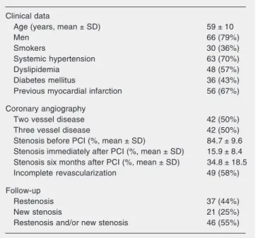
ETT before and 6 month after PCI. The study protocol was approved by our institu-tional Ethics Committee and written informed consent was obtained from all patients. Table 1 summarizes the clinical data of the pa-tients.
Coronary angiography and percutaneous coronary intervention
CA and PCI were performed according to standard techniques. All patients under-went CA before and 6 months after PCI. Selective left and right coronary arteriogra-phies and ventriculograarteriogra-phies were performed according to the techniques of Sones Jr. and Shirey (10) and Seldinger (11). All CA were reviewed independently of the clinical data by 2 investigators who were blind to the results of the physiological tests. Coronary arteries were submitted to revascularization only in the event of an initial narrowing of
≥70%. A successful PCI was defined as dilation of the stenosis, resulting in a final narrowing of <50%. Patients were consid-ered to have undergone incomplete revascu-larization if they had stenosis of ≥50% in any coronary artery not submitted to revas-cularization. Stent implantation and multivessel PCI were performed in all pa-tients. At follow-up, restenosis was defined as a ≥50% luminal diameter stenosis and progress of coronary artery disease when new stenosis was observed in the CA after PCI. The angiographic data are summarized in Table 1.
Exercise test
base-line and R wave of equal amplitude. A hori-zontal depression of ≥0.1 mV for men and of
≥0.2 mV for women occurring with or with-out chest pain was considered to be a posi-tive electrocardiographic result. End points for terminating exercise were severe angina, dyspnea, extreme fatigue, ST-segment de-pression of ≥0.3 mV, maximal aged-related heart rate, and the occurrence of other harm-ful conditions such as hypotension, severe arrhythmia, and dyspnea. Electrocardio-graphic recordings of all tests were evalu-ated independently in a blind fashion by two cardiologists; in case of disagreement, the matter was resolved by consensus.
The following parameters were meas-ured: heart rate, blood pressure (systolic and diastolic), rate-pressure product (heart rate x systolic blood pressure), presence of angina, maximal ST-segment depression, and exer-cise duration in seconds.
Statistical analysis
Categorical values are reported as fre-quencies and continuous variables as means ± SD. The paired Student t-test or unpaired
Wilcoxon test was used to compare quanti-tative variables before and after PCI. Uni-variate profile analyses were performed to determine the presence of restenosis or of a new stenosis by the ETT. Results of the ETTs were compared with CA data, and calculations of sensitivity, specificity posi-tive and negaposi-tive predicposi-tive values, and di-agnostic accuracy were performed. For these last variables, patients who did not achieve an 85% age predictive heart rate were not considered. A P value <0.05 was considered to be statistically significant.
Results
Clinical and coronary angiography data
The baseline clinical and angiographic characteristics of the study are listed in Table
1. A total of 84 patients (66 men) with an average age of 59 ± 10 years were studied. Fifty-six patients (67%) had Q-wave infarct-related vessel PCI in 56 culprit vessels (≥1 month myocardial infarction). Double ves-sel disease occurred in 42 patients (50%) and triple vessel disease in another 50%. Stent implantation and multivessel PCI were performed in all patients. The diameters of stenosis before and immediately and 6 months after PCI were 84.7 ± 9.6, 15.9 ± 9.6, and 34.8 ± 18.6%, respectively. The num-bers of stenoses treated per patient were 2.33 ± 0.87. The treated artery was the left ante-rior descending one in 68 (41%), the right coronary artery in 55 (33%) and left circum-flex artery in 44 (26%) cases. Revasculari-zation was complete (no stenosis >50% in any epicardial coronary artery) in 35 pa-tients (42%). PCI was successful in all of our patients. Angiographic restenosis and new stenosis were detected in 37 patients and 53 vessels (32%) and in 21 patients (25%) and 24 vessels, respectively. Restenosis and/or new stenosis was detected by CA in 46 patients (55%) 6 months after PCI.
Table 1. Clinical and angiographic data of the patients studied.
Clinical data
Age (years, mean ± SD) 59 ± 10
Men 66 (79%)
Smokers 30 (36%)
Systemic hypertension 63 (70%)
Dyslipidemia 48 (57%)
Diabetes mellitus 36 (43%) Previous myocardial infarction 56 (67%)
Coronary angiography
Two vessel disease 42 (50%) Three vessel disease 42 (50%) Stenosis before PCI (%, mean ± SD) 84.7 ± 9.6 Stenosis immediately after PCI (%, mean ± SD) 15.9 ± 8.4 Stenosis six months after PCI (%, mean ± SD) 34.8 ± 18.5 Incomplete revascularization 49 (58%)
Follow-up
Restenosis 37 (44%)
New stenosis 21 (25%)
Restenosis and/or new stenosis 46 (55%)
cian. In these patients, antianginal medica-tion at the time of ETT consisted of ß-block-ing agents (N = 21) or calcium-blockß-block-ing agents (N = 16).
Comparative analysis of the characteris-tics at peak exercise between the ETT before and after PCI did not show differences in heart rate (133 ± 23 and 135 ± 18 bpm, respectively, P = 0.556), but showed differ-ences in systolic blood pressure at peak ex-ercise (177 ± 28 and 186 ± 28 mmHg, respec-tively, P = 0.022), rate-pressure product (23.8 ± 5.6 and 25.3 ± 5.4 bpm · mmHg · 10-3,
respectively, P = 0.045), and duration of exercise (378 ± 155 and 417 ± 154 s, respec-tively, P = 0.003). Angina was reported by 27 patients (32%) before PCI and by 16 (19%) after PCI. The magnitude of ST-seg-ment depression tended to decrease after PCI, but with no statistically significant dif-ference (-0.07 ± 0.1 before and -0.05 ± 0.09 mV after PCI, P = 0.09).
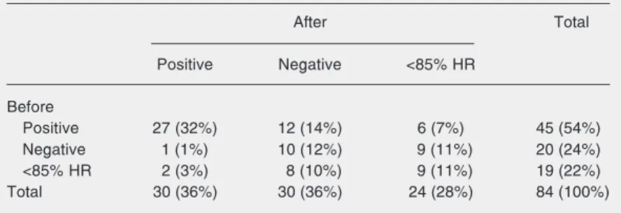
Regarding the results of ETT, 45 (54%) and 30 (36%) patients were positive, 20 (24%) and 30 (36%) were negative, and 19 (22%) and 24 (28%) did not achieve 85% of age-predictive heart rate before and after PCI, respectively (Table 2). The present study shows a significant association between these results (P = 0.01). In the majority of cases (54%) the results of ETT were positive be-fore PCI, while the number of the negative tests increased (24 to 36%) after PCI. The 27 patients with a positive test before and after the procedure showed a later appearance of ST-segment depression (250 and 312 s, re-spectively, P = 0.01) and a tendency to a decrease in the time of duration (354 and 269 s, respectively, P = 0.09) after PCI.
The main parameters of the ETT before and after PCI in patients with and without restenosis and/or new stenosis are summa-rized in Table 3. No difference was found in heart rate at peak exercise between the first and second tests. Both systolic blood pres-sure and rate-prespres-sure product at peak exer-cise were significantly higher during the
Exercise test
Oral antianginal medication was not stopped before or after PCI in 15 patients because discontinuation was considered to be contraindicated by the patient’s
physi-Table 2. Results of the exercise treadmill test before and after percutaneous coronary intervention (P = 0.014; McNemar test).
After Total
Positive Negative <85% HR
Before
Positive 27 (32%) 12 (14%) 6 (7%) 45 (54%) Negative 1 (1%) 10 (12%) 9 (11%) 20 (24%) <85% HR 2 (3%) 8 (10%) 9 (11%) 19 (22%) Total 30 (36%) 30 (36%) 24 (28%) 84 (100%)
Positive: abnormal electrocardiographic results; negative: normal electrocardiographic results; <85% HR: did not achieve predictive heart rate.
Table 3. Results of the exercise treadmill test (univariate profile analyses).
All patients Restenosis and/or new stenosis (N = 84)
Yes (N = 46) No (N = 38)
Heart rate at peak (bpm)
Before 133 ± 23 133 ± 23 135 ± 23 After 135 ± 18 135 ± 17 135 ± 20 P = 0.59# P = 0.59# P = 0.79+
Systolic blood pressure at peak (mmHg)
Before 177 ± 28 168 ± 25 188 ± 29 After 186 ± 28 182 ± 28 191 ± 27 P = 0.02# P = 0.02# P = 0.48+
Rate-pressure product at peak (bpm · mmHg · 10-3)
Before 23.8 ± 5.6 22.4 ± 5.1 25.5 ± 5.9 After 25.3 ± 5.4 24.6 ± 5.2 26.0 ± 5.5 P = 0.04# P = 0.04# P = 0.53+
Duration of exercise (s)
Before 378 ± 155 353 ± 136 407 ± 173 After 417 ± 154 381 ± 145 460 ± 154 P = 0.00# P = 0.11# P = 0.01+
Results of ETT after PCI
Angina 16 4 (9%) 12 (32%)
Positive 30 20 (44%) 10 (25%)
Negative 30 13 (28%) 17 (45%)
<85% age-predictive heart rate 24 13 (28%) 11 (30%)
ETT = exercise treadmill test; PCI = percutaneous coronary intervention.
ETT after PCI than before PCI in patients with restenosis and/or new stenosis (P = 0.022 and 0.049, respectively). Similarly, duration of exercise after PCI was signifi-cantly longer than before PCI, but only for patients without restenosis and/or new ste-nosis during follow-up (P = 0.008). Of the 16 patients with angina in the ETT after PCI, nine (56%) had restenosis and/or new le-sions in the coronary artery.
After PCI, the ETT was positive in 20 patients with restenosis and/or new stenosis and in 10 patients (4 with complete revascu-larization and 6 with incomplete revascular-ization) without these conditions. The sensi-tivity was 61%, specificity was 63%, posi-tive predicposi-tive value was 67%, negaposi-tive pre-dictive value was 57%, and accuracy was 62%.
Discussion
This study investigated a subgroup of patients with 2- and 3-vessel coronary artery disease treated with PCI, in an attempt to determine the behavior of ETT variables in these patients. Since the ETT is a noninva-sive procedure of low operational cost, we also tried to demonstrate the usefulness of this technique as an auxiliary tool for the diagnosis of restenosis and/or new lesions to be used by physicians in clinical practice.
The literature on the value of ETT at late (6 months) follow-up of patients with the same characteristics as our population, treated with multivessel PCI, is scarce since investi-gators prefer to study myocardial perfusion scintigraphy or pharmacological stress echo-cardiography, the best methods for a late diagnosis of restenosis, undoubtedly more sensitive and more specific for these cases. Nevertheless, keeping in mind the low cost of ETT compared to these other methods, we decided to undertake a thorough assessment of its variables under various circumstances to obtain as optimal results as possible.
In our study population, PCI resulted in
total revascularization in 35 (42%) patients. In patients with multivessel coronary artery disease it is often impossible to obtain com-plete myocardial revascularization with PCI because of the unfavorable morphological features of the coronary tree, such as chronic occlusion, twisting and marked calcifica-tion. Some relevant studies, such as the Rita (12), Cabri (13) and Bari (14) trials, have shown that in patients with multivessel in-volvement PCI was not successfully per-formed in all the vessels with obstructive lesions, thus characterizing incomplete re-vascularization. Therefore, the outcome of myocardial revascularization through PCI may turn out differently from what was planned, although an incomplete myocar-dial revascularization may be considered to be functionally complete from the stand-point of anatomy.
Of note, it is important to point out that, even considering the technological develop-ment of endoprostheses, the incidence of restenosis within the first 6 months after PCI is still elevated, between 20 and 40% (15,16). Among the patients studied here, the rate of restenosis was 44%, slightly above the value reported in the literature. However, our popu-lation was a particular one consisting of patients with multivessel involvement who were treated for up to five lesions, therefore with a higher chance of suffering restenosis. In addition, the procedure in question spe-cifically treats the vessel lesion but does not prevent coronary artery disease from pro-gressing. Our study detected 21 patients (25%) presenting progression of coronary artery disease within the first 6 months.
procedure and 20 (67%) of them suffered restenosis and/or new lesion.On the other hand, a significant number of patients still present ST downsloping even after complete revascularization is obtained (17,18). In our study, 33% of patients with a positive ETT did not present restenosis and/or coronary artery disease progression. This can be ex-plained by an abnormality of the coronary reserve and by the presence of an inflamma-tion process that only time can resolve. Zijlstra et al. (19) reported that normaliza-tion of the coronary reserve after PCI only occurs weeks or months after the procedure. On the basis of the present ETT results, there was both a clear reduction of the stress-induced ischemic response (from 45 to 30 patients) and relief from angina (from 27 to 16 patients) 6 months after the procedure, which reflected the improvement of coro-nary perfusion independent of the therapy used.
In our study population, stress-induced angina was detected in 16 patients during the exercise test carried out after PCI, 4 of them having had all of their diseased vessels treated. However, the absence of angina is not the best criterion for predicting coronary blood flow. Angina in fact disappears with the improvement of coronary perfusion; how-ever, it can also occur in patients with little or no change in myocardial blood flow at all. Hartman (20) also observed that 79% of his patients who had undergone myocardial re-vascularization were free from angina. This was the case for those with at least one graft patent, which is evidence of the improve-ment in myocardial perfusion being respon-sible, to some extent, for the reduction of angina, independent of the patency of the grafts. Holubkov et al. (21) evaluated pa-tients 1 year after PCI and observed that young people and female patients reported more symptoms, as also did those patients with diabetes mellitus, previous myocardial infarction, diseased grafts, and extensive coronary artery disease.
In our study population, systolic blood pressure, rate-pressure product at peak and exercise duration were significantly higher in the ETT after PCI, independent of the results of the latter. Considering that these variables are determinants of myocardial oxygen uptake, we can presume that there was some degree of improvement of myo-cardial perfusion even in those patients with incomplete revascularization, as well as in those with evidence of restenosis or new lesion after 6 months.
It is well known that the main exercise test parameters with diagnostic, as well as prognostic value after revascularization pro-cedures are: relief from angina, rate-pres-sure product, normalization of the ST seg-ment, and exercise duration.
Gohlke et al. (22) reported that values of rate-pressure product at peak exercise lower than 25 bpm · mmHg · 10-3 were associated
with either a poor result or left ventricular dysfunction, and when higher than 30 bpm · mmHg · 10-3 they might have been related to
complete or sufficient revascularization. In the present study, the rate-pressure product at peak increased significantly after PCI (from 23.8 ± 5.6 to 25.3 ± 5.4 bpm · mmHg · 10-3.
However, this variable and systolic blood pressure at peak did not discriminate be-tween patients with and without restenosis or coronary artery disease progression when these two groups were analyzed separately. Probably, these variables are correlated with improvement of myocardial perfusion be-cause restenosis or new lesion usually oc-curs in one vessel and the size of the is-chemic myocardium is smaller than before the procedure. Another possible reason for rate-pressure product and systolic blood pres-sure at peak not to discriminate the two groups was the use of antianginal medica-tion at the time of ETT. The behavior of these variables was in agreement with other studies (23,24).
popu-lation was exercise duration, but the same did not apply to a positive test, the presence of angina and rate-pressure product at peak, when these were analyzed separately. With certainty, some patients with antianginal medication at the time of ETT contributed to these findings. Some studies (25,26) have reported that double product, time of toler-ance to exercise and heart rate during the postoperative period of myocardial revascu-larization, when considered separately, are not good predictors of graft patency. Ab-sence of angina and increased functional ability are, however, good predictors when considered in combination (27). A meta-analysis study (28) reported that the sensi-tivity, specificity and accuracy of exercise electrocardiography for the detection of re-stenosis after PCI were 46% (33 to 58%), 77% (67 to 86%) and 62%, respectively. In the present study, we found a sensitivity slightly above average (61%), a lower speci-ficity (63.0%) and the same accuracy (62%).
Study limitations
A limitation of the present study is the controversy on the definition of restenosis. Angiographic restenosis is considered to be a continuous variable in large patient
popu-lations (29). Even though stenosis with a diameter of >50% has been reported as an appropriatedefinition of restenosis (30-32), it does not consider factors such as length and geometry of the lesion or endothelial dysfunction, which could cause genuine is-chemia. Another limitation is the relatively high rate of submaximal ETT, probably re-lated to drugs. Finally, no additional imag-ing, echocardiography or myocardial scin-tigraphy was used with the ETT. On the other hand, our results are specific for pa-tients with preserved left ventricular func-tion, tested without specific drug treatment when possible.
Sequential and comparative analysis of exercise electrocardiography variables be-fore and after PCI permitted us to reach the following conclusions: normalization of the ST segment after PCI usually indicates a successful procedure; increased exercise duration was indicative of permeability of the procedure-related coronaries, with sta-tistical significance (P = 0.008); presence or absence of angina, and the behavior of heart rate, blood pressure and rate-pressure prod-uct variables of the ETT after PCI were not good predictors of restenosis and/or new stenosis.
References
1. Gibbons RJ, Balady GL, Beasley JW et al. (1997). ACC/AHA guide-lines for exercise testing: a report of the American College of Cardi-ology/American Heart Association Task Force on practice guide-lines (Committee on Exercise Testing). Journal of the American College of Cardiology, 30: 260-311.
2. Davies RF, Goldberg AD, Forman S et al. (1997). Asymptomatic Cardiac Ischemia Pilot (ACIP) study two-year follow-up: outcomes of patients randomized to initial strategies of medical therapy versus revascularization. Circulation, 95: 2037-2043.
3. Stone PH, Chaitman BR, Dorman S et al. (1997). Prognostic signifi-cance of myocardial ischemia detected by ambulatory electrocar-diography, exercise treadmill testing and electrocardiogram at rest to predict cardiac events by one year. American Journal of Cardiol-ogy, 80: 1395-1401.
4. Breitenbucher A, Pfisterer M, Hoffmann A et al. (1990). Long-term follow-up of patients with silent ischemia during exercise radionu-clide angiography. Journal of the American College of Cardiology,
15: 999-1003.
5. Younis LT, Byers S, Shaw L et al. (1989). Prognostic importance of silent myocardial ischemia detected by intravenous dipyridamole thallium myocardial imaging in asymptomatic patients with coronary artery disease. Journal of the American College of Cardiology, 14: 1635-1641.
6. Dori G, Denekamp Y, Fishman S et al. (2003). Exercise stress testing, myocardial perfusion imaging and stress echocardiography for detecting restenosis after successful percutaneous transluminal coronary angioplasty: a review of performance. Journal of Internal Medicine, 253: 253-262.
7. Beygui F, Le Feuvre C, Maunoury C et al. (2000). Detection of coronary restenosis by exercise electrocardiography thallium-201 perfusion imaging and coronary angiography in asymptomatic pa-tients after percutaneous transluminal coronary angioplasty. Ameri-can Journal of Cardiology, 86: 35-40.
exercise testing in the detection of silent restenosis after successful coronary angioplasty. American Heart Journal, 129: 452-459. 9. The Medicine, Angioplasty, or Surgery Study (MASS-II) (2004). A
randomized controlled clinical trial of 3 therapeutic strategies for multivessel coronary artery disease: 1-year results. Journal of the American College of Cardiology, 43: 1743-1751.
10. Sones Jr FM & Shirey EK (1962). Cinecoronary arteriography. Mod-ern Concepts of Cardiovascular Disease, 31: 735-738.
11. Seldinger SY (1953). Catheter replacement of the needle in percuta-neous arteriography. A new technique. Acta Radiologica, 39: 368-376.
12. Rita Trial Participants (1993). Coronary angioplasty versus coronary artery bypass surgery. Lancet, 341: 573-580.
13. Cabri Trial Participants (1995). First-year results of Cabri (coronary angioplasty versus bypass revascularization investigation). Lancet, 346: 11-84.
14. The Bari Investigators (1997). Five-year clinical and functional out-come comparing bypass surgery with angioplasty in patients with multivessel coronary disease. Journal of the American Medical As-sociation, 277: 715-721.
15. Malekianpour M, Rodés J, Côté G et al. (1999). Value of exercise electrocardiography in the detection of restenosis after coronary angioplasty in patients with one-vessel disease. American Journal of Cardiology, 84: 258-263.
16. Galassi A, Foti R, Azzarelli S et al. (2000). Usefulness of exercise tomographic myocardial perfusion imaging for detection of resteno-sis after coronary stent implantation. American Journal of Cardiol-ogy, 85: 1362-1364.
17. McConahay DR (1977). Accuracy of treadmill testing in assessment of direct myocardial revascularization. Circulation, 56: 548-552. 18. Glasser SP & Clark PL (1980). The Clinical Approach to Exercise
Testing. Harper & Row, New York.
19. Zijlstra F, Reiber JC, Julliere Y et al. (1988). Normalization of coro-nary flow reserve by percutaneous transluminal corocoro-nary angio-plasty. American Journal of Cardiology, 61: 55-60.
20. Hartman CW (1976). Aortocoronary bypass surgery: correlation of angiographic, symptomatic and functional improvement at 1 year. American Journal of Cardiology, 37: 352-357.
21. Holubkov R, Laskey WK, Haviland A et al. (2002). Angina 1 year after percutaneous coronary intervention: A report from the NHLBI dynamic registry. American Heart Journal, 144: 826-833.
22. Gohlke H, Goklke-Barwolf C, Samek L et al. (1983). Serial exercise testing up to 6 years after coronary bypass surgery: behavior of exercise parameters in groups with different degrees of revasculari-zation determined by postoperative angiography. American Journal of Cardiology, 51: 1301-1306.
23. Chalela WA, Lima EV & Moffa PJ (1996). Exercise stress testing after coronary bypass surgery and percutaneous transluminal an-gioplasty. Arquivos Brasileiros de Cardiologia, 67: 59-62.
24. Wilson RF, Marcus ML & White CW (1988). Effects of coronary bypass surgery and angioplasty on coronary blood flow and flow reserve. Progress in Cardiovascular Diseases, 31: 95-114. 25. Lapin ES (1973). Changes in maximal exercise performance in the
evaluation of saphenous vein bypass surgery. Circulation, 47: 1164-1173.
26. Merril Jr AJ (1975). Value of maximal exercise testing in assess-ment of results. Circulation, 52 (Suppl 2): 173-177.
27. Block TA, Murray JA & English MT (1977). Improvement in exercise performance after unsuccessful myocardial revascularization. American Journal of Cardiology, 40: 673-680.
28. Garzon P & Eisenberg MJ (2001). Functional testing for the detec-tion of restenosis after percutaneous transluminal coronary angio-plasty: A meta analysis. Canadian Journal of Cardiology, 17: 41-48. 29. Kuntz RE & Baim DS (1993). Defining coronary restenosis. Newer
clinical and angiographic paradigms. Circulation, 88: 1310-1323. 30. Gottsauner-Wolf M, Sochor H, Moerti D et al. (1996). Assessing
coronary stenosis. Quantitative coronary angiography versus visual estimation from cine-film or pharmacological stress perfusion im-ages. European Heart Journal, 17: 1167-1174.
31. Di Carli M, Czernin J, Hoh CK et al. (1995). Relation among stenosis severity, myocardial blood flow, and flow reserve in patients with coronary artery disease. Circulation, 91: 1944-1951.