Synthesis and secretion of transferrin
by a bovine trabecular meshwork
cell line
Departamento de Biologia Celular e Molecular e Bioagentes Patogênicos, Faculdade de Medicina de Ribeirão Preto, Universidade de São Paulo, Ribeirão Preto, SP, Brasil
R. Bertazolli-Filho, E.M. Laicine and A. Haddad
Abstract
The trabecular meshwork (TM) is the main outflow pathway in the mammalian eye. Oxidative damage to TM cells has been suggested to be an important cause of impairment of TM functions, leading to deficient drainage of aqueous humor, with deleterious consequences to the eye. Transferrin, a metalloprotein involved in iron transport, has been characterized as an intrinsic eye protein. Since transferrin is implicated in the control of oxidative stress, the objective of the present study was to determine if a bovine TM cell line (CTOB) synthesizes and secretes transferrin. The CTOB cell line was cultured in the presence of 35S-methionine and the incubation medium was submitted to immunoprecipitation. Total RNAs from CTOB and isolated bovine TM (freshly isolated, incubated or not) were subjected to the reverse transcription-polymerase chain reaction and the ampli-fication products were sequenced. Also, both CTOB and histological TM preparations were processed for transferrin immunolocalization. A labeled peptide of about 80 kDa, the expected size for transferrin, was immunopurified from CTOB samples obtained from the incuba-tion assays. The reverse transcripincuba-tion-polymerase chain reacincuba-tion and sequencing experiments detected the presence of transferrin mRNA in CTOB and isolated bovine TM. Reactivity to antibodies against transferrin was observed both in CTOB and TM. The results obtained in all of these experiments indicated that the TM is capable of synthesizing and secreting transferrin. The possible implications for the physiology of the eye are discussed.
Correspondence R. Bertazolli-Filho
Departamento de Biologia Celular e Molecular, FMRP, USP 14049-900 Ribeirão Preto, SP Brasil
Fax: +55-16-3633-1786 E-mail: [email protected]
Research supported by CNPq (for A. Haddad and E.M. Laicine) and FAPESP (for E.M. Laicine and R. Bertazolli-Filho).
Received January 16, 2007 Accepted June 11, 2007
Key words
•Bovine eye
•Trabecular meshwork •Transferrin expression •Cell and tissue culture •Immunocytochemistry
Introduction
The trabecular meshwork (TM), located at the angle of the anterior chamber, plays a role in the drainage of the aqueous humor (AH) of the anterior chamber, both in the eyes of primates (which also exhibit the Schlemm canal) and of non-primate mam-mals (1). After circulating through the TM,
spur and uvea. Damage to the TM can re-duce the drainage of AH outflow, ultimately leading to an increase of intraocular pres-sure, the major causal risk factor of glau-coma (2).
Transferrin is an 80-kDa metalloprotein which is the major iron transporter glyco-protein in plasma (3). Even though the main source of transferrin in the body is the liver, many extra-hepatic sources exist. Several eye sites are capable of synthesizing and secreting transferrin, including the retina (4), lens (5) and ciliary epithelium (6). Both the lens and the ciliary epithelium face the pos-terior chamber, which means that the se-creted transferrin becomes a constituent of the AH. The presence of transferrin in AH as apotransferrin (iron-free) could be impor-tant in preventing the generation of free radicals since it could remove the free iron molecules in the AH. In the past few years, several investigations have indicated that oxidative stress is a possible cause of dam-age to the trabecular meshwork, impairing its functions in AH outflow (7). Since trans-ferrin has a potential protective effect against the free radicals present in the AH, it is of interest to investigate the ability of CTOB, a bovine TM cell line, to synthesize and se-crete transferrin.
Material and Methods
Cell culture
TM was obtained from a steer (Bos taurus) at a local slaughterhouse. Trabeculotomy was performed as follows: the enucleated eyes were washed in sterile PBS (137 mM NaCl, 2 mM KCl, 0.9 mM CaCl2, 1.54 mM
MgCl2, 6.5 mM Na2HPO4, 1.47 mM KH2PO4,
pH 7.2) and the posterior portion of each eye was removed; the anterior portions were either processed for immunohistochemistry (see below) or had their ciliary body-iris detached. Bovine TM was dissected under a surgical microscope by an ophthalmic
sur-geon with expertise in trabeculotomy. Once it was confirmed that the TM was free from any surrounding tissue, TM fragments were chopped into very small pieces, which were used for the primary cultures. Alternatively, other fragments were digested with trypsin-EDTA solution to obtain free cells. Both the small pieces and the TM-free cells were plated onto flasks (Corning, New York, NY, USA) containing Dulbecco’s modified Eagle’s medium (DMEM, Gibco, Grand Is-land, NY, USA), supplemented with 10% fetal calf serum (Gibco) in a humidified incubator under a 5% CO2-air atmosphere at
37ºC until reaching confluence. Three suc-cessful cell cultures were obtained and fro-zen after 7 to 10 passages. One of these cultures has been continuously replicated more than 200 times. Several tests have been carried out to check the origin of the cells and their ability to phagocytize the protein of the crystalline. Additionally, TM frag-ments were cultured as explants in a cham-ber of a Transwell® 6MM plate (Costar, New
York, NY, USA) filled with 1 mL serum-free DMEM for 24 h, with at least 3 medium changes during incubation before RNA ex-traction and immunocytochemistry process-ing.
Reverse transcription-polymerase chain reaction assays
indicated by the manufacturer. The oligo-nucleotide set of primers used in PCR ampli-fication and sequencing were synthesized by Imprint. The set of primers (upper-tgtta gcctgcgcggttctgg 23-43; lower-gacatttaaa ggcccctgagtagc 628-652) was designed in the expectation that it would span at least one intron, permitting the detection of pos-sible genomic contamination. Primers were chosen on the basis of published bovine liver transferrin cDNA nucleotide sequences (NM_177484) and were selected using the DNASTAR program (DNASTAR Inc., Madison, WI, USA, licensed to Hemocentro de Ribeirão Preto). cDNA was amplified by polymerase chain reaction using 5 µL of the products from RT, 25 pmol of each primer, 50 µL of reaction buffer, 1 mM of dNTP mixture, and 0.5 µL of Taq polymerase (Gibco 2.5 U/µL) in a final volume of 50 µL. A thermal cycler (GeneAmp® PCR System
9700, PE Applied Biosystems, Foster City, CA, USA) was used with the following tem-perature profiles: initial melting at 94ºC for 2 min, 32 1-min melting cycles at 94ºC, 1 min annealing at 58ºC, and 1 min extension at 72ºC. After the last cycle, polymerization was extended for 10 min, when the chains were complete. The products of amplifica-tion (10 µL) were analyzed on 1.2% agarose gels and the amplified bands were gel-puri-fied using the GFX™ kit (Pharmacia, Little Chalfont, Buchinghamshire, UK).
Nucleotide sequencing
PCR DNA products were sequenced on both strands using the automated DNA se-quencer ABI PRISM 377 Genetic Analyzer (Foster City, CA, USA) and the BigDye Rhodamine Terminator Cycle sequencing kit (PE Applied Biosystems), at the Hemo-centro of Ribeirão Preto facilities.
Isotopic labeling experiments
For the isotopic labeling experiments the
semi-confluent cells, grown in a 25-cm2 flask,
were washed several times in sterile PBS and 2 mL DMEM without methionine plus 25 µCi 35S-methionine (Amersham, Little
Chalfont, Buckinghamshire, UK), specific activity >800 Ci/mmol, were then added to each flask. The preparations were incubated for 24 h, the media were recovered, pre-cleared by centrifugation at 16,000 g for 30 min at 4ºC, and submitted to immunopre-cipitation.
Immunoprecipitation and SDS-PAGE analysis
The media resulting from the incubation of CTOB cells were immunoprecipitated using 5 µL of goat anti-rabbit transferrin antibody (Cappel ICN Pharmaceuticals Inc., Aurora, OH, USA) plus protein A bound to Sepharose CL4B, as described (8). After immunoprecipitation, the samples were ana-lyzed by 10% SDS-PAGE under reducing conditions (9) plus fluorography (10). After SDS-PAGE, the gels were stained with Coo-massie brilliant blue R-250, photographed, impregnated with Amplify™ (Amersham), dried under vacuum and exposed to X-OMAT-AR5 film (Kodak, Rochester, NY, USA) for different times at -80ºC.
Immunocytochemistry
an Axiophot microscope (Zeiss, Oberkochen, Germany) and the images captured with a CCD-IRIS camera (Sony, Tokyo, Japan) and digitized (Snappy 3.0, Play Inc., London, UK).
Results
Expression of transferrin mRNA in the CTOB cell line and isolated trabecular meshwork
The RT-polymerase chain reaction was used to detect the expression of transferrin mRNA both in the CTOB cell line and iso-lated TM before and after incubation. Based on the known transferrin gene structures (e.g., human and rabbit ones), the primer sets used in our experiments were expected to cover the region of the transferrin mes-sage spanning exons 2 to 6.
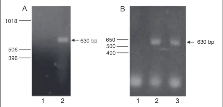
Figure 1A shows the representative am-plifications of the transferrin message from CTOB. Figure 1B shows a representative amplification of the transferrin message from freshly isolated TM (2) and from TM incu-bated for 21 h (3). The amplification prod-ucts were of expected size and the sequence analysis of the amplified products indicated 100% homology with the published transfer-rin sequence (NM-177484). These experiments indicate that both the CTOB cell line and the isolated TM express transferrin mRNA.
Immunoprecipitation assays
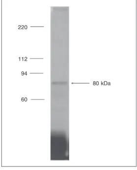
The incubation experiments using the CTOB cell line followed by immunoprecipi-tation with the specific antiserum against rabbit transferrin were performed to deter-mine the ability of CTOB to synthesize and secrete transferrin into the culture medium. Figure 2 shows a representative immunopre-cipitation experiment of the isotopically la-beled transferrin from the CTOB culture media. Fluorography revealed a band of about 80 kDa, corresponding to the expected size of transferrin.
incubated with the first antibody (goat anti-rabbit transferrin; Cappel) diluted 1:200 in blocking buffer for 2 h at room temperature. The coverslips were rinsed in TTBS and incubated with the secondary antibody, rho-damine-conjugated anti-goat IgG, diluted 1:200, for 1 h at room temperature. After washing in TTBS and mounting in a solution of Fluoromount™ (Southern Biotechnology Associated, Inc., Birmingham, AL, USA) medium, the cells were analyzed with a Con-focal microscope (Leica TCS NT, Wetzlar, Germany) coupled to an inverted DM IRBE Leica microscope. The images were cap-tured using a Fuji Pictrography 3000 printer (Tokyo, Japan) connected to the confocal device. The controls were run either omit-ting the primary antibody or using the non-immune goat serum. The anterior portion of the eye and the explants of isolated and incubated TM were fixed in 10% formalin in 0.1 M Sorensen’s buffer for 24 h at 4ºC, embedded in paraffin and cut into 6-µm thick sections. The immunohistochemical reactions were performed as described for the CTOB, except that it was used the Vectastain ABC Kit (Anti-goat IgG, Vector, Burlingame, CA, USA) to detect the primary antibody. The specimens were examined with
630 bp 650
500 630 bp 1018
506 396
1 2 1 2 3
A B
Figure 1. RT-PCR of transferring. A, 1) Negative control (water), 2) CTOB, a bovine trabecular meshwork cell line. B,1) Negative control (water), 2) isolated bovine trabecular meshwork, 3) bovine 21-h incubated trabecular meshwork explant. Samples of 10 µL were analyzed on 1.2% agarose gels. The arrows point at the amplification products with the expected length (630 bp). Molecular weight markers are at left: 1 kb Ladder (A) and 1 kb Ladder Plus (B) (Gibco).
Immunohistochemistry
The specific antibody against transferrin was used in an indirect immunofluorescence assay to examine the cellular distribution of transferrin in the CTOB cell line. The samples were analyzed using confocal microscopy.
Figure 3 shows a representative labeling pattern of transferrin antigenicity in a CTOB cell. Antigenicity was detected throughout the cytoplasm.
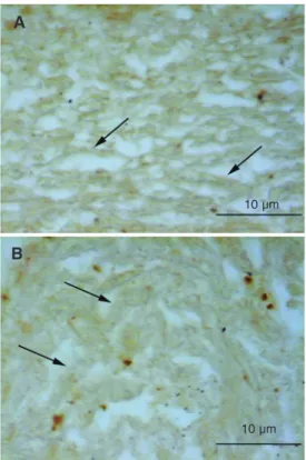
A similar labeling pattern was observed in intact (Figure 4A) and incubated TM (Fig-ure 4B), with diffuse staining of trabecular cells. A slight swelling of the trabeculae can be seen in Figure 4B compared with intact TM (Figure 4A).
Discussion
We report here the expression of the metalloprotein transferrin based on mRNA and protein analysis by CTOB, a cell line derived from bovine TM. The expression of transferrin mRNA was analyzed both in the cell line and in isolated tissue, incubated or not. Trabecular cell lines are extensively used in studies of TM function since it is difficult to obtain large amounts of TM samples. However, as in any in vitro analy-sis, it is important to check whether cell line expression reflects the phenotype of the in-tact tissue or if it is a spurious modification of the secretion pattern due to incubation conditions. There are reports indicating that a tissue in culture may not behave as it does in its proper environment in a living animal. This is the case for TM concerning the ex-pression of several genes which are expressed
in vivo but not in vitro and vice versa (for a comprehensive review, see Ref. 11). The existence of an intrinsic vitreous transferrin has been demonstrated (12) and many stud-ies were subsequently carried out to deter-mine its origins. Many eye components have been considered to be potential sources of vitreous transferrin, including the retina (4),
220
112
94
60
80 kDa
Figure 2. SDS-PAGE plus fluo-rography of the transferrin. The labeled fraction present in the extracts of CTOB (bovine tra-becular meshwork cell line) cul-ture medium was immunopre-cipitated after incubation for 24 h in methionine-free DMEM con-taining 25 µCi 35S-methionine. The immunoprecipitation was carried out using antitransferrin antibody plus protein A bound to Sepharose CL4B and the result-ing immunoprecipitated sample was processed for SDS-PAGE plus fluorography on 10% poly-acrylamide gel. The arrow indi-cates the transferrin band. Mo-lecular weight markers are at left.
n
20 µm
Figure 3. Cellular distribution of transferring and immunolabeling of CTOB (bovine trabecular meshwork cell line). The posi-tive reaction is visible through-out the cytoplasm of a single cell. Nucleus (n). The control (primary antibody omission) was negative (not shown). The sec-ondary antibody was TRITC-bound. Bar = 20 µm.
lens (5) and ciliary epithelium (6). Regard-ing the aqueous humor, the intrinsic trans-ferrin could reach it either by migration from the vitreous and/or by secretion from the lens and ciliary epithelium. The migration of vitreous protein to the aqueous humor has been demonstrated (13).
potentiate oxygen toxicity by the generation of a wide range of free radical species, in-cluding hydroxyl radicals, the most reactive of the free radicals, which have the ability to react with a wide range of cellular compo-nents. The deleterious effects of iron in the vitreous have been demonstrated (14). Since the aqueous humor has a high concentration of hydrogen peroxide (15), it is of particular importance to remove any free transition metal present in it.
It has been shown that oxidative stress
affects the morphology and physiology of the outflow pathway in aging as well as in glau-coma (16). Also, it has a negative effect on the TM cell adhesiveness and leads to a cytoskel-eton rearrangement in addition to NF-κB acti-vation (17). NF-κB was the first transcrip-tional factor shown to respond directly to oxi-dative stress. In fact, it was demonstrated that glaucomatous TM expresses selectin-E by the activation of NF-kB and selectin-E is consid-ered to be the first phenotype marker for glau-coma (18). The proteasoma of TM cells sub-mitted to chronic oxidative stress is inhibited (19) and the oxidative DNA damage in the patients with glaucoma is significantly higher than in controls (20).
Inside the eye, apotransferrin has proven to be effective in the prevention of some inflammatory conditions (21,22), possibly acting synergistically with other antioxidants present in the eye (15). Keeping the aqueous humor free of the transition metals is prob-ably important for the long-term mainte-nance of homeostasis of TM, preventing the excessive generation of free radicals con-stantly related to aging and degenerating disease (23).
Acknowledgments
We are grateful to Vani M.A. Correa and Domingos S. de Souza-Filho for technical help. We also wish to thank Dr. Ryoko Chokiu (Volta Redonda, RJ, Brazil) for her expert assistance with trabeculotomy.
Figure 4. Tissue distribution of transferring on 6-µm paraffin sections. Immunolabeling of the intact (in situ) bovine trabecular meshwork (A) and cultured tra-becular meshwork explant (B). The positive reaction is visible on the trabecular cells and sur-rounding thick matrix sections (arrows). The control (primary antibody omission) was negative (not shown). The reactions were developed with DAB and 0.003% hydrogen peroxide us-ing the Vectastain ABC kit (Vec-tor). No counterstaining. Bar = 10 µm.
10 µm
10 µm
A
B
References
1. Lutjen-Drecoll E. Functional morphology of the trabecular meshwork in primate eyes. Prog Retin Eye Res 1999; 18: 91-119.
2. Kass MA, Heuer DK, Higginbotham EJ, Johnson CA, Keltner JL, Miller JP, et al. The Ocular Hypertension Treatment Study: a ran-domized trial determines that topical ocular hypotensive medication delays or prevents the onset of primary open-angle glaucoma. Arch Ophthalmol 2002; 120: 701-713 (discussion 829-830).
3. de Jong G, van Dijk JP, van Eijk HG. The biology of transferrin. Clin Chim Acta 1990; 190: 1-46.
4. Davis AA, Hunt RC. Transferrin is made and bound by
photorecep-tor cells. J Cell Physiol 1993; 156: 280-285.
5. McGahan MC, Harned J, Goralska M, Sherry B, Fleisher LN. Trans-ferrin secretion by lens epithelial cells in culture. Exp Eye Res 1995; 60: 667-673.
6. Bertazolli-Filho R, Laicine EM, Haddad A. Synthesis and secretion of transferrin by isolated ciliary epithelium of rabbit. Biochem Biophys Res Commun 2003; 305: 820-825.
7. Babizhayev MA, Bunin AY. Lipid peroxidation in open-angle glau-coma. Acta Ophthalmol 1989; 67: 371-377.
tech-niques. Boston: Birkhäuser; 1996.
9. Laemmli UK. Cleavage of structural proteins during the assembly of the head of bacteriophage T4. Nature 1970; 227: 680-685. 10. Waterborg JH, Matthews HR. Fluorography of polyacrylamide gels
containing tritium. In: Walker JM (Editor), Methods in molecular biology proteins. Clifton: Humana Press; 1984. p 147-152. 11. Borrás T. Gene expression in the trabecular meshwork and the
influence of intraocular pressure. Prog Retin Eye Res 2003; 22: 435-463.
12. Laicine EM, Haddad A. Transferrin, one of the major vitreous pro-teins, is produced within the eye. Exp Eye Res 1994; 59: 441-445. 13. Haddad A, Laicine EM, de Almeida JC. Origin and renewal of the
intrinsic glycoproteins of the aqueous humor. Graefes Arch Clin Exp Ophthalmol 1991; 229: 371-379.
14. Hui YN, Sorgente N, Ryan SJ. Liquefaction of rabbit vitreous by ferrous ions. Curr Eye Res 1988; 7: 655-660.
15. Rose RC, Richer SP, Bode AM. Ocular oxidants and antioxidant protection. Proc Soc Exp Biol Med 1998; 217: 397-407.
16. Green K. Free radicals and aging of anterior segment tissues of the eye: a hypothesis. Ophthalmic Res 1995; 27 (Suppl 1): 143-149. 17. Zhou L, Li Y, Yue BY. Oxidative stress affects cytoskeletal structure
and cell-matrix interactions in cells from an ocular tissue: the trabec-ular meshwork. J Cell Physiol 1999; 180: 182-189.
18. Wang N, Chintala SK, Fini ME, Schuman JS. Activation of a tissue-specific stress response in the aqueous outflow pathway of the eye defines the glaucoma disease phenotype. Nat Med 2001; 7: 304-309.
19. Caballero M, Liton PB, Epstein DL, Gonzalez P. Proteasome inhibi-tion by chronic oxidative stress in human trabecular meshwork cells.
Biochem Biophys Res Commun 2003; 308: 346-352.
20. Izzotti A, Sacca SC, Cartiglia C, De Flora S. Oxidative deoxyribo-nucleic acid damage in the eyes of glaucoma patients. Am J Med
2003; 114: 638-646.
21. McGahan MC, Grimes AM, Fleisher LN. Hemoglobin exacerbates the ocular inflammatory response to endotoxin. Graefes Arch Clin Exp Ophthalmol 1996; 234: 643-647.
22. McGahan MC, Grimes AM, Fleisher LN. Transferrin inhibits the ocular inflammatory response. Exp Eye Res 1994; 58: 509-511. 23. Tezel G. Oxidative stress in glaucomatous neurodegeneration: