Abstract
Submitted: April 14, 2017
Modiication: August 12, 2017 Accepted: August 21, 2017
The effects of different restorative
materials on periodontopathogens
in combined restorative-periodontal
treatment
Objective: The aim of the study was to evaluate the association between subgingival restorations and the target periodontopathogenic bacteria (Pg, Td and Pi) in subgingival bioilm during one year after combined restorative-periodontal treatment. Material and Methods: Seventeen systemically healthy subjects, who were positive for the presence of three cervical lesions associated with gingival recessions in three different adjacent teeth, were included in the study. A total of 51 combined defects were treated with connective tissue graft plus a nanoilled composite resin (NCR+CTG), a resin-modiied glass ionemer cement (RMGI+CTG) and a luoride-releasing resin material with pre-reacted glass (PRG), called giomer (Giomer+CTG). Periodontal clinical measurements and subgingival plaque samples were obtained from all combined defects at baseline and at 6 and 12 months after the surgery. The number of bacteria were evaluated by the real-time polymerase chain reaction (qPCR) method. Results: No statistically signiicant difference in the amount of DNA copies of Pg, Td and Pi was observed in any of the groups at any time points (p>0.05). In addition, there was no statistically signiicant difference in the amount of DNA copies of the bacteria at baseline and at 6 and 12 months postoperatively, regardless of treatment group (p>0.05). Conclusion: This study suggests that subgingivally placed NCR, RMGI and giomer restorations can show similar effects on periodontopathogenic bacteria in the treatment of gingival recessions that are associated with noncarious cervical lesions (NCCLs).
Keywords: Bacteria. Tooth abrasion. Connective tissue. Dental
restoration. Bioilms. Sila Çagri ISLER1
Gonen OZCAN1 Gülcin AKCA2 Zahide KOCABAS3
1Gazi University, Faculty of Dentistry, Department of Periodontology, Ankara, Turkey. 2Gazi University, Faculty of Dentistry, Department of Medical Microbiology, Ankara, Turkey. 3Ankara University, Faculty of Agriculture, Biometry and Genetics Unit, Ankara, Turkey. Corresponding address:
Introduction
Gingival recessions and noncarious cervical lesions (NCCLs) are frequently associated with the same tooth surface, forming a combined defect, and are
closely related34. These combined defects result in
numerous aesthetic and functional problems, and
a comprehensive treatment approach is required to
address the issue. A combined restorative-periodontal
therapy, in which the restorative therapy is completed
before mucogingival surgery, has been proposed for
the treatment of gingival recession that is associated with NCCLs14,27,36. Following the healing period after surgery, the soft tissue is positioned over a part of
the restorative material and the apical border of the
restoration is in the subgingival area. However, the
response of the gingival tissues to the restorative
materials is very important, and this relationship has
been thoroughly investigated over many years18. It
has been reported that subgingival restorations are
associated with greater plaque accumulation, bleeding
on probing, and attachment loss17,while other studies
have indicated that the restorations do not result in
greater bioilm formation, bacterial accumulation and
clinical attachment loss, compared with non-restored
areas7,23,28.
Bacterial composition on subgingival restorations
can trigger the development of periodontal disease. It has been suggested that some members of this composition, known as “keystone pathogens”, could regulate bioilm virulence and modulate the host immune response9,11,13. Longitudinal studies have
shown that periodontal disease progression can be
predicted by the levels of Porphyromonas gingivalis
(Pg) and Treponema denticola (Td) in subgingival
plaque3,9,12. Moreover, it has been reported that Pg
and Prevotella intermedia (Pi) are more frequently
associated with deeper periodontal pockets31.
Various dental materials and surgical approaches
have been used to manage these combined defects,
in order to provide the most predictable combined
restorative-periodontal treatment14. In this treatment
method, resin composites or resin-modiied glass ionomer cements (RMGIs) have been commonly used to restore NCCLs19, and gingival recessions
have been treated using the coronally advanced lap (CAF) technique, either alone or in combination with a connective tissue graft (CTG)14,21,24,28. Some of the
previous studies evaluated the effects of subgingivally
placed restorative materials on periodontopathogenic
bacteria in the combined restorative-periodontal
treatment23,28. However, there is a lack of information
in the current literature regarding the effect of
subgingival restorations that are carried out using nanoilled composite resin (NCR), RMGI and giomer on periodontopathogenic bacteria in the treatment of gingival recessions associated with NCCLs.
The primary objectives of this study were to evaluate
the association between subgingival NCR, RMGI and
giomer restorations and three periodontopathogenic
bacteria (Pg, Td and Pi) in subgingival bioilm during
one year after combined restorative-periodontal
treatment, and to examine the correlations between
these pathogens and the clinical data.
Material and methods
Study design and population
This was a prospective, 12-month split-mouth
clinical study. A total of 23 individuals, who were
admitted to the Department of Periodontology, at the Faculty of Dentistry, Gazi University, Ankara, Turkey, were referred for treatment of gingival recessions associated with NCCLs. The study protocol was approved by the Ethics Committee of the Faculty of Medicine, Gazi University (Protocol ID: 25901600-7587). The participants were then informed of this
protocol and their written informed consent was
obtained.
The participants were included on the basis of
the following inclusion criteria: (1) age >18 years,
(2) positive for the presence of three cervical lesions
associated with multiple gingival recessions in three
different adjacent teeth, excluding molars, (3) Miller Class I gingival recession defects (≥2 and ≤5 mm) associated with buccal NCCL Class B + step20, (4) NCCL
depth of 1–2 mm, (5) non-smoker, (6) systemically healthy (7) and probing depth (PD) ≤3 mm.
NCCLs were randomly allocated to three treatment groups using a computer-generated randomization scheme, as follows: NCR+CTG group, in which the combined defects were restored with NCR and treated by CTG; RMGI+CTG group, in which the combined defects were restored with RMGI and treated by CTG; Giomer+CTG, in which the combined defects were restored with giomer and treated by CTG.
log difference in counts of DNA copies of bacteria within the treatment groups, with estimated standard deviation of 0.5 in each group, an effect size of 0.4 and a power of 82.3%, using a factorial
repeated-measures analysis of variance test. This would require
16 combined defects in each group.
Clinical measurements
Periodontal clinical measurements and subgingival
plaque samples were obtained by an examiner that
was blinded to the treatment allocation. All of the
following clinical measurements were recorded
immediately before surgery (baseline), and at 6 and 12 months after surgery: (1) plaque index (PI)16; (2)
bleeding on probing (BOP)1; (3) probing depth (PD)
measured as the distance from the gingival margin to the bottom of the probe-able pocket; (3) relative recession height (rRH) measured as distance from the most apical point of gingival margin to the incisional border of the tooth; (4) relative clinical attachment level (rCAL) deined as (PD+rRH); (5) combined defect height (CDH), measured as the distance from the coronal to the apical margins of the noncarious cervical lesion; (6) percentage of combined defect coverage (CDC), calculated as ([preoperative CDH – postoperative CDH]/preoperative CLH) x 100 for all groups.
A calibration exercise was performed to determine
acceptable intra-examiner reproducibility. The
calibration was achieved by examination of 15 defects in ive participants twice in a period of 72 h. Calibration was accepted if clinical measurements taken at baseline and at 72 h were similar to 0.5 mm
at the 90% level2.
Plaque sample collection
Sampling was performed at baseline (before the
restorative-surgical treatments), and 6 and 12 months
after surgery. Prior to sampling, supragingival plaque
was gently removed from the gingival margin of the tooth using sterilized cotton rolls. The subgingival plaque samples were collected from the mid-buccal
aspect of the gingival sulcus. Two paper points were
inserted into the gingival sulcus and left in the area
for 20 s8. They were then placed into sterile tubes
containing 200 µl 1x Tris-acetate-EDTA (TAE) buffer in sterilized screw-capped cryotubes and stored at -80°C prior to DNA extraction of the samples.
Microbiological analysis
Sample preparation and bacterial culture
As positive controls, Pg ATCC#33227 and Pi
ATCC#2561 strains were cultured in Fastidious Anaerobe Agar (Merck, Darmstadt, Germany) supplemented with 10% sheep blood agar, Vit K (1µg/ ml) and hemin (5 µg/ml) in an automatic anaerobic chamber (Electrotek, Devon, United Kingdom) with an atmosphere of 80% N2, 10% H2 and 10% CO2 at
37°C for 2–7 days. One loopful of a colony of each cultured strain was suspended in 1 ml of sterile distilled water and used for genomic DNA extraction after measuring the amount of DNA quantiied by NanoDrop (ThermoFisher Scientiic, Helsinki, Finland). These were used as controls.
DNA extraction of the sample
The subgingival samples, suspended in 200 µl 1x
TAE buffer, were homogenized by vigorous mixing on a vortex. After homogenization, the genomic DNA from all of the samples was extracted using the QIAamp DNA mini Kit (cat no. 51306, Qiagen, Hilden, Germany), working with a spin colon technique and DNA puriication kit (GE Health Care Bio-Science Corp., Piscataway, NJ, USA) in accordance with the manufacturer’s instructions. In order to quantify the bacterial DNA of the samples, the template control DNAs were designed according to the chosen primers
and probes and purchased from the Primer Design
company as adjusted in the concentration of 109
copies/µl. For Pg, a16S ribosomal RNA gene with the accession number AF414809; for Pi, a 16S ribosomal RNA gene with the accession number L16468 and the template DNA of the Td strain JCM 8153, with the
accession number AB621358, were used as controls in the PCR and qPCR assays.
PCR ampliication
PCR ampliication was carried out in a reaction volume of 20 µl consisting of 7 µl template DNA and 13 µl reaction mixture containing 0.75 µl of each of the primers, 10 µl 2X SYBR master mix (cat no:801-520, Lot no: QP116G25001, Roche, Basel, Switzerland),1 µl Rox dye and 0.5 μl
RNAse/DNAse-free water. PCR cycling was carried out in a thermal
cycler (ThermoFisher Scientiic, Helsinki, Finland).
Quantiication of speciic bacterial species by
quantitative real-time PCR
as described below, using bacterial species-speciic primers: Pg forward, 5’-GTAGATGACTGATGGTGA-3’;
Pg reverse, 5’-TTATGGCACTTAAGCCGA-3’; Pg probe,
5’-FAM-AGAAGCCCCGAAGGGAAGA-TAMRA-3’; Pi
forward, 5’-TTTGTTGGGGAGTAAAGCGGG-3’; Pi
reverse, 5’-TCAACATCTCTGTATCTGCGT-3’; Pi probe,
5’-FAM-CGGTCTGTTAAGCGTGTTGTG-TAMRA -3’;
Td forward, 5’-GAATGTGCTCATTTACATAAAGGT-3’;
Td reverse, 5’-GATACCCATCGTTGCCTTGGT-3’; and
Td probe,
5’-FAM-ATGGGCCCGCGTCCCATTAGCT-TAMRA -3’. In addition, a basic local alignment search was used to ind any regions of local similarity to check the specificity of the primers, and no similarities were found. Quantitative real-time PCR ampliication protocols for each bacterium were tested to conirm the protocol for the optimal performance in alignments. The protocol was then adjusted as follows for all primers: initial denaturation at 95°C for 10 min, followed by 45 PCR cycles at 95°C for 15 s for denaturation, 60°C for 60 s for primer annealing, and 72°C for 30 s for extension. The reactions were performed using Applied Biosystems SimpliAmp qPCR (Applied Biosystems, Foster City, CA, USA). The dynamic range of quantiication of the PCR analysis was determined by serial dilution of plasmid generated
standards for each of the chosen bacteria in the range
of 109–102 copies/ml.
Restorative procedures
Professional oral hygiene procedures were
performed in the initial therapy in each participant,
including dental scaling, polishing and occlusal
adjustment, when necessary, at least 2 weeks prior
to the restorative treatment. Before the restorative procedures were carried out, the NCCLs were randomly assigned using sealed-coded opaque envelopes containing the type of restorative materials. In the NCR group, cavities were illed with a nanoilled-composite (FiltekTM Supreme Plus-3M ESPE, St. Paul, MN, USA). A two-step etch-and-rinse adhesive (Adper Single Bond Plus SB, 3M ESPE, St. Paul, MN, USA) was applied to the NCCLs, and light cured for a minimum of 20 s. The NCCLs were restored with FiltekTM Supreme using a layering technique. Each layer was light-cured for 20 s. In the RMGI group, cavities were illed with Fuji Ionomer Type II LC (GC Corporation, Tokyo, Japan). Firstly, GC Dentin Conditioner was applied to the NCCLs and light cured for 20 s. Encapsulated Fuji II LC was mixed according to the manufacturer’s instructions,
placed into the NCCLs and then light cured again for 20 s. In the giomer group, cavities were illed with Beautiil (Shofu Inc., Kyoto Japan). A two-step self-etching procedure, consisting of self-self-etching primer and luoride-releasing bonding agent (FL-Bond II, Shofu Inc., Kyoto, Japan), was used for the NCCLs and light cured for 20 s. Beautiil, which is supplied in syringe form, was lowed into the NCCLs and then light cured for 20 s. After polymerization of the restorative materials, inishing was carried out using aluminum oxide disks of decreasing abrasiveness (Sof-Lex XT, 3M ESPE, St. Paul, MN, USA).
Surgical procedures
Two weeks after the restorative appointment, the
participants underwent surgical procedures, all of
which were performed by the same expert periodontist
(S.C.I.). Following local anaesthesia, all recessions in each participant were treated with modiication of the CAF technique35. The lap design is an envelope type
without vertically releasing incisions. A split-full-split thickness lap was elevated to expose at least 3 mm of the marginal bone apical to the dehiscence area.
The restoration margin was then established using a
diamond bur. The exposed root surface apical to the
restoration was planed with curettes, and a CTG was
obtained with a single incision technique10. Grafts
were positioned to cover the exposed roots and then
sutured to interdental papillae using 5-0 resorbable
coated polyglactin sutures (Dogsan Surgical Sutures,
Trabzon, Turkey). The laps were coronally positioned,
completely covering the combined defects. Vertical
double-crossed sutures37 were used to stabilize the
lap. No periodontal dressing was used.
Statistical analysis
Statistical analyses were carried out using statistical software (PASW Statistics 18.0; SPSS, Inc., Chicago, IL, USA). Descriptive data [counts of the bacterial DNA copies (copies/µl)and clinical data] were reported as the mean ± standard error of the mean ( ), and counts of the bacterial DNA copies were transformed to logarithmic (base 10) values. A two-factor repeated-measures analysis of variance (RM-ANOVA) was used to evaluate the counts of the bacterial DNA copies for treatment methods and time. Comparison of the frequency detection of each periodontopathogenic
copies as the independent variable was performed, and Pearson’s correlation coeficient was used to evaluate the relationships between counts of the bacterial DNA copies and rCAL, CDH and CDC. For all analyses, the level of signiicance was p=0.05.
Results
As subgingival plaque samples could not be
obtained from all patients during a 12-month observation period, a inal microbiological evaluation of 17 patients (9 men and 8 women; mean age 42±6.8 years) with a total of 51 Miller class I recessions was carried out. Nine maxillary incisors, eleven maxillary canines, seventeen maxillary premolars, three mandibular incisors, ive mandibular canines, six mandibular premolars were treated and plaque
samples were obtained from those teeth.
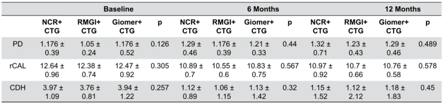
Regarding BOP and PI, none of the treatment groups showed any statistically signiicant changes in the number of positive sites between baseline and
the 12-month observation period (p>0.05). However, all groups presented statistically signiicant reductions in the CDH values (p<0.05). The percentage of CDC were 71.18±23.16% for the NCR+CTG group;
71.33±22.33% for the RMGI+CTG group; and 64.23±20.33% for the giomer+CTG group at 12 months postoperatively. There was no signiicant difference in terms of both CDH and CDC values in any of the groups at the 12-month follow-up. The clinical
parameters are presented in Table 1.
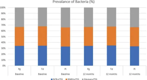
No statistically signiicant difference in the amount of DNA copies of Pg, Td and Pi wasobserved in any of the groups at any time points (p>0.05). In addition, there were no statistically signiicant differences in the counts of the bacterial DNA copies at baseline and at 6 and 12 months postoperatively, regardless of the treatment group (p>0.05; Figures 1 and 2). A reduction was observed in the detection frequency
of Td over time, although this was not statistically signiicant, while the frequency of Pg and Pi was slightly higher at 12 months postoperatively compared
with baseline for all groups (Figure 3).
The two-way ANOVA did not show any signiicant relationship between the amount of DNA copies of all bacteria and the changes in PD values (12 months after surgery and baseline) (PD<1 mm and PD≥1 mm) (Table 2).
The association between changes (12 months
after surgery and baseline) in counts of the bacterial DNA copies and clinical measurements (rCAL, CDH
Baseline 6 Months 12 Months
NCR+ CTG
RMGI+ CTG
Giomer+ CTG
p NCR+
CTG
RMGI+ CTG
Giomer+ CTG
p NCR+
CTG
RMGI+ CTG
Giomer+ CTG
p
PD 1.176 ±
0.39 1.05 ± 0.24 1.176 ± 0.52 0.126 1.29 ± 0.46 1.176 ± 0.39 1.21 ± 0.33 0.44 1.32 ± 0.71 1.23 ± 0.43 1.29 ± 0.46 0.489
rCAL 12.64 ±
0.96 12.38 ± 0.74 12.47 ± 0.92 0.305 10.89 ± 0.7 10.55 ± 0.6 10.83 ± 0.75 0.567 10.97 ± 0.92 10.7 ± 0.66 10.76 ± 0.58 0.578
CDH 3.97 ±
1.09 3.76 ± 0.81 3.94 ± 1.22 0.257 1.12 ± 0.89 1.06 ± 1.15 1.13 ± 1.42 0.32 1.15 ± 1.52 1.12 ± 2.12 1.18 ± 1.83 0.45
p<0.05 considered statistically signiicant for intergroup comparisons, repeated-measures analysis of variance test. PD=Probing Depth; rCAL=relative Clinical Attachment Level; CDH=Combined Defect Height
Table 1- Clinical parameters of the treatment groups in the study follow-up periods
Figure 1- Comparisons of the counts of DNA copies of Pg, Td and Pi (copies/µl) in the treatment groups regardless of time, (a),
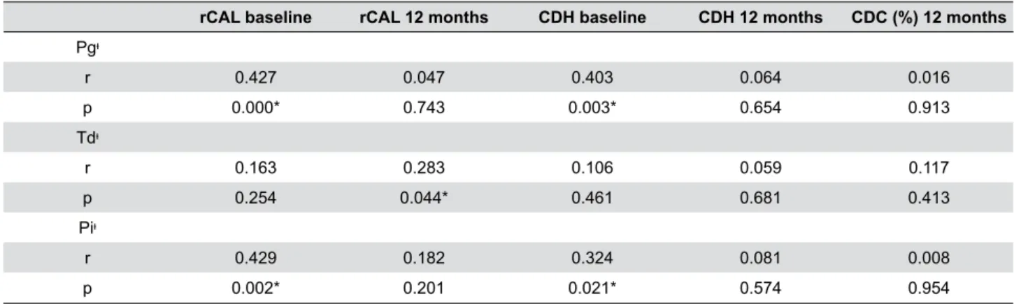
and CDC %) using Pearson’s correlation coeficient is shown in Table 3. The change in counts of Pg’s DNA
copies was positively correlated with rCAL values at baseline in the NCR+CTG group (r=0.507, p<0.05), the RMGI+CTG group (r=0.482, p<0.05) and the giomer+CTG group (r=0.527, p<0.05). Regardless of treatment methods, positive correlations between the amount of DNA copies of Pg and Pi and rCAL and
CDH values at baseline were observed at 12 months postoperatively (p<0.05).
Discussion
In this study, the inluence of different subgingival restorations on periodontal health was evaluated
via combined restorative-periodontal treatment.
The results indicate that, irrespective of the type of
restorative materials, the subgingival restorations did not produce signiicant changes in clinical and microbiological examinations 12 months after surgery.
Previous studies have reported that subgingival
PD*<1 mm PD*≥1 mm
( ) ( )
NCR+CTG RMGI+CTG Giomer+CTG p NCR+CTG RMGI+CTG Giomer+CTG p
Pgᶧ -0.322 ± 0.22 -0.189 ± 0.23 -0.35 ± 0.13 0.447 0.371 ± 0.21 0.139 ± 0.16 0.269 ± 0.14 0.15
Tdᶧ 0.047 ± 0.18 -0.198 ± 0.12 0.135 ± 0.09 0.052 0.078 ± 0.2 -0.166 ± 0.12 0.086 ± 0.09 0.27
Piᶧ -0.372 ± 0.51 -0.23 ± 0.21 0.154 ± 0.2 0.297 -0.345 ± 0.34 0.174 ± 0.25 0.479 ± 0.35 0.891
* changes between 12 months and baseline in probing depth values; ᶧ changes between 12 months and baseline in counts of bacterial DNA copies; p<0.05, two-way ANOVA test
Pg=Porphyromonas gingivalis; Td=Treponema denticola; Pi=Prevotella intermedia
Table 2- Change in counts of DNA copies of Pg, Td and Pi (copies/µl) between treatment groups with the changes of probing depth
between 12 months postoperatively and baseline
Figure 2- Comparisons of the counts of DNA copies of Pg, Td and Pi (copies/µl) at baseline and at 6 and 12 months postoperatively,
irrespective of treatment groups, (a), Porphyromonas gingivalis, (b), Treponema denticola, (c), and Prevotella intermedia, respectively. p<0.05 considered statistically signiicant, repeated-measures analysis of variance test
Figure 3- The frequency detection of Pg, Td and Pi at baseline and at 12 months postoperatively in the treatment groups. p<0.05
placement of restorative materials was more
susceptible to the initation of periodontal disease by
plaque accumulation and release of toxic products5,17,29.
However, studies with a similar study design to ours showed that well-inished subgingival restorations were not associated with periodontal inlammation in combined restorative-periodontal treatment14,21,22,24-26.
Evaluation of differences in the frequency and
quantity of the pathogens is critical in identifying the
relationship between periodontopathogenic bacteria
and periodontal disease. Previous studies have indicated that an increased amount of DNA copies of Pg
and Td tended to be related to worsening periodontal
health status4,15. During the 12-month observation
period of our study, subgingival restorations performed with NCR, RMGI and giomer did not signiicantly affect the amount of DNA copies of Pg, Td and Pi in subgingival plaques. These indings are in accordance with those of previous studies23,28. Santamaria, et al.23
(2013) indicated that the presence of restorations in
the subgingival region could not interfere with the
subgingival microbiata. These authors showed that the target periodontopathogenic bacteria colonization levels were similar between a group that had been restored using RMGI and a non-restored group during the observation periods23. Another previous
study that compared different restorative materials
reported that composite resin showed some negative
effects on the composition of subgingival microbiata compared with RMGI, and also that the decrease in periodontal pathogens was more evident in the RMGI group than in the composite resin group at 6 months
after surgery28. In this study, all groups showed similar
levels of bacterial colonization during the observation periods. This difference can be explained by the fact that a different type of composite material (microilled composite resin) was used to restore the NCCLs in the study by Santos, et al.28 (2007). However, in our study,
a nanocomposite material was used. Furthermore,
the microbiological analysis method could be another
reason for the disagreement between the study by Santos and our study. Counts of bacterial species were determined in samples using the checkerboard DNA-DNA hybridization technique in the study by Santos, et al.28 (2007). The concentration of the target bacteria in
the RMGI group was lower than in the other groups in this study, with no statistically signiicant differences (p>0.05). This is in accordance with the results of an
in vitro study conducted by Tarasingh, et al.32 (2015),
and could be explained by the fact that RMGIs can inhibit the growth of some bacterial species, due to their initial low pH. In contrast with previous studies, the similarity between the results of both RMGI and composite resin in our study was due to the use of
a nanocomposite material. Flausino, et al.6 (2014)
asserted that the incorporation of nanoparticles in
composite resin improved surface topography with less bioilm formation in their study. In addition, giomer, which has the properties of luoride release and luoride recharge potential, was associated with results that are similar to the other restorative materials in
this study.
The trigger for the initiation of periodontal diseases
is the presence of complex microbial biofilms. A
complex consisting of Pg, Tannerella forsythia and Td,
which is called “red complex”, plays important roles in
rCAL baseline rCAL 12 months CDH baseline CDH 12 months CDC (%) 12 months
Pgᶧ
r 0.427 0.047 0.403 0.064 0.016
p 0.000* 0.743 0.003* 0.654 0.913
Tdᶧ
r 0.163 0.283 0.106 0.059 0.117
p 0.254 0.044* 0.461 0.681 0.413
Piᶧ
r 0.429 0.182 0.324 0.081 0.008
p 0.002* 0.201 0.021* 0.574 0.954
* p<0.05, statistically signiicant correlation between Pg, Td, Pi and periodontal measurements; ᶧ changes between 12 months and baseline in counts of bacterial DNA copies;
Pg=Porphyromonas gingivalis; Td=Treponema denticola; Pi=Prevotella intermedia;
rCAL: relative clinical attachment level; CDH: combined defect height; CDC: combined defect coverage
Table 3- Correlations between the changes in counts of DNA copies of Pg, Td and Pi (copies/µl) and the rCAL, CDH and CDC (%) values
the pathogenesis of periodontal disease33 and is highly
related to clinical parameters, such as periodontal
pocket depth and bleeding on probing3. It has also
been found that the second group of bacterial species,
known as the “orange complex” and including Pi, is
also associated with clinical parameters of disease.
Both complex microorganisms are generally found together, and evidence shows that colonization by the red complex species is preceded by colonization and proliferation of the orange complex30. In our study, no
signiicant relationship was found between the amount of DNA copies of all bacteria and PD values in the treatment groups at any of the study periods (p>0.05). However, positive correlations between rCAL values at baseline and the change in counts of Pg’ DNAcopies
between the baseline and 12 months after surgery were found for all groups (p<0.05). Similarly, several studies have reported that red complex species are highly correlated with CAL values13. Another parameter
that correlated with counts of the bacterial DNA copies in this study was CDH values; signiicant positive correlations between rCAL and CDH values at baseline and both counts of DNA copies of Pg and Pi were observed at 12 months postoperatively, regardless of treatment (p<0.05). This can be explained by the fact that the higher CDH values, the more bacteria counts are found in subgingival bioilms.
In our study design, absence of a control group (CTG alone) can be considered a limitation. However, previous studies have demonstrated that the surgical procedures alone could not sufice to reduce dentin hypersensitivity and to provide better aesthetic
results21,22,24,26,36. Moreover, well-inished subgingival
restorations have not been reported to trigger development of periodontal inlammation in combined restorative-periodontal treatment14,21-28. Therefore, this
study was hypothesized to reveal the most satisfactory type of restorative material via microbiological evaluation in the combined periodontal/restorative treatment.
Conclusion
Within the limitations of this study, it was shown
that subgingival placement of restorative materials did not negatively affect the subgingival microlora during the 12-month period after performing combined restorative-periodontal treatment. In addition, the
study indicated that NCR, RMGI and giomer showed similar effects on periodontopathogenic bacteria in the
treatment of gingival recessions that are associated with NCCLs.
Acknowledgments
The study was financially supported by Gazi University Research Board.
Conlicts of interest
The authors report no conlicts of interest related to this study.
References
1- Ainamo J, Bay I. Problems and proposals for recording gingivitis and plaque. Int Dent J. 1975;25(4):229-35.
2- Aroca S, Keglevich T, Barbieri B, Gera I, Etienne D. Clinical evaluation of a modiied coronally advanced lap alone or in combination with a platelet-rich ibrin membrane for the treatment of adjacent multiple gingival recessions: a 6-month study. J Periodontol. 2009;80(2):244–
52.
3- Byrne SJ, Dashper SG, Darby IB, Adams GG, Hoffmann B, Reynolds EC. Progression of chronic periodontitis can be predicted by the levels
of Porphyromonas gingivalis and Treponema denticola in subgingival
plaque. Oral Microbiol Immunol. 2009;24(6):469-77.
4- Carrouel F, Viennot S, Santamaria J, Veber P, Bourgeois D.
Quantitative molecular detection of 19 major pathogens in the
interdental biofilm of periodontally healthy young adults. Front
Microbiol. 2016;7:840.
5- Ferracane JL. Elution of leachable components from composites. J Oral Rehabil. 1994;21(4):441-52
6- Flausino JS, Soares PB, Carvalho VF, Magalhães D, Silva WM, Costa HL, et al. Bioilm formation on different materials for tooth restoration: analysis of surface characteristics. J Mater Sci. 2014;49(19):6820-9. 7- Gurgel BC, Solera NG, Peixoto RF, Assis AO, Calderon PD, Medeiros MC. Evaluation of the periodontal conditions of teeth with restored and non-restored non-carious cervical lesions. Quintessence Int. 2016;47(10):825-31.
8- Haffajee AD, Yaskell T, Torresyap G, Teles R, Socransky SS. Comparison between polymerase chain reaction-based and checkerboard DNA hybridization techniques for microbial assessment of subgingival plaque samples. J Clin Periodontol. 2009;36(8):642-9. 9- Hajishengallis G, Darveau RP, Curtis MA. The keystone-pathogen hypothesis. Nat Rev Microbiol. 2012;10(10):717-25.
10- Hürzeler MB, Dietmar W. A single-incision technique to harvest subepithelial connective tissue grafts from the palate. Int J Periodontics Restorative Dent. 1999;19(3):279-87.
11- Jun HK, Jung YJ, Choi BK. Treponema denticola, Porphyromonas gingivalis, and Tannerella forsythia induce cell death and release of
endogenous danger signals. Arch Oral Biol. 2017;73:72-8.
12- Kakuta E, Nomura Y, Morozumi T, Nakagawa T, Nakamura T, Noguchi K, et al. Assessing the progression of chronic periodontitis
using subgingival pathogen levels: a 24-month prospective multicenter
cohort study. BMC Oral Health. 2017;17(1):46.
14- Lucchesi JA, Santos VR, Amaral CM, Peruzzo DC, Duarte PM. Coronally positioned lap for treatment of restored root surfaces: a 6-month clinical evaluation. J Periodontol. 2007;78(4):615-23. 15- Mineoka T, Awano S, Rikimaru T, Kurata H, Yoshida A, Ansai T, et al. Site-speciic development of periodontal disease is associated
with increased levels of Porphyromonas gingivalis, Treponema
denticola, and Tannerella forsythia in subgingival plaque. J Periodontol.
2008;79(4):670-6.
16- O'Leary TJ, Drake RB, Naylor JE. The plaque control record. J Periodontol. 1972;43(1):38.
17- Padbury A Jr, Eber R, Wang HL. Interactions between the gingiva and the margin of restorations. J Clin Periodontol 2003;30:379-85 18- Paolantonio M, D’ercole S, Perinetti G, Tripodi D, Catamo G, Serral E, et al. Clinical and microbiological effects of different restorative
materials on the periodontal tissues adjacent to subgingival class V
restorations. J Clin Periodontol. 2004;31(3):200-7.
19- Peumans M, Kanumilli P, De Munck J, Van Landuyt K, Lambrechts P, Van Meerbeek B. Clinical effectiveness of contemporary adhesives: a systematic review of current clinical trials. Dent Mater.
2005;21(9):864-81.
20- Pini-Prato G, Franceschi D, Cairo F, Nieri M, Rotundo R. Classiication of dental surface defects in areas of gingival recession. J Periodontol. 2010;81(6):885-90.
21- Santamaria MP, Ambrosano GB, Casati MZ, Nociti Júnior FH, Sallum AW, Sallum EA. Connective tissue graft plus resin- modiied glass
ionomer restoration for the treatment of gingival recession associated
with non-carious cervical lesion: a randomized-controlled clinical trial. J Periodontol. 2009;36(9):791-8.
22- Santamaria MP, Ambrosano GM, Casati MZ, Nociti FH Jr, Sallum AW, Sallum EA. Connective tissue graft and resin glass ionomer
for the treatment of gingival recession associated with noncarious
cervical lesions: a case series. Int J Periodontics Restorative Dent. 2011;31(5):57-63.
23- Santamaria MP, Casati MZ, Nociti FH Jr, Sallum AW, Sallum EA, Aukhil I, et al. Connective tissue graft plus resin-modiied glass ionomer
restoration for the treatment of gingival recession associated with
non-carious cervical lesions: microbiological and immunological results. Clin Oral Investig. 2013;17(1):67-77.
24- Santamaria MP, Queiroz LA, Mathias IF, Neves FL, Silveira CA, Bresciani E, et al. Resin composite plus connective tissue graft to treat
single maxillary gingival recession associated with non-carious cervical
lesion: randomized clinical trial. J Clin Periodontol. 2016;43(5):461-8. 25- Santamaria MP, Silva Feitosa D, Casati MZ, Nociti FH Jr, Sallum AW, Sallum EA. Randomized controlled clinical trial evaluating connective tissue graft plus resin-modiied glass ionomer restoration for the
treatment of gingival recession associated with non-carious cervical
lesion: 2-year follow-up. J Periodontol. 2013;84(9):e1-8.
26- Santamaria MP, Suaid FF, Casati MZ, Nociti FH, Sallum AW, Sallum EA. Coronally positioned flap plus resin-modified glass ionomer
restoration for the treatment of gingival recession associated with
non-carious cervical lesions: a randomized controlled clinical trial. J Periodontol. 2008;79(4):621-8.
27- Santamaria MP, Suaid FF, Nociti FH Jr, Casati MZ, Sallum AW,
Sallum, EA. Periodontal surgery and glass ionomer restoration in the
treatment of gingival recession associated with a non-carious cervical
lesion. Report of three cases. J Periodontol. 2007;78(6):1146-53. 28- Santos VR, Lucchesi JA, Cortelli SC, Amaral CM, Feres M, Duarte PM. Effects of glass ionomer and microilled composite subgingival restorations on periodontal tissue and subgingival bioilm: a 6-month evaluation. J Periodontol 2007;78(8):1522-8
29- Schätzle M, Land NP, Anerud A, Boysen H, Bürgin W, Löe H. The inluence of margins of restorations of the periodontal tissues over 26 years. J Clin Periodontol. 2001;28(1):57-64.
30- Socransky SS, Haffajee AD, Cugini MA, Smith C, Kent RL Jr. Microbial complexes in subgingival plaque. J Clin Periodontol. 1998;25(2):134-44.
31- Socransky SS, Haffajee AD, Smith C, Dibart S. Relation of counts of microbial species to clinical status at the sampled site. J Clin Periodontol. 1991;18(10):766-75.
32- Tarasingh P, Reddy JS, Suhasini K, Hemachandrika I. Comparative evaluation of antimicrobial eficacy of resin-modiied glass ionomers,
compomers and giomers - an invitro study. J Clin Diagn Res. 2015;9(7):ZC85-7.
33- Thurnheer T, Belibasakis GN, Bostanci N. Colonization of gingival epithelia by subgingival bioilms in vitro: role of “red complex” bacteria.
Arch Oral Biol. 2014;59(9):977-86.
34- Toffenetti F, Vanini L, Tammaro S. Gingival recessions and noncarious cervical lesions: a soft and hard tissue challenge. J Esthet Dent. 1998;10(4):208-20.
35- Zucchelli G, De Sanctis M. Treatment of multiple recession-type defects in patients with esthetic demands. J Periodontol. 2000;71(9):1506-14.
36- Zucchelli G, Gori G, Mele M, Stefanini M, Mazotti C, Marzadori M, et al. Non-carious cervical lesions associated with gingival recessions: a decision-making process. J Periodontol. 2011;82(12):1713-24. 37- Zuhr O, Rebele SF, Thalmair T, Fickl S, Hürzeler MB. A modiied
suture technique for plastic and periodontal and implant surgery - the