Prevalence of factor V Leiden in patients
with venous thrombosis
Prevalência do fator V de Leiden em pacientes com trombose venosa
Cristiane Aparecida Michel; Julia B. Rocha; Daiane C. Costa; Carlos Augusto Lima; Anna Paula B. Batschauer
Universidade do Vale do Itajaí, Santa Catarina, Brazil.
First submission on 27/04/16; last submission on 07/07/16; accepted for publication on 13/07/16; published on 20/08/16
ABSTRACT
Introduction: Venous thrombosis is a multifactor disease with high incidence in the population. The development of this disease is closely linked to the presence of environmental and genetic factors and may occur in combination or alone. Among the various genetic mutations that may predispose to a thrombotic event, the most frequent in the population is the G1691A mutation in clotting factor V, known as factor
V Leiden (FVL). This mutation brings the phenotype known as activated protein C resistance, leading to a hypercoagulable state, which increases the risk of thrombosis. Objective: To investigate the presence of FVL in individuals with history of venous thrombosis, and as the control group, individuals with no history of the disease. Method: The method used was the polymerase chain reaction (PCR) followed by restriction fragment length polymorphism (RFLP). Results: The results showed mutation prevalence of 21.5% in heterozygosity in patient
group; no individuals with mutated homozygous were identiied. The results also showed a high recurrence rate among the mutation carriers. Conclusion: In conclusion, the research of mutation on factor V has strong impact on investigation of venous thromboembolism, in order to elucidate the etiology of the event, and in treatment and in prophylaxis against the recurrence.
Key words: polymerase chain reaction; venous thrombosis; thrombophilia.
INTRODUCTION
Venous thrombosis is a pathology with high incidence in the population and is a serious health problem due to its high morbidity, if not recognized and treated early and effectively. Its major complication is when the thrombus detaches from the vessel and reaches the proximal venous system, this may cause pulmonary embolism. The risk of thrombosis increases in patients undergoing orthopedic surgery or major surgery, during pregnancy, postpartum period, in cancer patients, or after trauma and prolonged immobilizations. In addition to these environmental factors, genetic factors may induce a systemic hypercoagulable state, causing venous thrombosis(1, 2).
The term hypercoagulable is deined as state of activation of blood clotting without the formation of the ibrin clot. This state can be transitory, such as during pregnancy or in the postpartum period, or permanent, in cases of hereditary thrombophilia(3).
This may be caused by insuficient inhibition of the blood clotting cascade, either by mutations that result in deiciency of natural
blood coagulation inhibitors, either by mutations leading to the increase in the level or function of clotting factors(4, 5).
In late 1993, Dahlbäck et al., investigating a family with a history of venous thrombosis, discovered through blood clotting tests that some family members showed a poor anticoagulant response to activated protein C. The researchers named this phenotype as activated protein C resistance (APCR)(6). In the
following year, Dahlbäck and Hildebrand (1994) showed that the factor V not only expressed anticoagulants functions, but also participated as a cofactor in the anticoagulant system controlled by the active protein C (APC), and the mutations in the factor V gene could lead to resistance to APC(7).
Shortly after, in the city of Leiden, Netherlands, a group of researchers found that the phenotype of APCR was associated with heterozygosity or homozygosity for one point-mutation in the factor V gene, at position 1691, where occurred a substitution
of a guanine (G) to adenine (A) – G1691A. The mutation
causes an improper synthesis of the factor V molecule, in which the arginine is replaced by glutamine at position 506 of the
protein, one of the factor V cleavage sites. Changing the cleavage site, the mutated factor V becomes resistant to the neutralization by APC, resulting in the APCR phenotype. Since the discovery, this mutation is known as Factor V Leiden (FVL) or factor V R506Q(8).
The factor V has a dominant inheritance, that is, heterozygosity for the FVL increases the risk of thrombosis as much as ive to ten times, and homozygosity (when both alleles are mutated), at ifty
to one hundred times(2). This mutation is common in Caucasians
and almost absent among blacks and Asians(4, 5).
Although many studies have been developed since the discovery of the mutation, data on its prevalence in Brazilian population are still scarce. The investigation of the presence of FVL in the general population and especially in patients with venous thrombosis is very important to clarify the causes and trace possible carriers of the mutation in asymptomatic members of the family, in order to assess the associated risks and determine a preventive medical monitoring(9).
OBJECTIVE
This research aimed at determining the prevalence of G1691A
(FVL) mutation by the allelic discrimination of individuals who had episodes of venous thrombosis and individuals with no history of these events, besides other environmental factors associated with thrombosis, using the polymerase chain reaction followed by restriction fragment length polymorphism (PCR-RFLP) method.
METHOD
Participants were referred from a vascular clinic in the city Itajaí, Santa Catarina, who met the inclusion criteria: thromboembolic events history, conirmed by clinical and imaging diagnosis, and/or had family history of these events. As the control group, individuals who did not have thrombotic events and had no family history of venous thrombosis were evaluated. All subjects agreed on participating in this research upon reading and signing the free and informed consent form, which was approved by the Research Ethics Committee of the Universidade do Vale do Itajaí (Univali), under the opinion 737.684, August, 2014.
Data collection was conducted through a questionnaire in order to investigate the presence of other environmental risk factors associated with venous thrombosis, namely: trauma, prolonged immobilizations, use of oral contraceptive pills, pregnancies and abortions, chronic diseases, cancer, among others.
The material collection was performed from a sample of 3 ml of peripheral blood in a sterile tube containing anticoagulant (ethylenediaminetetraacetic acid [EDTA]), for analysis of the FVL
mutation.
Blood samples had the genomic deoxyribonucleic acid (DNA) extracted from the peripheral blood leukocytes, using the extraction commercial kit Purelink® (Invitrogen®), obtaining, at the end, a
volume of 100 μl of DNA solution. The extracted DNA samples were quantiied and assessed for purity by spectrophotometry, assessing the optical density (OD) at 260/280 ratio, in which OD values between 1.7-1.9 were considered successful for ampliication.
Allelic discrimination of patients regarding the factor V was performed using the polymerase chain reaction (PCR). For the reaction we used 200 ng of DNA, 50 pmol of each oligonucleotide (DNAexpress®) (sense FV1: 5’-TCTCTTGAAGGAAATGCCCCATTA-3’
and anti sense FV2: 5’-GGGCTAATAGGACTACTTCTAATC-3’), 2.5 nmol of each deoxynucleotide triphosphate (dNTP) (Invitrogen®), 1 U of Taq DNA polymerase (Invitrogen Life
Technologies®, Grand Island, NY, USA) and buffer in a inal
volume of 50 μl. For PCR, we used the protocol for 35 cycles with a variation in annealing temperature: 30 seconds at 94ºC, 30 seconds at 60ºC and 1 minute at 72ºC. PCR were performed in the Thermal Cycler from Mastercycler personal (Eppendorf, Hamburg,
Germany).
The analysis of PCR products was obtained after agarose gel electrophoresis and 2% tris-borate-EDTA buffer (TBE) 1×. For this purpose, agarose gel was applied to a volume of 6 µl (5 ul of each sample homogenized with 1 µl bromophenol blue), the electrophoresis run was performed and the gel was stained with ethidium bromide for ultraviolet transilluminator viewing.
The ampliied PCR products were digested overnight at 37º by Mnl I restriction enzymes (New England Biolab Beverly, MA, USA), using a inal volume of 20 μl, 10 μl of the PCR product and 10 μl of enzyme/buffer mix, according to the manufacturer’s instructions.
The size analysis of the fragments obtained by enzymatic fragmentation was performed again after electrophoresis in 2% agarose gel under the same conditions listed above. For proper analysis of the size of the fragments obtained in this run, we applied a standard molecular weight of 100 pb (DNA ladder 100 pb RTU Kasvi®) to the electrophoresis gel, and the samples were
stained with the Safer Dye (Kasvi®) for later viewing in ultraviolet
transilluminator.
loss of the cleavage site by the Mnl I restriction enzyme and thus, individuals heterozygous for the mutation present fragments of 120 pb, 42 pb and 162 pb(10).
RESULTS
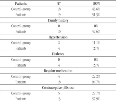
FVL research was performed on 37 individuals of both genders, aged between 9 and 74 years, and mean age of 42.1 years. From the 37 samples analyzed, 51.3% were from patients with personal history of thrombotic event and/or family history and 48.6% constituted the control group of the study. Table 1 shows the
clinical and personal characteristics of the groups surveyed, comorbidities and use ofmedication.
Among the individuals in the control group we did not identify carriers of FVL mutation. In this group, 83.3% were women and 16.7%, men, mean age 41.5 years. Among the factors that predispose to deep vein thrombosis, 11.1% of patients were hypertensive, 16.6%, smokers and none reported diabetes mellitus.
In this group, 22.2% of subjects reported use of regular medication and, among women 27.7% use oral contraceptives.
Among the patients with thrombotic events history, 21.5% were carriers of FVL mutation in heterozygosity. Homozygosity mutation carriers were not identiied, and not mutated homozygous individuals (wild type) amounted to 88.5% of patients, according to data shown in Table 2. From the mutation carriers, 75% had
thrombotic events with recurrence or complications, and 25% reported family history of the disease, with no associated personal
TABLE 2 − Molecular profiling of patients regarding FVL
Genotype n %
Heterozygous 4 21.5
Homozygous mutated 0 0
Wild type (homozygous non-mutated) 15 88.5
FVL: factor V Leiden.
TABLE 1 − Clinical and personal characteristics of the participants
Patients 37 100%
Control-group 18 48.6%
Patients 19 51.3%
Family history
Control-group 0 0%
Patients 10 52.6%
Hypertension
Control-group 2 11.1%
Patients 4 21%
Diabetes
Control-group 0 0%
Patients 4 21%
Regular medication
Control-group 4 22.2%
Patients 18 94.7%
Contraceptive pills use
Control-group 5 27.7%
Patients 13 57.9%
event. Regarding gender, 75% were female and were 25% male.
Also in the group with thrombosis history, the mean age was 42.7 years. The female was represented by 84.2% of subjects and 15.8% were male. In the interview, 52.6% of patients reported family history of venous thrombosis, in addition to the associated personal event. Diabetics and hypertensive amounted to 42% of patients in this group. Regarding the use of drugs, 31.6% conirmed the use of coumarin anticoagulants, 36.8%, use drugs to control metabolic disorders such as hypertension, diabetes, hypercholesterolemia and hypothyroidism, and 26.3%, use antidepressants. Among women, 57.9% of patients reported use of oral contraceptives. Only 10.5% of individuals reported trauma before the thrombotic event; none reported major surgery or prolonged immobilization before
the event.
DISCUSSION
With the characteristics described in the studied group of patients, we found a higher frequency of thromboembolic events
in females. Moreira et al. (2009), evaluating the presence and inluence of risk factors associated with thrombosis in patients from the state of Ceará, also found higher prevalence of venous thrombosis in women (75%). According to the research conducted by these authors, women are statistically more likely to develop thrombosis, with a three times greater risk(11).
According to the literature, the higher frequency of thrombosis in women is due to the greater exposure to environmental risk factors for the development of thrombosis, such as oral contraceptives, pregnancy and abortion, hormone replacement therapy, among others(12). With regard to these factors, 57.6% of
the participating women who developed thrombosis made use of oral contraceptives. Estrogens induce signiicant changes in blood coagulation, such as increasing coagulation factors and decreasing the natural inhibitors of coagulation, resulting in a light procoagulant effect, increasing the resistance to protein C
and generation of thrombin(13-15). Oral contraceptives with
progestogens, offering greater risk for women with FVL(16). The
World Health Organization (WHO) predicts that patients with deep venous thrombosis personal history, pulmonary embolism and those carrying mutations associated to thrombosis, should not make use of combined oral contraceptives(17).
In this study, the risk factors for venous thrombosis, such as hypertension, diabetes and smoking, were not associated to thrombotic event of participating patients, because these disorders have been reported long after the occurrence of the irst thrombotic event and smokers were not identiied in this group. There is no description in the literature involving the use of drugs for treatment of metabolic diseases with venous thrombosis or its complications. The FVL is the most common genetic mutation that leads to thrombophilia with a prevalence in Caucasian populations of approximately 5%, and is extremely rare in other populations(18).
Rosendaal et al. (2005) describe that 3% of the general population are heterozygous and, homozygous individuals, have expected prevalence of two per 10,000 births. The same authors evidence that in heterozygous carriers of the mutation, the risk of venous thrombosis increases three to eight times and in homozygous it may increases 50 to 80 times(14).
According to the literature, the mutation is found in 20% of patients with venous thrombosis, and in over 50% of patients with thrombophilia(14). This igure corroborates the results found
in this study in which the prevalence of the FVL mutation found was 21.5%. Moreover, in a study by Ramos et al. (2006), the
frequency found was 13.3% for the FVL mutation(10). In another
study conducted in Minas Gerais, Guimarães et al. (2009) investigated the presence of mutations in FVL and in prothrombin
(G20210A) in 1,103 individuals suspected of thrombophilia. The FVL mutation was detected in 83 heterozygous subjects (7.52%) and four homozygous (0.36%), results lower to those found in this. However, homozygous individuals were not identiied in this
study(19). A recent study conducted in the Czech Republic with 575
patients found a frequency of 20.9% of patients with FVL mutation, corroborating the results(20).
In this study, approximately 30% of patients with thrombotic episodes followed on anticoagulants use. According to the recommendations for anticoagulation therapy, the presence of inherited thrombophilia is not considered a major risk factor for determining the treatment duration, however the literature is disparate regarding the duration of anticoagulant therapy to prevent a future thrombosis event(21).
Ridker et al. (2003), in a randomized, double-blind,
placebo-control study, demonstrated that extended therapy with coumarin drugs resulted in signiicant reduction in the risk of recurrent venous thrombosis, including patients with hereditary
thrombophilia(22). Agnelli and Becattini (2008), evaluating the
duration of treatment for venous thrombosis and how to predict recurrence, argue that all patients should receive anticoagulant therapy for three months, but patients with intrinsic risk for recurrence of thrombosis and bleeding events should be reviewed as the beneit of extended therapy(23).
With regard to the recurrence of thrombosis, literature data are conlicting, both the use of an appropriate prophylactic therapy for these patients, and the risk of recurrence for heterozygosity FVL mutation carriers. In the present study, the thrombosis recurrence rate was 75% among patients with this mutation. A study published in 2002 compared the incidence of recurrent venous thromboembolism among individuals heterozygous for the mutation and those without the mutation, inding a higher frequency of recurrence of those without the mutation (20% and 21.6%, respectively)(24).
In a study developed in 2006, which evaluated 259 patients carriers of FVL and prothrombin (G20210A) mutations with recurrent thrombosis, the authors found that individuals homozygous for one of the two mutations or those carriers of both mutations in heterozygosity have an increased risk of recurrence, when compared to non-mutated individuals. The same authors suggest that long-term anticoagulation therapy after the irst thrombotic event seems to be beneicial in these cases(25). Moreover,
Lijfering et al. (2010), evaluated 788 patients with recurrent
venous thrombosis and carriers of FVL and G20210A mutation, the authors states that these individuals do not have increased risk of developing recurrent thrombotic condition(26).
In a case report published recently, White et al. conclude that
subjects heterozygous for the FVL mutation are at increased risk for recurrence, especially in the presence of modiiable risk factors. Therefore, awareness of patients about these risk factors are essential in order to reduce the risk of recurrent thromboembolism(21).
From a clinical point of view, it is dificult to calculate the risks to predict a recurrent thrombotic event because many factors involve the etiopathogenesis of thrombosis. A study published by Vlieg et al. (2015) combines high levels of D-dimer to the risk
of recurrence of venous thrombosis and pulmonary embolism, in an attempt to establish a laboratory marker with high predictive value to aid in calculating the risk of a new thrombotic event(27).
CONCLUSION
REFERENCES
1. Fauci AS, Braunwald E, Isselbacher KJ, et al. Harrison medicina interna. 14 ed. Rio de Janeiro: McGraw-Hill; 1998.
2. Goldmann L, Ausiello D. Cecil tratado de medicina interna. 22 ed. Rio de Janeiro: Elsevier; 2004.
3. Lopes AC. Tratado de Clínica Médica. 2 ed. São Paulo: Roca; 2009. 4. Duque FLV, Mello NA. Trombogênese – tromboilia. J Vasc Bras, Rio de Janeiro. 2003; 2(2): 105-18.
5. Schiffman FJ. Fisiopatologia hematológica. São Paulo: Santos; 2004. 6. Dahlbäck B, Carlsson M, Svensson PJ. Familial thrombophilia due to a previously unrecognized mechanism characterized by poor anticoagulant response to activated protein C: prediction of a cofactor to activated protein C. Proc Natl Acad Sci USA. 1993; 90: 1004-8.
7. Dahlbäck B, Hildebrand B. Inherited resistance to activated protein C is corrected by anticoagulant cofactor activity found to be a property of fator V. Proc Natl Acad Sci USA. 1994; 91: 1396-400.
8. Bertina MR, Koeleman BPC, Koester T, et al. Mutation in blood coagulation fator V associated with resistance to activated protein C. Nature. 1994; 369: 64-7.
9. Bonim AS, Favoreto NMS, Dilva RNM, Brum CA, Valadão AF. Investigação de mutação nos genes do fator V, da trombina, e na metilenotetrahidrofolatoredutase (MTHFR) em pacientes com história de trombose. Farmácia & Ciência, Ipatinga. 2011; 2: 23-8.
thromboembolism and reassessment of anticoagulation in recurrence. Furthermore, the use of oral contraceptive seems to be strongly associated to the thrombotic events developed by the patients. It is noteworthy that the impact of our conclusion is
limited by the sample size of the study, requiring, therefore, more research involving the FVL and deep vein thrombosis in order to better deine the genetic proile of this population in relation to the presence of mutation in factor V.
RESUMO
Introdução: A trombose venosa é uma doença de caráter multifatorial e de alta incidência na população. O desenvolvimento dessa patologia está intimamente ligado à presença de fatores ambientais e genéticos, podendo ocorrer em associação ou isoladamente. Entre as diversas mutações genéticas que podem predispor a um evento trombótico, a de maior ocorrência na população é a mutação G1691A no fator V da coagulação, conhecida como fator V de Leiden (FVL). Essa mutação provoca o fenótipo conhecido como resistência à proteína C ativada, levando a um quadro de hipercoagulabilidade, que aumenta o risco de trombose. Objetivo: Investigar a presença do FVL em indivíduos com histórico de trombose venosa e, como grupo-controle, indivíduos sem histórico. Método: A metodologia utilizada foi a técnica de reação em cadeia da polimerase (PCR), seguida de polimorfismo no comprimento dos fragmentos de restrição (RFLP). Resultados: Os resultados mostraram prevalência da mutação de 21,5% em heterozigose entre o grupo de pacientes; não foram identificados indivíduos homozigotos mutados. Os resultados também apresentaram alto índice de recorrência entre os portadores da mutação. Conclusão: Conclui-se que a pesquisa da mutação do fator V tem forte impacto na investigação do tromboembolismo venoso a fim de elucidar a etiologia do evento, além de auxiliar no tratamento e na profilaxia diante da recorrência.
Unitermos: reação em cadeia da polimerase; trombose venosa; trombofilia.
10. Ramos CCPS, Campos JF, Melo FCBC, et al. Frequência do fator V Leiden em indivíduos sob investigação de tromboilia, Recife, Pernambuco, Brasil. Rev Bras Hematol Hemoter, São José do Rio Preto. 2006; 28(2): 131-4.
11. Moreira AM, Rabenhorst SHB, Holanda RARR, Pitombeira MH. Fatores de risco associados a trombose em pacientes do estado do Ceará. Rev Bras Hematol Hemoter, São José do Rio Preto. 2009; 31(3): 132-6.
12. Siqueira C. Trombose na mulher. Rev SOCERJ, Rio de Janeiro. 2002; 15(1): 34-8.
13. Brito MB, Nobre F, Vieira CS. Contracepção hormonal e sistema cardiovascular. Arq Bras Cardiol. 2011; 96(4): 81-9.
14. Rosendaal FR. Venous thrombosis: the role of genes, environment, and behavior. Hematol Am Soc Hematol Educ Program. 2005; p. 1-12. 15. Vieira CS, Oliveira LCO, Sá MFS. Hormônios femininos e hemostasia. Rev Bras Ginecol Obstet. 2007; 29(10): 538-47.
16. Kemmeren JM, Algra A, Meijers JC, et al. Effect of second-and thrid-generation oral contraceptives on the protein C system in the absence or presence of the fator V Leiden mutation: a randomized trial. Blood. 2004; 103(3): 927-33.
17. World Health Organization (WHO): medical eligibility criteria for contraceptive use. 5th ed. Geneva; 2015.
18. Rees DC, Cox M, Clegg JB. World distribution on factor V Leiden. Lancet. 1995; 346: p. 1133-4.
Gerais-Brasil com suspeita clínica de trombose. Rev Bras Hematol Hemotera, São José do Rio Preto. 2009; 31(1): 19-24.
20. Hirmerova J, Seidlerova J. The association of fator V Leiden with various clinical patterns of venous thomboembolism – the fator V Leiden paradox. Q J Med. 2014; 107: 715-20.
21. White CW, Thomason AR, Prince V. Recurrent venous thromboembolism in a patient with heterozygous factor V leiden mutation. Hosp Pharm. 2014; 49(8): 748-51.
22. Ridker PM. Long-term, low-dose warfarin among venous thrombosis patients with and without factor V Leiden mutation: rationale and design for the Prevention of Recurrent Venous Thromboembolism (PREVENT) trial. Vasc Med. 2003; 3: 67-73.
23. Agnelli G, Becattini C. Treatment of DVT: how long is enough and how do you predict recurrence. J Thromb Thrombolysis. 2008; 25: 37-44.
24. Eichinger S, Weltermann A, Mannhalter C, et al. The Risk of recurrent venous thrombosis in heterozygous carriers of fator V Leiden and a irst spontaneous venous thromboembolism. Arch Intern Med. 2002; 162(20): 2357-60.
25. González-Porras JR, García-Sanz R, Alberca I, et al. Risk of recurrent venous thrombosis inpatients with G20210A mutation in the prothrombin gene or factor V Leiden mutation. Blood Coagul Fibrin. 2006; 17(1): 23-8. 26. Lijfering WM, Middeldorp S, Veeger NJ, et al. Risk of recurrent venous thrombosis in homozygous carriers and double heterozygous carriers of factor V Leiden and prothrombin G20210A. Circulation. 2010; 121: 1706-12. 27. Vlieg AVH. The risk of a irst and a recurrent venous thrombosis associated with na elevated D-dimer level and an elevated thrombin potential: results of the THE-VTE study. J Thromb Haemost. 2015; 13: 1642-52.
CORRESPONDING AUTHOR
Cristiane Aparecida Michel