First record of the genus
Tropidia
Lindl. (Orchidaceae) for Brazil
Ana Kelly Koch1,4, Climbiê Ferreira Hall1, Eric Smidt2, Audia Brito Rodrigues de Almeida2, Mônica Bolson2 and Celice Alexandre Silva3
Received: 28.04.2016; accepted: 15.08.2016
ABSTRACT - (First record of the genus Tropidia Lindl. (Orchidaceae) for Brazil). The genus Tropidia is composed of ca. 20-30 species distributed in southern Asia, South Pacific islands and northern Australia, and a single species occurring in America, from United States to Ecuador. The first record of the genus Tropidia for Brazil is presented here. It consists of Tropidia polystachya, a species found in the Cerrado biome, Midwestern Brazil. Description, illustration, photos, and distribution map of the species are provided. Additionally, the leaf anatomy of the species was studied and it is very similar to the leaves of other species of Tropidieae described so far. DNA sequences of T. polystachya are presented (plastid matK and rbcL and nuclear ITS) in order to assist future phylogenetic studies with the genus.
Keywords: Anatomy, Cerrado biome, terricolous habit, youngpalm orchid
RESUMO - (Primeiro registro do gênero Tropidia Lindl. (Orchidaceae) para o Brasil). O gênero Tropidia é composto por ca. 20-30 espécies distribuídas pelo sul da Ásia, ilhas do Pacífico Sul, norte da Australia, e uma única espécie ocorrendo também na América, desde os Estados Unidos até o Equador. O primeiro registro do gênero Tropidia para o Brasil é apresentado aqui. A espécie é Tropidia polystachya, encontrada no bioma Cerrado, no Centro Oeste do Brasil. Neste trabalho são fornecidos para a espécie uma descrição, ilustração, fotos e um mapa de distribuição. Adicionalmente, foi estudada a anatomia foliar da espécie, que apresenta morfologia muito similar a das folhas de outras espécies de Tropidieae que já foram estudadas. Sequência de DNA de T. polystachya são apresentadas (plastidial, matK e rbcL, e nuclear, ITS) afim de complementar futuros estudos filogenéticos com o gênero.
Palavras-chave: Anatomia, bioma Cerrado, hábito terrícola, orquídea youngpalm
1. Museu Paraense Emílio Goeldi, Programa de Capacitação Institucional, Campus de Pesquisa, Coordenação de Botânica. Av. Perimetral, Terra Firme, 66077-830 Belém, PA, Brasil
2. Universidade Federal do Paraná, Setor de Ciências Biológicas, Centro Politécnico, Jardim das Américas, Caixa postal 19031, 81531-980 Curitiba, Paraná, Brasil
3. Universidade do Estado de Mato Grosso, Centro de Estudos, Pesquisa e Desenvolvimento Agro-Ambiental (CPEDA) Rod. MT 358, km 7,5, Jardim Aeroporto, Caixa postal 287, 78300-000 Tangará da Serra, Mato Grosso, Brasil
4. Corresponding author: anakbio@gmail.com
Introduction
The genus Tropidia Lindl (Orchidaceae) is
composed of ca. 20-30 species, distributed in southern Asia, South Pacific islands and northern Australia, and a single species occurring in America, from United States to Ecuador (Pridgeon et al. 2005, Govaerts et al.
2016). Early classifications (e.g. Dressler 1981) placed Tropidia as related to Spiranthoid orchids, especially
due to its erect dorsal anther and soft sectile pollinia (Pridgeon et al. 2005). However, cladistic analysis
based on anatomical and morphological characters positioned the genus within Epidendroideae (Stern
et al. 1993, Freudenstein & Rasmussen 1999). On the
other hand, molecular analyses, of the rbcL plastid
gene, positioned Tropidia as part of an unresolved
grade that is sister of most other Epidendroideae (Cameron et al. 1999). A recent phylogenetic study
on the subfamily based on mitocondrial, nuclear and plastid regions combined (Freudenstein & Chase 2015) placed Tropidia as sister to Corymborkis
Thouars, with strong support, among the other early-branching epidendroids.
Tropidia comprises terricolous herbs, green or
species of the genus (Pridgeon et al. 2005). Detailed
information about T. polystachya are scarce, however,
is distributed from Southern Florida through the West Indies, Venezuela, Central America and Galapagos Islands (Pridgeon et al. 2005).
In this study we expand the known distribution of T. polystachya, registering the first record of the
species for Brazil. Additionally, based on the analyses of the Brazilian specimen, we present a description, detailed illustration, photographs, distribution map, anatomical characterization of the leaves, and plastid genes (rbcL and matK) and nuclear (ITS) sequences
of the species.
Material and methods
Sampling - In 2012 and 2013, during field works in southwestern Mato Grosso State, in an area of the Cerrado biome, sterile individuals of T. polystachya
were found. The population occurs in the Reserva Particular do Patrimônio Natural (RPPN) Vale do Sepotuba, located in the São Marcelo Farm (municipality of Tangará da Serra). The reserve occupies 1.200 ha of native semideciduous forest on the margins of the Sepotuba River, and presents a rhodic hapludox soil (Martins et al. 2010). This
region is climatically classified as AW, according to the Köppen (1948) system, with an average annual temperature of 24.4 ºC, annual precipitation of 1.500 mm, and a relative humidity ranging from 50% to 80%.
Collected specimens were grown in the Catasetum Orquidarium at the campus Tangará da Serra of Universidade do Estado de Mato Grosso (UNEMAT). A specimen flowered in April 2013 and a voucher was pressed and deposited at Herbarium TANG (acronym according to Thiers 2015).
Species description - Flower measurements were taken from pickled flowers and all other measurements were made from the dried material. General morphological terms were based on Harris and Harris (1994), Radford
et al. (1974) and Stearn (1983). The illustration was
based on voucher material and pickled flowers. Map of distribution was made using the distribution presented in genera Orchidacearum (Pridgeon 2005) and from
compiled data available in Govaerts et al. (2016).
DNA sequencing - DNA sequences from three regions (matK and rbcL plastid genes, and the nuclear ITS)
according to Doyle & Doyle (1987), using 2× CTAB extraction buffer, without the addition of RNase A and scaled to 2 ml tubes. Polymerase chain reaction (PCR) of all fragments was performed in 20 µl reactions (1× enzyme buffer, 2.5 mM MgCl2, 0.2 mM dNTPs, 0.5 µM of each primer, 10 ng BSA, 1 U of Taq DNA Polymerase (Invitrogen, Life Technologies Corporation), and 20-50 ng of genomic DNA. For the nuclear region 5 M betaine, 2% DMSO were added to the reaction. The nrITS region was amplified and sequenced with the primers described in Desfeux & Lejeune (1996); matK with the primers matK 3F_Kim f and matK 1R_Kim r (Ki-Joong Kim, pers. comm.); and rbcL with the primers rbcLa_f (Levin et al. 2003) and rbcLa_r (Kress & Eriksson 2007).
Thermal cycling profile consisted of an initial step of 1 min at 94 °C and a final step of 5 min at 72 ºC, which was intercalated with 40 cycles of denaturation at 94 °C for 30 s, annealing at 51 °C for 40 s (nrITS), 53°C (matK and rbcL), and extension at 72 °C for 40 s.
The PCR products were purified with polyethylene glycol (PEG) 10%, and the sequencing reactions were performed in the ABI3739XL genetic analyzer (Macrogen Inc., South Korea), using the same primers of the PCR amplification. The sequences were superimposed and edited with the Staden Package software (Staden et al. 2003). Statistics of the three
genomic regions of T. polystachya were calculated
in the MEGA 6 software (Tamura et al. 2013). The
sequences obtained were compared with those from the GenBank database using the BLASTn tool from the National Center for Biotechnology Information (NCBI) (https://blast.ncbi.nlm.nih.gov/) with default parameters. Sequences of plastid matK, rbcL and nrITS nucleotide obtained during this study were deposited in GenBank under accession numbers KR604798, KR604799 and KR604800.
Anatomical characterization - Two leaves of one individual were pickled in 70% ethyl alcohol or FAA 50 (Johansen 1940). Cross and paradermic sections from the medium third of the leaf blade were obtained. Histological sections were freehand sectioned with razor blade. Cross-sections were stained with astra blue and safranin 9:1 (Bukatsch 1972) and the paradermic sections were stained with toluidine blue as described by O’ Brien et al. (1965). Semi-permanent
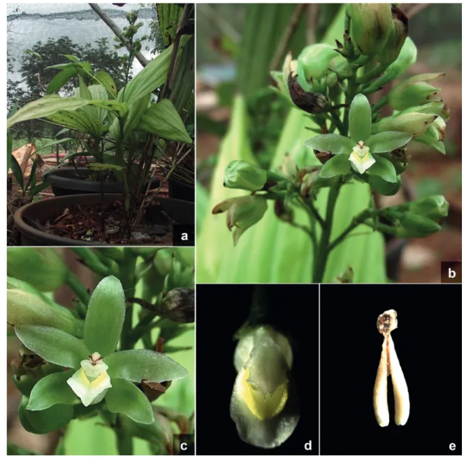
Figure 2. Tropidia polystachya. a. Habit. b. Inflorescence. c. Flower. d. Column with lip (front view). e. Pollinarium. Photos by Ana Kelly Koch and Celice Alexandre Silva.
Results and Discussion
Tropidia polystachya (Sw.) Ames, Orchidaceae 2:
262 (1908). Figures 1-2
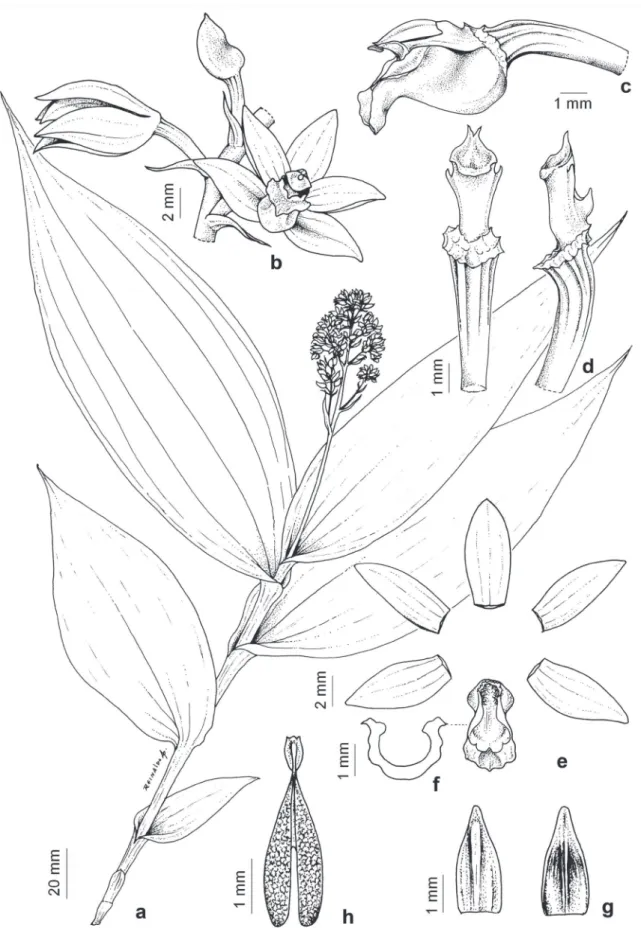
Terricolous herbs. Roots villous. Stem ca. 25 cm tall, 2-3 cm diam., erect, cylindrical, partially covered by tubular sheaths when juvenile, naked when older, distally covered by foliar sheaths. Sheaths thin, often
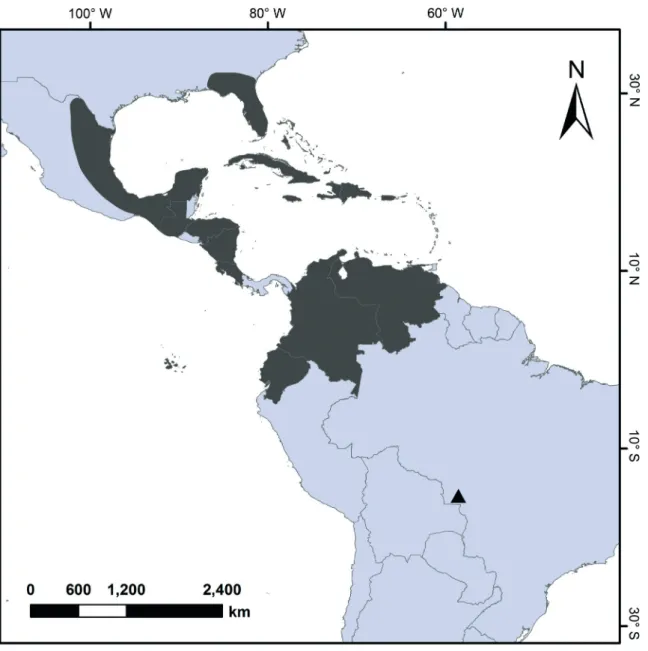
Figure 3. Distribution map of Tropidia polystachya including the new record for Brazil. lip ca. 7 × 4 mm, white with a yellow macula on the
disc, oblong, constricted at the middle, embracing the column, sometimes obscurely 4-lobed, apex retuse and recurved; disc 2-lamellate, distally flabellate; column ca. 3 mm long, greenish, subcylindrical; anther 2-3 mm long, ovoid; pollinarium 3-4 mm long, viscidium ca. 1 mm long, oblongate, hamulus stipe 1 mm long, slender; pollinia 2, ca. 2 mm long, white, clavate; stigma transversally elliptic; rostellum broadly triangular, apex bifid. Capsules not seen. Examined material: BRAZIL. Mato Grosso: Tangará
da Serra, Reserva Particular do Patrimônio Natural Vale do Sepotuba, Fazenda São Marcelo, bank of the Sepotuba River, 2-IV-2013, D.C. Dias 7 (TANG!).
Habitat and distribution - Tropidia polystachya has
been cited for Southern Florida through the West Indies, Venezuela, Central America and Galapagos Islands (Pridgeon et al. 2005), and in the present study its
distribution is extended to the Central Brazil (figure 3). The young palm orchid is a short plant with palm aspect, which usually grows in deep shade of forest floor, often on leaf litter, loamy soil or sandy humus (Hammer 1997, Pridgeon et al. 2005). In Brazil, T. polystachya
grows on loamy soil in open areas of Cerrado. These data increase not only the area of occurrence, but also the types of habitat where the species occurs.
Conservation status - Tropidia polystachya has a broad
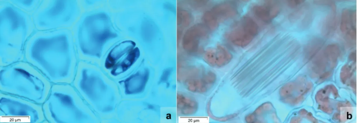
Figure 4. Anatomical characterization of the leaves of Tropidia polystachya. a. Abaxial epidermis with tetracytic stomata and irregular-shaped epidermal cells (front view); b. Idioblasts containing crystals in the shape of druses (front view); c. Unisseriate tector trichome (front view); d. General view of the leaf (cross section); e. Epidermal cells, homogeneous mesophyll, stomata with substomatal chamber (arrow) and collateral vascular bundle (cross section); f. Middle vascular bundle partially surrounded by sclerenchyma fibres (cross section). Categories and Criteria (IUCN 2014). On the other
hand, T. polystachya is known only in one locality
in Brazil, and although found within a protected conservation unit, the species should be considered as Critically Endangered (CE; B1) for the country. DNA sequencing - The genus Tropidia and
Corymborkis are recognizable within the Tropidieae
tribe, among the other early-branching Epidendroideae
another sequence of T. polystachya (EU490674.1)
deposited in the GenBank database of the NCBI; matK
plastid sequence (KR604800), with 897 bp, presented 99% identity with T. graminea (EF079304.1); and rbcL plastid sequence (KR604799), with 561 bp,
presented 100% identity with Tropidia sp. (Chase
O-211). Unfortunately, due to the lack of more sequences from species of Tropidia deposited in the
phylogenetic placement of T. polystachya within the
genus at the moment.
Anatomical characterization - The leaf of T. polystachya
presents, in front view, epidermal cells irregularly shaped, slightly curved anticlinal walls and tetracytic stomata (figure 4a). Additionally, stegmatas were observed close to the vascular bundles, as well as the presence of idioblasts with raphides in epidermal tissue (figure 4b), and uniseriate glandular trichomes scattered on the leaf surface (figure 4c). In cross section, the leaf blade shows a flat surface (figure 4d). The uniseriate epidermis cells are elliptical or isodiametric in shape, and the cells of the adaxial epidermis are higher than the cells of the abaxial epidermis (figure 4e). Presence of a thin cuticle covering the leaf blade was observed. Hypostomatic leaves show stomata at the same level of the other epidermal cells and present well developed substomatal cavity. Chlorenchyma is homogeneous, with isodiametric, rectangular or elliptical cells arranged from three to four layers horizontally disposed. Presence of collateral vascular bundles surrounded by a sclerenchymatic sheath was also observed (figure 4e). Midrib is constituted by a large collateral vascular bundle, whose adaxial region is surrounded by three to four layers of fibers, whereas the abaxial region of the bundle is surrounded by seven to eight layers of fibers arranged in an U shape (figure 4f).
The vegetative morphology and anatomy have proved to be great data sources to the understanding of relationships in groups within Orchidaceae, as opposed to reproductive characters, notably prone to selective pressures by pollinator groups and therefore considered more homoplastic (Cameron 2005, Salazar & Dressler 2011). Previously, leaf anatomy of Tropidia
was only known from T. curculigoides Lindl., species
distributed from the Himalaya to New Caledonia (Stern et al. 1993). Leaves of T. polystachya, from
the individuals found in open areas of Cerrado are anatomically very similar to the leaves of other species of Tropidieae described so far (Stern et al.
2013, 2014), including the genus Corymborkis Thou.
Thus, we believe that the leaf anatomical features may have low taxonomic significance for the tribe, since they remained unchanged even in very different habitats and species. The overall similarities with others basal Epidendroideae like Sobralia Ruiz &
Pavón, Palmorchis Barb. Rodr., Xerorchis Schltr.
and Eleanthus C. Presl indicated a conservative
leaf structure and would probably be useful for the recognition of these basal groups of the subfamily in a broader context.
Acknowledgements
The authors acknowledges the support from the Brazilian agencies: Conselho Nacional de Desenvolvimento Científico e Tecnológico (CNPq) for the fellowship of the Programa de Capacitação Institucional (PCI-DB; MPEG/MCTI) to the first and second authors, and the productivity grant CNPq-Nível 2 to the third author; Fundação de Amparo à Pesquisa do Estado de Mato Grosso (FAPEMAT) process 296039/2010 for the financial support to the last author. We also thank the administration of the São Marcelo Farm; and Reinaldo Aguiar for the illustration.
Literature cited
Bukatsch, F. 1972. Bemerkungen zur Doppelfärbung
Astrablau-safranin. Mikrokosmos 61:33-36.
Cameron, K.M. 2005. Leave it to the leaves: a molecular
phylogenetic study of Malaxideae (Epidendroideae, Orchidaceae). American Journal of Botany 92: 1025-1032.
Cameron, K.M., Chase, M.W., Whitten, W.M., Kores, P.J., Jarrell, D.C., Albert, V.A., Yukawa, T., Hills, H.G. & Goldman, D.H. 1999. A phylogenetic analysis
of the Orchidaceae: evidence from rbcL nucleotide sequences. American Journal of Botany 86:208-224.
Desfeux, C. & Lejeune, B. 1996. Systematics of
Euromediterranean Silene (Caryophyllaceae): evidence from a phylogenetic analysis using ITS sequences. Comptes rendus de l’Académie des sciences Série III, Sciences de la vie 319: 351-358.
Doyle, J. & Doyle, J. 1987. A rapid DNA isolation procedure
for small amounts of leaf tissue. Phytochemical Bulletin 19: 810-815.
Dressler, R. 1981. The orchids - natural history and
classification. Harvard University Press, Cambridge.
Ford, C.S., Ayres, K.L., Toomey, N., Haider, N., Stahl, J.V.A., Kelly, L.J., Wikström, N., Hollingsworth, P.M., Duff, R.J., Hoot, S.B., Cowan, R.S., Chase, M.W. & Wilkinson, M.J. 2009. Selection of candidate
coding DNA barcoding regions for use on land plants. Botanical Journal of the Linnean Society 159:1-11.
Freudenstein, J.V. & Chase, M.W. 2015. Phylogenetic
relationships in Epidendroideae (Orchidaceae), one of the great flowering plant radiations: progressive specialization and diversification. Annals of Botany 115: 665-681.
Freudenstein, J.V. & Rasmussen, F.N. 1999. What does
morphology tell us about orchid relationshis? A cladistic analysis. American Journal of Botany 86: 225-248.
Govaerts, R., Bernet, P., Kratochvil, K., Gerlach, G., Carr, G., Alrich, P., Pridgeon, A.M., Pfahl, J., Campacci, M.A., Holland Baptista, D., Tigges, H., Shaw, J., Cribb, P., George, A., Kreuz, K. & Wood, J. 2016. World Checklist of Orchidaceae. Facilitated by
terminology - an illustrated glossary. Spring Lake Publishing, Spring Lake.
IUCN. 2014. The IUCN red list of threatened species,
version 3.1. IUCN Red List Unit, Cambridge U.K. Available in http://www.iucnredlist.org/ (access in 15-XI-2014).
Johansen, D.A. 1940. Plant microtechnique. McGraw Hill
Book, New York.
Kaiser, E. 1880. Verfahren zur Herstellung einer tadellosen
glyceringelatine. Bot Zentralb 180:25-26.
Köppen, W. 1948. Climatologia: con un estudio de los climas
de la Tierra. Fondo de Cultura Economica, Mexico.
Kress, J. & Erickson, D.L. 2007. A two-locus global
DNA barcode for land plants: the coding rbcL gene complements the non-coding trnH-psbA spacer region. PLoS ONE 6: 1-10.
Levin, R.A., Wagner, W.L., Hoch, P.H., Nepokroeff, M., Pires, J.C., Zimmer, E.A. & Sytsma, K.J. 2003.
Family-level relationships of Onagraceae based on chloroplast rbcL and ndhF data. American Journal of Botany 90: 107-115.
Martins, A., Dallacort, R., Inoue, M.H., Santi, A., Kolling, E.M. & Coletti, A.J. 2010. Probabilidade de precipitação
para a microrregião de Tangará da Serra, Estado do Mato Grosso. Pesquisa Agropecuária Tropical 40: 291-296.
NCBI. 2016. National Center for Biotechnology
Information. Bethesda, U.S.A. Available in https:// blast.ncbi.nlm.nih.gov (access in 20-III-2016).
O’Brien, T.P., Feder, N. & Mccully, M.E. 1965.
Polychromatic staining of plant cell walls by toluidine blue O. Protoplasma 59: 368-373.
Oxford.
Radford, A.E., Dickison, W.C., Massey, J.R. & Bell, C.R. 1974. Vascular Plant Systematics. Harper & Row,
New York.
Salazar, G.A. & Dressler, R.L. 2011. The leaves got it
right again: dna phylogenetics supports a sister-group relationship between Eurystyles and Lankesterella (Orchidaceae: Spiranthinae) Lankesteriana 11: 337-347. Staden. R., Judge, D.P. & Bonfield, J.K. 2003. Analysing
sequences using the Staden Package and EMBOSS. In: S.A. Krawetz & D.D. Womble (eds.). Introduction to bioinformatics. Humana Press, Totowa, pp. 393-410.
Stearn, W.T. 1983. Botanical Latin. David & Charles,
London.
Stern, W.L. 2014. Orchidaceae. In: M. Gregory & D.F. Cutler (eds.). Anatomy of the monocotyledons. vol. X. Oxford University Press, Oxford, pp 1-288.
Stern, W.L., Morris, M.W. & Judd, W.S. 1993.
Comparative vegetative anatomy and systematics of Spiranthoideae (Orchidaceae) Botanical Journal of the Linnean Society 113: 161-197.
Tamura, K., Stecher, G., Peterson, D., Filipski, A. & Kumar, S. 2013. MEGA6: Molecular Evolutionary
Genetics Analysis version 6.0. Molecular Biology and Evolution 30: 2725-2729.
Thiers, B. [2016 continuously updated]. 2014. Index