Dental Press J. Orthod. 13 v. 15, no. 1, p. 13-15, Jan./Feb. 2010
Three-dimensional face morphometry
Facial anthropometry plays a key role in clini-cal assessments, providing an accurate diagnosis for different syndromes. Clinicians working with the head and face (maxillo-facial, plastic and aesthetic surgeons; orthodontists and prosthodontists) are the mostly interested in this three-dimensional in-formation, being able to estimate the normal and abnormal growth, planning and evaluating surgi-cal or orthodontic treatment, plastic surgeries and anthropometric studies.1,6
Currently, classic direct anthropometry is be-ing replaced with various three-dimensional im-age (3D) analyzers, and the knowledge and appli-cation of this technology is essential for clinicians to analyze the information for planning and evalu-ating medical procedures and treatments.
Facial landmarks (previously marked on the face of the subject) represent the link between
conventional and digital anthropometry7:
con-ventional anthropometry identifies soft-tissue landmarks, and places some instrument (calipers, protractors) over them. Fundamentally, digital anthropometry collects a set of digital landmarks from the soft-tissue surface, and uses their spatial x, y, z coordinates as end-points for calculations based on Euclidean geometry: linear distances and angles. Together with these classic measurements, mathematics and geometrics allow the assessment of more complex characteristics from the same set of landmarks used by conventional anthropom-etry: estimations of volumes and surfaces, analyses of symmetry, and detailed assessments of shape.2-5,8
THREE-DIMENSIONAL MORPHOMETRY
METHODS
Two main groups of instruments can be used for soft-tissue three-dimensional facial anthro-pometry: contact instruments (electromagnetic and electromechanical digitizers, ultrasound probes) and optical/non contact instruments (la-ser scanners, optoelectronic instruments, stereo-photogrammetry, Moiré topography). All these instruments are not invasive and do not provoke pain or discomfort to the subjects. But both
WH A T’S N E W I N DE N T I S T R Y
Márcio de Menezes*, Chiarella Sforza**
* Postgraduate student, Department of Human Morphology, University of Milan, Milano, Italy. ** Professor, Department of Human Morphology, University of Milan, Milano, Italy.
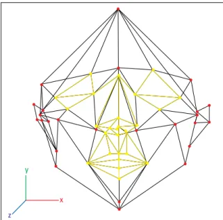
FIGURE 1 - Landmarks digitized by the electromagnetic three-dimen-sional tablet.
y
x
Three-dimensional face morphometry
Dental Press J. Orthod. 14 v. 15, no. 1, p. 13-15, Jan./Feb. 2010
categories of instruments have advantages and limitations, which should be considered accord-ing to the investigated problem and the human resources.
An ideal method for the quantitative evalua-tion of the patients should combine:
- non-invasive; - low-cost;
- a fast (simple data collection technique that provides three-dimensional digital data of the fa-cial morphology);
- possibility to create a digital database; - possibility to use computerized techniques for visualization, simulation and quantitative as-sessment of the treatment.
CONTACT INSTRUMENTS
Ultrasound probes, electromagnetic and elec-tromechanic digitizers are among the most used contact instruments. The ultrasound method is widely used for prenatal, intrauterine imaging and diagnosis, and three-dimensional reconstruc-tions of fetal face are becoming a current clinical practice. However its application for postnatal facial morphometry is limited and it’s being used just to evaluate the facial soft tissue thickness. Electromagnetic and electromechanic digitiz-ers provide the three-dimensional coordinates of landmarks that are previously marked on the face of the subject, which directly correspond to anatomical and anthropometric structures. The facial landmarks are digitized one by one using a stylus connected to the digitizer.2,4,7
Ultrasound probes use acoustic waves in the Megahertz frequency domain, while electromag-netic and electromechanic digitizers are based on electromagnetic waves. The main limitations of the instruments are the reduction of information obtained, and the data acquisition time, which is extremely long when compared to optical meth-ods. Indeed, movements of facial muscles (in par-ticular around mouth and eyes), as well as global head movements, may occur during digitization
producing errors. The acquisition of only single, selected landmarks impedes to produce life-like facial models (Fig 1).
OPTICAL INSTRUMENTS
The optical instruments need no physical contact with the skin, thus eliminating the risk of cutaneous compression, and of potential injuries during measurements.
The instruments of this category are laser scanners and stereophotogrammetric systems. Laser scanners illuminate the face with a laser light source, and digital cameras capture the re-flected light; the depth information is obtained by triangulation geometry.
Stereophotogrammetry uses a light source (ei-ther patterned or conventional) to illuminate the face, and two or more synchronized cameras re-cord images from different points of view. A pre-vious calibration, made with objects with known geometric characteristics, supplies the mathemati-cal information to obtain a stereoscopic recon-struction of the face. The system can record also facial texture, and combines the three-dimension-al information with an accurate reproduction of all facial characteristics (Fig 2, A and B).
de Menezes M, Sforza C
Dental Press J. Orthod. 15 v. 15, no. 1, p. 13-15, Jan./Feb. 2010
Contact address Márcio de Menezes
Praça Cônego Joaquim Alves 79 CEP: 14.300-000 – Batatais / SP E-mail: [email protected] REFERENCES
1. de Menezes M, Rosati R, Allievi C, Sforza C. A photographic system for the three-dimensional study of facial morphology. Angle Orthod. 2009 Nov;79(6):1070-7.
2. Ferrario VF, Sforza C, Poggio CE, Cova M, Tartaglia G. Preliminary evaluation of an electromagnetic three-dimensional digitizer in facial anthropometry. Cleft Palate Craniofac J. 1998 Jan;35(1):9-15.
3. Hajeer MY, Ayoub AF, Millett DT. Three-dimensional assessment of facial soft-tissue asymmetry before and after orthognathic surgery. Br J Oral Maxillofac Surg. 2004 Oct;42(5):396-404.
4. Ozsoy U, Demirel BM, Yildirim FB, Tosun O, Sarikcioglu L. Method selection in craniofacial measurements: advantages and disadvantages of 3D digitization method. J Craniomaxillofac Surg. 2009 Jul;37(5):285-90.
5. Plooij JM, Swennen GR, Rangel FA, Maal TJ, Schutyser FA, Bronkhorst EM, Kuijpers-Jagtman AM, Bergé SJ. Evaluation of reproducibility and reliability of 3D soft tissue analysis using 3D stereophotogrammetry. Int J Oral Maxillofac Surg. 2009 Mar;38(3):267-73.
6. Rosati R, Dellavia C, Colombo A, de Menezes M, Sforza C. Nasal base symmetry: a three dimensional anthropometric study. Minerva Stomatol. 2009 Jul-Aug;58(7-8):347-57.
FIGURE 2 - A) Vectra-3D system, CanfieldScientific, Inc., Fairfield, NJ, USA. B) Facial image obtained with stereophotogrammetry system.
FIGURE 3 - Photogrammetry system (Photomodeler).
Different methods for 3D analysis are being de-veloped and studied, but in general the elevate costs restrict the use in clinical practice.
7. Sforza C, Ferrario VF. Soft-tissue facial anthropometry in three dimensions: from anatomical landmarks to digital morphology in research, clinics and forensic anthropology. J Anthropol Sci. 2006;84:97-124.
8. Shaner DJ, Peterson AE, Beattie OB, Bamforth JS. Assessment of soft tissue facial asymmetry in medically normal and syndrome-affected individuals by analysis of landmarks and measurements. Am J Med Genet. 2000 Jul;93(2):143-54.