Transverse effect of Haas and Hyrax appliances on the
upper dental arch in patients with unilateral complete
cleft lip and palate: A comparative study
Anna Júlia de Oliveira Façanha1, Tulio Silva Lara2, Daniela Gamba Garib3, Omar Gabriel da Silva Filho4
How to cite this article: Façanha AJO, Lara TS, Garib DG, Silva Filho OG. Transverse effect of Haas and Hyrax appliances on the upper dental arch in pa-tients with unilateral complete cleft lip and palate: A comparative study. Dental Press J Orthod. 2014 Mar-Apr;19(2):39-45. doi: http://dx.doi.org/10.1590/2176-9451.19.2.039-045.oar
» The authors report no commercial, proprietary or financial interest in the products or companies described in this article.
Contact address: Tulio Silva Lara
Rua Sílvio Marchione, 3-20 – Vila Universitária – Bauru/SP — Brazil. CEP: 17012-900 – E-mail: [email protected]
» Patients displayed in this article previously approved the use of their facial and in-traoral photographs.
1 Specialist in Orthodontics, Hospital for Rehabilitation of Craniofacial
Anomalies – São Paulo University (HRAC-USP).
2 Professor of Interceptive Orthodontics (HRAC-USP).
3 Full professor, School of Dentistry — University of São Paulo/Bauru. Professor
of Orthodontics (HRAC-USP).
4 MSc in Orthodontics, State University of São Paulo (UNESP).
Submitted: March 14, 2012 - Revised and accepted: September 03, 2012
Objective:The aim of the present study was to evaluate the transverse effect of rapid maxillary expansion in patients with unilateral complete cleft lip and palate while comparing the Haas and Hyrax appliances. Methods: The sample consisted of 48 patients divided into two groups: Group I – 25 patients treated with modified Haas appliance (mean age: 10 years 8 months); and Group II – 23 patients treated with Hyrax appliance (mean age: 10 years 6 months). Casts were taken dur-ing pre-expansion and after removal of the appliance at the end of the retention period. The models were scanned with the aid of the 3 Shape R700 3D scanner. Initial and final transverse distances were measured at cusp tips and cervical-palatal points of maxillary teeth by using the Ortho Analyzer™ 3D software. Results: The mean expansion obtained between cusp tips and cervical-palatal points for inter-canine width was 4.80 mm and 4.35 mm with the Haas appliance and 5.91 mm and 5.91 mm with the Hyrax appliance. As for first premolars or first deciduous molars, the values obtained were 6.46 mm and 5.90 mm in the Haas group and 7.11 mm and 6.65 mm in the Hyrax group. With regard to first mo-lars, values were 6.11 mm and 5.24 mm in the Haas group and 7.55 mm and 6.31 mm in the Hyrax group. Conclusion: Rapid maxillary expansion significantly increased the transverse dimensions of the upper dental arch in patients with cleft palate, with no significant differences between the Hass and Hyrax expanders.
Keywords:Palatine expansion technique. Cleft palate. Dental arch.
Objetivo:avaliar o efeito transversal na arcada dentária superior do procedimento de expansão rápida da maxila em pacien-tes com fissura transforame incisivo unilateral, comparando os expansores tipo Haas modificado e de Hyrax. Métodos: a amostra constou de 48 pacientes divididos em dois grupos: grupo I, 25 pacientes que utilizaram o aparelho expansor tipo Haas modificado, com média de idade de 10 anos e 8 meses; e grupo II, 23 pacientes que utilizaram o Hyrax, com média de idade de 10 anos e 6 meses. Modelos de gesso foram realizados na fase pré-expansão e após 6 meses de contenção, após a remoção do aparelho. Os modelos foram digitalizados com auxílio do scanner 3Shape R700 3D e as distâncias transversais iniciais e finais foram medidas entre as pontas de cúspides e pontos cervicopalatinos de dentes superiores pelo método digital no software OrthoAnalyserT 3D. Resultados: a média de expansão obtida entre as pontas de cúspides e entre os pontos cervicopalatinos, respectivamente, para a distância intercaninos, foi de 4,80mm e 4,35mm para o Haas e de 5,91mm e 5,91mm para o Hyrax; 6,46mm e 5,90mm para os primeiros molares decíduos ou primeiros pré-molares no grupo Haas, 7,11mm e 6,65mm no grupo Hyrax; e 6,11mm e 5,24mm para os primeiros molares no grupo Haas e 7,55mm e 6,31mm no grupo Hyrax. Conclusão: o procedimento de expansão rápida da maxila produziu aumentos sig-nificativos das dimensões transversais da arcada dentária superior em pacientes com fissura, sem diferenças significativas entre os expansores Haas modificado e Hyrax.
INTRODUCTION
Unilateral complete clet lip and palate simultane-ously involves the primary and secondary palate and accounts for 30% of all clets. This condition requires more extensive treatment, as the clet divides the max-illa and the alveolar arch into two completely distinct segments.1 Treatment initially involves primary
func-tional and esthetic surgeries for the closing of the lip and palate, which have a long-term impact on mid face growth.2,3 The patient is then followed up throughout
the growth period until entering the orthodontic phase (end of the deciduous dentition phase).4
Primary surgeries of the lip and palate usually po-tentiate reductions in the transverse and sagittal di-mensions of the upper arch as a consequence of the re-stricted growth of the mid face and the approximation of the initially separated maxillary segments.3 These
sagittal and transverse deiciencies of the upper alveolar arch is expressed already at the mixed dentition phase and tend to become aggravated in adolescence.5 Thus,
the task of orthodontists is to counteract the harmful efects of the altered facial growth also characterized by posterior and anterior cross bite — oten found in patients with clet lip and palate.6
Besides other occlusion issues,7 posterior cross
bite is the most common malocclusion in these pa-tients, involving a single tooth or the entire dental arch, showing a tendency toward exacerbation from the mixed to the permanent dentition. Interceptive orthodontic interventions should be performed dur-ing the mixed dentition phase for correction of the compromised transverse dimension.8 Moreover, the
expansion of the upper arch also plays an important role in preparing the arch and cleft region for the secondary alveolar bone graft, which is performed at the end of the mixed dentition prior to the eruption of the permanent canine adjacent to the cleft region.4
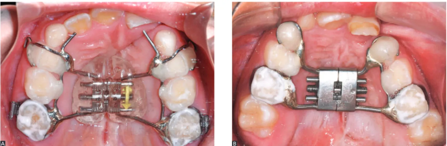
Rapid maxillary expansion performed by means of a Haas or Hyrax appliance is the most common method employed at the Hospital for Rehabilitation of Craniofacial Anomalies (USP, Brazil) to increase the width of the maxilla. The main difference be-tween the two appliances is the type of anchorage: supported with the Hyrax appliance and tooth-mucosa-supported with the Haas appliance.
The active expansion phase in patients with
unilat-of the maxillary segments and widening unilat-of the cleft.8
Expansion in these patients is not followed by bone formation at the median palatine suture, as it occurs in patients without cleft palate,9 because the
distanc-ing of the maxillary halves occurs in the region of the cleft. Thus, retention should remain after the appli-ance is removed and until bone graft is performed. Expansion in such patients involves similar restric-tions to those found in patients without cleft palate, as the other maxillary sutures offer considerable resis-tance to widening, requiring orthopedic appliances.
Motivated by the interest in evaluating the results of the expansion philosophy of the team at the Hospi-tal for Rehabilitation of Craniofacial Anomalies/USP, the aim of the present prospective study was to assess alterations in the transverse dimension of the upper dental arch in patients with unilateral complete clet lip and palate who have undergone rapid maxillary ex-pansion, comparing the results achieved with the Haas and Hyrax appliances with the aid of digital models.
MATERIAL AND METHODS
This study was approved by the Hospital for Reha-bilitation of Craniofacial Anomalies/USP Institutional Review Board (protocol 255/2010-SVAPEPE-CEP).
The sample consisted of 48 patients enrolled in the orthodontic sector of the hospital. All patients had unilateral complete cleft lip and palate, had under-gone primary surgeries at an early age and were in the mixed dentition phase, exhibiting maxillary atresia with an indication for rapid maxillary expansion.
cusps of the lower molars). Hooks for a facial mask were welded to the inverse traction of the maxilla for patients that also had sagittal discrepancy with good prognosis for orthopedic treatment.
After achieving overcorrection, the screw was stabilized with acrylic resin. The expander was maintained passive with a retention protocol for six months, after which the expander was removed and a fixed retainer was installed.
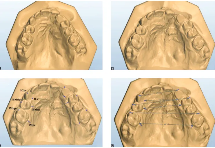
Dental cast of the maxillary arch were obtained at two different times: immediately before banding for the expander (T1), and after the six-month period of retention immediately following the removal of the expander (T2). The impressions were made with al-ginate and filled with Paris plaster. The dental casts were then scanned by a 3Shape R700 3D scanner (3Shape A/S, Copenhagen, Denmark), which re-produces a three-dimensional digital image based on laser beams scans projected over the plaster models in different directions. The Ortho Analyzer ™ 3D program was used to obtain the three-dimensional image of the scanned models, allowing frontal, lat-eral, posterior and occlusal visualization. The refer-ence points were marked on the model in the occlu-sal view for the calculation of the distances between teeth. Figure 2 illustrates the measurements made on the digital models of the upper dental arches.
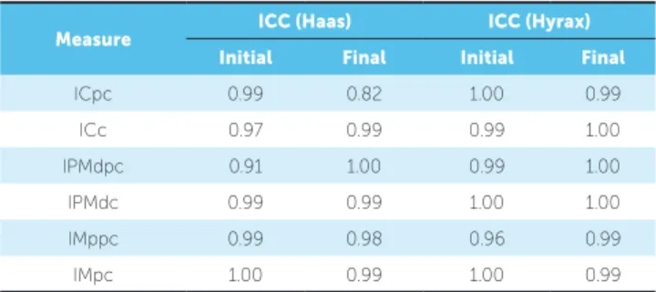
Analyses were performed by a single examiner. Sixty percent of the sample was analyzed a second time ater a seven-day interval in order to determine the measure-ments reliability using the intraclass correlation coei-cient (ICC) for which the following scores were used: 0.80 to 1.00 = excellent agreement; 0.60 to 0.80 = sub-stantial; 0.40 to 0.60 = moderate; 0.20 to 0.40 = fair; 0 to 0.20 = discreet; and –1 to 0 = poor.
Mean, standard deviation (SD), maximum and minimum values were calculated. The paired Student’s t-test was employed to determine statisti-cally significant differences between the initial and final measurements of each group. The independent t-test was used for inter-group comparison. The level of significance was set at 5% (p < 0.05).
RESULTS
Tables 1 to 4 display the results of the measure-ment reliability and transverse distances measured on the models.
DISCUSSION
Clet lip and palate occurs in the mid face and causes structural problems in the alveolar bone and maxilla. The urgent relationship with the malocclusion is dem-onstrated by the anatomic rupture that compromises the alveolar ridge as well as dental problems, such as
Figure 1 - Occlusal photographs of two patients from the sample illustrating both expanders: Modified Haas (A) and Hyrax (B)
Figure 2 - Initial and final digital models showing the results of rapid maxillary expansion (A and B); reference points (C) and measurements made (D) on post-expansion models
agenesis and malpositioning, as well as sagittal and transverse maxillary diiciencies.10 The treatment
protocol adopted by the Hospital for Rehabilitation of Craniofacial Anomalies/USP for patients with uni-lateral complete clet lip and palate emphasizes early surgical procedures (cheiloplasty and palatoplasty), with no orthopedic intervention in the preoperative or immediate postoperative periods. As a general rule, orthodontic treatment is initiated at the onset of the mixed dentition phase. Two reasons justify the lack of orthodontic intervention in the deciduous dentition phase: the instability of the early correction of cross bites, which leads to an excessively long treatment and retention time; and the fact that alterations in the shape of the dental arch and the occlusion are exacerbated in
Expansion is the irst step of orthodontic treatment (pre-alveolar bone grat). It aims at reestablishing the transverse dimensions of the atresic maxilla. The max-illary expander designed by Haas is the main appliance used for lateral repositioning of the collapsed maxil-lary processes6 and follows the same activation protocol
used for patients without clet palate. In this phase, the inverse traction of the maxilla may be associated with the post-expansion period to revert cases of negative sagittal discrepancy, expressed by anterior crossbite.12
The Hyrax appliance is also commonly used for maxil-lary expansion. The main diference between the two expanders is the acrylic support on the palate in the Haas appliance. However, both expanders are efective in increasing the transverse dimension of the maxilla.
A
C
B
Table 3 - Transverse distances measured on initial and final digital models and mean amount of expansion (Dif.) in millimeters for patients using the Hyrax expander.
* statistically significant difference (p < 0.05)
Measure Initial model Final model Dif. p
Mean ± S.D. Mean ± S.D.
ICpc 24.63 ± 3.98 30.54 ± 2.95 5.91 < 0.01*
ICc 20.37 ± 2.96 26.28 ± 3.91 5.91 < 0.01*
IPMdc 33.10 ± 3.71 40.21 ± 3.96 7.12 < 0.01*
IPMdc 22.72 ± 3.59 29.37 ± 3.50 6.65 < 0.01*
IMppc 44.68 ± 4.26 52.23 ± 3.73 7.55 < 0.01*
IMpc 31.32 ± 3.90 37.63 ± 3.79 6.31 < 0.01*
Table 4 - Comparison of modified Haas and Hyrax appliances groups regard-ing mean increase in transverse dimensions of the maxilla followregard-ing rapid maxillary expansion.
ns = non-significant.
* statistically significant difference (p < 0.05).
Measure Group
Haas (n)
Group
Hyrax (n)
Diference
(mm) P
ICpc 4.80 (18) 5.91 (18) 1.11 0.16 ns
ICc 4.35 (11) 5.91 (16) 1.56 0.06 ns
IPMdpc 6.46 (19) 7.11 (16) 0.65 0.48 ns
IPMdc 5.90 (19) 6.65 (16) 0.75 0.29 ns
IMppc 6.11 (24) 7.55 (22) 1.44 0.02*
IMpc 5.24 (24) 6.31 (21) 1.07 0.08 ns
of the patient’s palate. Whenever the transverse width and depth allow, the Haas expander is the appliance of choice due to the anchorage provided by its acryl-ic portion. In the present study, the modified Haas expander was used, which differs from the original by the presence of two orthodontic bands instead of four, and by bonded orthodontic clips on the decidu-ous molars.
Following the tendency of using digital records in Orthodontics,13 the evaluation of the transverse
ef-fect of the Haas and Hyrax expanders was performed with digital models, which ofer advantages in terms of storage, retrieval, durability, diagnostic versatility and transmitting information.14 Moreover, studies
com-paring digital and conventional models report consid-erable accuracy and reproducibility in the measure-ments of tooth width, overjet and overbite.15 The ICC
of the measurements obtained from the digital models by a single examiner on two diferent occasions dem-onstrate the reliability of the method (Table 1).
Both appliances were capable of restoring the ad-equate upper arch morphology and correcting the pos-terior cross bite (Figs 1 and 2). The results demonstrate signiicant increase (p < 0.0001) in all transverse di-mensions measured (Tables 2 and 3), which is in agree-ment with indings reported in the literature.16
The increases in the inter-molar, inter-premolar or inter-deciduous molar and inter-canine widths were statistically significant in both groups. The group treated with the Haas expander had an in-crease in inter-molar distance of 6.11 mm on the tips of the mesiovestibular cusps, 5.24 mm on the pala-tine-cervical portion of the molars, and an increase in the inter-canine distance of 4.79 mm when mea-sured on the cusps and 4.35 mm when meamea-sured on the palatine-cervical portion (Table 2). These values are similar to those reported by previous studies.17,19
In a study involving digital models of 32 children without cleft palate and with unilateral or bilateral cross bite (16 children treated with the Haas expand-er and 16 treated with the Hyrax expandexpand-er),the Haas group had an increase in inter-molar distance of 6.33 mm on the tips of the mesiovestibular cusps and 6.04 mm on the central sulcus of the first molars; an in-crease in the inter-canine distance of 2.27 mm when measured on the cusps and 4.74 mm when measured on the cervical portion.20
Table 1 - Error of the method (Intraclass Correlation Coefficient).
Measure ICC (Haas) ICC (Hyrax)
Initial Final Initial Final
ICpc 0.99 0.82 1.00 0.99
ICc 0.97 0.99 0.99 1.00
IPMdpc 0.91 1.00 0.99 1.00
IPMdc 0.99 0.99 1.00 1.00
IMppc 0.99 0.98 0.96 0.99
IMpc 1.00 0.99 1.00 0.99
Table 2 - Transverse distances measured on initial and final digital models and mean amount of expansion (Dif.) in millimeters for patients using the modi-fied Haas expander.
* statistically significant difference (p < 0.05)
Measure Initial model Final model Dif. p
Mean ± S.D. Mean ± S.D.
ICpc 26.81 ± 3.13 31.60 ± 3.35 4.79 < 0.01*
ICc 21.15 ± 3.39 25.50 ± 3.76 4.35 < 0.01*
IPMdpc 35.39 ± 3.67 41.85 ± 4.22 6.46 < 0.01*
IPMdc 24.25 ± 3.03 30.15 ± 3.36 5.9 < 0.01*
IMppc 49.20 ± 3.91 55.31 ± 3.28 6.11 < 0.01*
In the present study, the patients treated with the Hyrax expander had an increase in inter-molar dis-tance of 7.55 mm on the tips of the mesiovestibular cusps and 6.31 mm in the palatine-cervical portion; an increase in the inter-canine distance of 5.91 mm when measured on the cusps or on the palatine-cer-vical portion (Table 3). These results are similar to findings reported in the literature for patients in the mixed dentition phase treated with the Hyrax ex-pander.19,21,23 However, another study reports larger
increases (9.97 mm in inter-molar distance on the tips of the mesiopalatine cusps and 9.51 mm in the in the mesial portion of the central sulcus of the and 7.93 mm in the inter-canine distance when measured on the cusps and 6.29 mm when measured on the cervical portion).20
No statistically significant differences were found between groups in the comparison of the inter-molar and inter-canine distances obtained with the Haas or Hyrax appliances, except for the inter-molar distance measured on the tips of the cusps, which was greater in the Hyrax group (Table 4). This was likely due to the greater tooth tipping caused by this appliance.
Discrepant results are reported in the literature, with some studies reporting a greater increase in inter-molar and inter-canine distances using the Hyrax ex-pander20 and others reporting a greater tendency of
vestibular tipping of molars using the Haas expand-er.16 Both appliances generally demonstrate similar
behavior regarding the expansion of the dentoalveolar region of the maxilla.24 However, the Haas expander
may cause greater vestibular tipping of the anchoring teeth (3.5o for the first molar) in comparison to the
Hyrax expander (1.6o), although this is not a clinically
relevant difference.24
CONCLUSIONS
- Rapid maxillary expansion using the modi-fied Haas and Hyrax expanders proved efficient in increasing the transverse dimensions of the upper dental arch in patients with unilateral complete cleft lip and palate.
1. Silva Filho OG, Ferrari Júnior FM, Carvalho RM, Mazzottini R. A cirurgia ortognática na reabilitação do paciente portador de issura unilateral completa de lábio e palato. Rev Dental Press Ortod Ortop Facial. 1998;3(4):51-70.
2. Silva Filho OG, Freitas JAS. Caracterização morfológica e origem embriológica. In: Trindade IEK, Silva Filho OG, organizadores. Fissuras labiopalatinas: uma abordagem interdisciplinar. São Paulo: Ed. Santos; 2007. cap. 2, p. 16-49.
3. Silva Filho OG, Ramos AL, Abdo RC. The inluence of unilateral cleft lip and palate on maxillary dental arch morphology. Angle Orthod. 1992;62(4):283-90.
4. Cavassan AO, Silva Filho OG. Abordagem ortodôntica. In: Trindade IEK; Silva Filho OG. organizadores. Fissuras labiopalatinas: uma abordagem interdisciplinar. São Paulo: Ed. Santos; 2007. cap. 12, p. 213-38. 5. Athanasiou AE, Mazahery M, Zarrinnia K. Dental arch dimensions
in patients with unilateral cleft lip and palate. Cleft Palate J. 1988;25(2):139-45.
6. Capelozza Filho L, Almeida AM, Ursi WJS. Rapid maxillary expansion in cleft lip and palate patients. J Clin Orthod. 1994;28(1):34-9.
7. Reis AC, Capelozza Filho L, Ozawa TO, Cavassan AO. Avaliação da angulação e inclinação dos elementos dentários em pacientes adultos jovens portadores de issura transforame incisivo bilateral. Rev Dental Press Ortod Ortop Facial. 2008;13(1):113-23.
8. Long RE Jr, Semb GS, Shaw WC. Orthodontic treatment of the patient with complete lip and palate: lessons of the past 60 years. Cleft Palate Craniofac J. 2000 [Access in 2010 set 10];37(6). Available from: http:// www.cpcjournal.org/doi/pdf/10.1597/1545569%282000%29037%3C0533 %3AOTOTPW%3E2.0.CO%3B2.
9. Silva Filho OG, Lara TS, Silva HC, Bertoz FA. Comportamento da sutura palatina mediana em crianças submetidas à expansão rápida da maxila: avaliação mediante imagem de tomograia computadorizada. Rev Dental Press Ortod Ortop Facial. 2007;12(3):94-103.
10. Garib DG, Silva Filho OG, Janson G, Pinto JHN. Etiologia das más oclusões: perspectiva clínica (parte III) – issuras labiopalatinas. Rev Clín Ortod Dental Press. 2010;9(4):30-6.
11. Abdo RCC, Silva Filho OG, Ramos AL. Comportamento do arco dentário superior de crianças issuradas de lábio e palato, operadas – estudo longitudinal de 3 a 6 anos. Ortodontia. 1992;25(2):15-26.
12. Silva Filho OG, Capelozza Filho L, Wernech VA, Freitas JAS. Abordagem ortodôntica ao paciente com issura unilateral completa de lábio e palato. Ortodontia. 1998;31(3):32-44.
ReFeRenCeS
13. Creed B, Kau CH, English JD, Xia JJ, Lee RP. A comparison of the accuracy of linear measurements obtained from cone beam computerized tomography images and digital models. Semin Orthod. 2011;17(1):49-56.
14. Jofe L. OrthoCAD: digital models for a digital era. J Orthod. 2004;31(4):344-7.
15. Santoro M, Galkin S, Teredesai M, Nicolay OF, Cangialosi TJ. Comparison of measurements made on digital and plaster models. Am J Orthod Dentofacial Orthop. 2003;124(1):101-5.
16. Weissheimer A. Efeitos imediatos da expansão rápida da maxila no sentido transversal, com os disjuntores tipo Haas e Hyrax, em tomograia computadorizada cone beam [dissertação]. Porto Alegre (RS): Pontifícia Universidade Católica do Rio Grande do Sul; 2008.
17. McNamara JA, Baccetti T, Franchi L, Herberger TA. Rapid maxillary expansion followed by ixed appliances: a long-term evaluation of changes in arch dimension. Angle Orthod. 2003;73(4):344-53. 18. Lima AL, Lima Filho RMA, Bolognese AM. Long-term clinical outcome
of rapid maxillary expansion as the only treatment performed in class I malocclusion. Angle Orthod. 2005;75(3):416-20.
19. Oliveira NL, Da Silveira AC, Kusnoto B, Viana G. Three-dimensional assessment of morphologic changes of the maxilla: a comparison of 2 kinds of palatal expanders. Am J Orthod Dentofacial Orthop. 2004;126(3):354-62.
20. Mundstock KS. Estudo dos efeitos da expansão rápida de maxila em pacientes com mordida cruzada posterior tratados com aparelhos de Haas e de Hyrax [tese]. Araraquara (SP): Universidade Estadual Paulista; 2006.
21. Adkins MD, Nanda RS, Currier GF. Arch perimeter changes on rapid palatal expansion. Am J Orthod Dentofacial Orthop. 1990;97(3):194-9. 22. Chiavini PCR. Efeitos da expansão rápida da maxila com aparelho
expansor tipo Hyrax: avaliação cefalométrica póstero-anterior em modelos de estudo [tese]. Araraquara (SP): Faculdade de Odontologia de Araraquara; 2004.
23. Ciambotti C, Ngan P, Durkee M, Kohli K, Kim H. A comparison of dental and dentoalveolar changes between rapid palatal expansion and nickel-titanium palatal expansion appliances. Am J Orthod Dentofacial Orthop. 2001;119(1):11-20.