*Correspondence: M. V. C. Lonardoni. Department of Clinical Analysis, Health Sciences Center, Maringá State University, Avenida Colombo 5790, 87020-900 – Maringá - PR, Brazil. E-mail: [email protected]
A
rti
Pharmaceutical Sciences vol. 45, n. 4, oct./dec., 2009
Variables associated with the post-treatment healing of
lesions in patients with American cutaneous leishmaniasis in
Paraná State, Brazil
Maria Teresinha Gomes Casavechia
1, Thaís Gomes Verzignassi Silveira
1, Ueslei Teodoro
2, Vanderly
Janeiro
1, Margareth Udo
3, Maria Valdrinez Campana Lonardoni
2*
Health Sciences Center, Maringá State University, 2Department of Clinical Analysis, Health Sciences Center, Maringá State
University, 3Department of Statistics, Exact Sciences Center, Maringá State University.
The purpose of this study was to investigate the relationship of several variables to the healing of lesions in patients with American cutaneous leishmaniasis (ACL). The patients with clinical and/or laboratorial diagnoses of the disease were followed up for varying periods after treatment by clinical evaluation and indirect immunoluorescence assay (IFA), fromSeptember 2000 to December 2003. The lesions of 85.3% of the 163 patients had healed by their last return for clinical evaluation, and of these, 82.7% had negative IFA results, indicating an association between the healing of lesions and IFA negativity (p=0.000). In patients evaluated up to 120 days after treatment, there was a signiicant association between negative IFA results and the healing of lesions (p=0.0000). Logistic regression analysis showed that negative IFA results on patients’ irst return after treatment predicted a 2.175 fold greater chance of lesion healing (p=0.0001). These results indicate an association between IFA negativity at the irst return up to a period of 120 days, and the healing of lesions, and that the chances of healing are signiicantly higher in patients with negative IFA results at their irst return after treatment.
Uniterms: Cutaneous leishmaniasis/treatment. Leishmania (Viannia) braziliensis. Cutaneous lesion/ healing.
O objetivo deste estudo foi investigar a associação de algumas variáveis para a cicatrização de lesões em pacientes com leishmaniose tegumentar americana (LTA). Os pacientes com diagnóstico clínico e laboratorial foram acompanhados depois do tratamento por avaliação clínica e reação de imunoluorescência indireta (IFI), de setembro de 2000 a dezembro de 2003. Dos 163 pacientes 85,3% apresentaram cicatrização das lesões no último retorno para a avaliação clínica e 82,7% destes tiveram a IFI negativa indicando uma associação entre a cicatrização das lesões e a negativação da IFI (p=0,000). Nos pacientes acompanhados até 120 dias depois do tratamento houve associação signiicativa entre os resultados negativos da IFI e a cicatrização das lesões (p=0,0000). A análise pela regressão logística mostrou que quando a IFI do primeiro retorno após o tratamento foi negativa, o paciente tinha 2,175 mais chance de cicatrização (p=0,0001). Os resultados mostram associação entre a negativação da IFI e a cicatrização das lesões quando o primeiro retorno foi até 120 dias e que as chances de cicatrização são signiicativamente maiores nos pacientes que apresentaram IFI negativa no primeiro retorno depois do tratamento.
Unitermos: Leishmaniose cutânea/tratamento. Leishmania (Viannia) braziliensis. Lesão cutânea/ cicatrização.
INTRODUCTION
American cutaneous leishmaniasis (ACL) is an
ACL has been recorded in the majority of cities in the state of Paraná (Roberto et al., 1997; Silveira et al., 1999; Lima et al., 2002), which accounts for 98% of cases in the southern region of Brazil (Brasil, 2000). The success of ACL treatment is limited. The clinical control of treatment is imprecise, and relapses, even after complete treatment and healing of the initial le-sions, have been documented (Guevara et al., 1993). The irst-choice drug in Brazil for treating ACL is N-methyl glucamine antimoniate (Glucantime®). According to the
World Health Organization (WHO, 1995), more than 90% of relapses occur within one year of treatment. Furthermore, the mucosal form may occur among these patients, generally appearing months or even years after the resolution of the skin lesions. Antibodies detected by indirect immunoluorescence assay (IFA) are negative after successful treatment of the disease (Furtado, 1980; Souza et al., 1982). The presence of anti-Leishmania
antibodies after treatment may signify the persistence of the parasite, and the reduction of antibody titers has been suggested as a tool for the control of treatment eficacy (Chiari, Mayrink, Magalhães, 1973; Mendonça et al., 1988). Therefore, the following-up of patients by clini-cal and serologiclini-cal evaluation is fundamental (Roberto
et al., 1997).
The objective of this research was to study the possi-ble association of the variapossi-bles sex, IFA at initial diagnosis, IFA at irst return, IFA at different post-treatment periods and IFA at last return for clinical evaluation for the healing of lesions in ACL patients. To this end, a group of ACL patients were followed-up for varying periods after clinical and laboratorial diagnosis, after which the contribution of each variable was analyzed.
MATERIAL AND METHODS
Patients
One hundred and sixty-three ACL patients, regis-tered in the 13th Health Region of Cianorte, in northwest
Paraná fromSeptember, 2000 to December, 2003 were selected. Treatment was prescribed for all patients after positive clinical and/or laboratorial diagnoses. All patients were instructed to return for clinical evaluation and collec-tion of blood samples for the IFA. The follow-up period of the patients varied from 22 to 696 days from the start of treatment, with all patients being evaluated at least once after treatment. The total number of evaluations (including initial diagnosis and subsequent returns) varied from two to ive for each patient.
Laboratorial diagnosis
The laboratorial tests for ACL used in this study were Direct Parasite Search (DPS), Montenegro Skin Test (MST), and Indirect Immunoluorescence Assay (IFA). In the DPS, lesion samples were collected by scraping. Smears were made on glass slides, stained by Giemsa, and examined by microscope for the presence of amas-tigote forms. The MST reagent consisted of Leishmania (L.) amazonensis promastigotes containing 40 µg/mL of protein nitrogen, kindly supplied by the Immunobiological Production and Research Center of the Paraná State Health Ofice. The test was considered positive when an indura-tion diameter greater than or equal to 5mm was observed 48 hours after inoculation. The IFAfor IgG was carried out using promastigote forms of Leishmania (V.) braziliensis.
The serum was diluted, beginning at 1/20. Samples with titers equal to or greater than 40 were considered positive (Silveira et al., 1999).
Treatment protocol
The patients were intravenously treated with N-me-thyl glucamine antimoniate at 15 mg Sb5+/kg per day
(Bra-sil, 2000). Five patients did not respond to this treatment protocol. Therefore, three of these non-responders were also treated with Pentamidine® (pentamidine
isothiocya-nate), one with Pentamidine and intralesional iniltration with pentavalent antimonial, and one with intralesional iniltration only. The patients were considered clinically cured after complete healing of their lesions.
Ethical issues
This study complied with Resolution No. 196/96 of the National Health Council of the Brazilian Ministry of Health, and was approved by the Permanent Committee for Ethics in Research Involving Humans of the State University of Maringá (No. 006/2004).
Statistical analysis
contributed to healing. Variables were considered signii-cant at the 5% signiicance level.
The following variables were considered in the lo-gistic regression model: sex, age, IFA at initial diagnosis (IFA_ini), IFA at irst return (IFA_ret) and the number of days until the irst return (days). The dependent variable was lesion healing by irst return (Cret). The following categorizations were used: sex: 1 for male and –1 for female; IFA_ini: 1 if negative and –1 if positive; IFA_ret: 1 if negative and –1 if positive; days: 1 if the irst return occurred within a maximum of 120 days and –1 if the irst return occurred after 120 days; Cret: 1 if healed and 0 if not healed.
The results found through the statistical analysis showed that the age variable was not signiicant for the model (p=0.7507). Therefore, the adjusted model was given as:
where: β0= 1.6002, β1= - 0.5272, β2= 0.6203, β3= 0.7769
and β4= 0.2843.
RESULTS
Of the 163 cases of ACL studied, 127 (77.9%) were male and 36 (22.1%) female. Patient age ranged between 2 and 79 years (median = 33 years), 8.6% were aged up to 15 years and 6.8% were over 65. All the patients had cutaneous forms of ACL. One hundred and ten patients (67.5%) had a single lesion, 31 (19.0%) had two lesions and 12 (7.4%) had three or more. It was not possible to obtain this information for 10 patients. Two patients had negative DPS results, with IFA and MST not being carried out, and were included in the sample due to their clinical diagnoses.
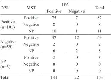
Table 1 shows that the laboratorial diagnoses of 161 (98.8%) patients were positive for ACL on at least one of the methods used. The DPS was positive in 101 of the 160 patients submitted to the examination (63.1%), while the MST was positive in 134 out of 144 patients (93.1%), and the IFA positive in 141 out of 163 patients (86.5%). The diagnosis was conirmed by all three laboratorial methods in 75 (46.0%) patients. Of the 62 patients with negative results on the DPS, or who were not submitted to the DPS, 40 (64.5%) had positive IFA and MST results, 12 (19.4%) had positive MST results only and 8 (12.9%) had positive IFA results only.
In the follow-up of the 163 patients, 139 (85.3%) showed the healing of lesions by their last return for clini-cal evaluation (Table II). In 115 (82.7%) of these patients,
IFA was negative, indicating an association between the healing of lesions and the negativity of IFA (p=0.000). Ho-wever, 24 patients (17.3%) continued to have positive IFA results despite having healed lesions. Of the 24 patients that did not show healing of lesions, 17 (70.8%) continued to have positive IFA results.
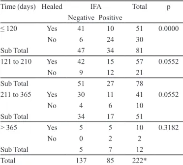
Table III shows the results of the clinical and sero-logic evaluations of the followed-up patients. Of the 81 evaluations performed within 120 days of treatment, 51 patients (63.0%) showed healing of lesions, 41 of whom (80.2%) had negative IFA results. Thirty patients (37.0%) showed no healing of lesions, six of whom (20.0%) had negative IFA results. A signiicant association (p=0.0000) was observed between IFA negativity and the healing of lesions within this evaluation period. Of the 78 patients followed-up from 121 to 210 days, 57 (73.1%) showed TABLE I - Results of direct parasite search (DPS), Montenegro skin test (MST) and indirect immunoluorescence assay (IFA), carried out in the diagnosis of American cutaneous leishmaniasis (ACL) in the 163 patients studied from 2000 to 2003
DPS MST IFA Total
Positive Negative
Positive (n=101)
Positive 75 7 82
Negative 8 0 8
NP 10 1 11
Negative (n=59)
Positive 37 12 49
Negative 2 0 2
NP 6 2 8
NP (n=3)
Positive 3 0 3
Negative 0 0 0
NP 0 0 0
Total 141 22 163
NP=Not Performed
TABLE II - Results on indirect immunofluorescence assay (IFA) and of lesion healing in the 163 patients with American cutaneous leishmaniasis (ACL) at last return for clinical evaluation during post-treatment follow-up from 2000 to 2003
Healed IFA Total
Negative Positive
Yes 115 24 139
No 7 17 24
Total 122 41 163
healing of lesions while 42 (73.7%) had a negative IFA. Twenty-one (26.9%) patients from this group showed no healing of lesions, nine of whom (42.9%) had a negative IFA (p=0.0552). Of the 51 patients followed-up from 211
to 365 days, 41 (80.4%) showed healing of lesions, and of these 30 (73.2%) had a negative IFA. Ten (19.6%) of these patients showed no healing of lesions and four (40.0%) had a negative IFA (p=0.0552). Finally, of the twelve patients followed-up after 365 days, ten (83.3%) showed healing of lesions, ive of whom (50.0%) had a negative IFA. Two (20.0%) patients from this group presented no healing of lesions and showed IFA positivity (p=0.3182).
The results of clinical and serologic evaluations performed during other periods (121 to 210 days, 211 to 365 days, and over 365 days) were also analyzed. However, no signiicant associations between IFA nega-tivity and the healing of lesions were observed for these periods (p>0.05).
As a signiicant association (p=0.0000) was obser-ved between IFA negativity and the healing of lesions in the period up to 120 days, logistic regression was perfor-med on the data from this period to analyze the possible factors associated to lesion healing. It was observed that patients who had a negative IFA on their irst return after treatment had a 2.175 greater likelihood of their lesions healing than when IFA was positive (p=0.0001; CI=1.490-3.174) (Table IV). Analysis of other factors such as gender, IFA result at initial diagnosis, and time of irst return after treatment (up to 120 days or after 120 days) found no associations with lesion healing (p>0.05).
DISCUSSION
The majority of the patients studied were from the male working-class population, suggesting an extra-domi-TABLE III -Results on indirect immunofluorescence assay
(IFA) and for lesion healing in the 163 patients with American cutaneous leishmaniasis (ACL) during different post-treatment periods from 2000 to 2003
Time (days) Healed IFA Total p
Negative Positive
≤ 120 Yes 41 10 51 0.0000
No 6 24 30
Sub Total 47 34 81
121 to 210 Yes 42 15 57 0.0552
No 9 12 21
Sub Total 51 27 78
211 to 365 Yes 30 11 41 0.0552
No 4 6 10
Sub Total 34 17 51
> 365 Yes 5 5 10 0.3182
No 0 2 2
Sub Total 5 7 12
Total 137 85 222*
*After initial diagnosis, 114 patients returned once, 42 twice, 5 three times, 1 four times and 1 ive times, amounting to a total of 222 blood samples subjected to IFA; p: value of Fisher’s exact test: IFA versus healing.
TABLE IV - Logistic regression analysis of factors associated with healing of lesions in 163 patients with American cutaneous leishmaniasis (ACL), followed-up from 2000 to 2003
Variables Healed (95% conidence interval)Odds Ratio Chi-squared p
Yes No
Gender
Male 81 46 0.590
(0.344 – 1.013) 3.6662 0.0555
Female 31 5
IFA on initial diagnosis
Positive 92 49 1.859
(0.848 – 4.076) 2.3993 0.1214
Negative 20 2
IFA on First Return$(within 120 days)
Positive 30 35 2.175
(1.490 – 3.174) 16.2201 0.0001
Negative 82 16
Time of First Return
Within 120 days 49 30 1.329
(0.915 – 1.930) 2.2320 0.1352
ciliary transmission of ACL, as was previously observed in the north of the state of Paraná (Silveira et al., 1999; Castro
et al., 2002). However, the occurrence of cases in the age ranges of up to 15 and over 65 years, for both sexes, also suggests intra- or peri-domiciliary transmission occurred, corroborating data from the north of Paraná State (Roberto
et al., 1997; Lima et al., 2002) and on an endemic area of the State of Minas Gerais, Brazil (Passos et al., 2001).
In the present study, IFA showed a higher rate of positivity than the MST or DPS in the diagnosis of ACL, contrasting with results obtained by Silveira et al. (1999), who found higher positivity for the MST in patients from the same geographical area. The sensitivity of IFA in the present study was similar to that found in studies on other endemic areas (Marzochi et al., 1980; Lugo de Yarbuh
et al., 1996). Together, the three methods used for ACL diagnosis were able to detect 98.8% of the ACL cases, showing it to be a useful combination for diagnosing the disease (Gontijo, Carvalho, 2003).
A highly signiicant association between the hea-ling of lesions and negative IFA results was observed in this study, conirming that successful treatment reduces levels of anti-Leishmania antibodies (Amato et al., 1998). Romero et al. (2005) also observed a reduction in antibody levels on enzyme-linked immunosorbent assay (ELISA) post treatment. Therefore, the reduction of anti-Leishmania antibody levels after treatment could be an important tool in the control of ACL treatment (Mendonça et al., 1988).
The data for different post-treatment periods was analyzed in order to identify the optimal time after treat-ment to evaluate the association between the healing of lesions and IFA seroconversion. The analysis showed that lesion healing was associated to negative IFA results when the evaluation was carried out in the period of up to 120 days after treatment. According to Passos et al. (2001), the treatment and elimination of the parasites induces lower antigenic stimulation, which contributes to IFA negativity, and Saravia et al. (1989) showed that there was an associa-tion between longer lesion evoluassocia-tion time and higher IFA titers in patients infected with L. braziliensis.
In the present study, a tendency of IFA negativity seen among those patients whose lesions had healed and who were analyzed during the periods of 121 to 210 and 211 to 365 days after treatment (p=0.0552). However, no association was observed between IFA negativity and the healing of lesions in those patients evaluated 365 days or more after treatment. This suggests that patients who continue to have antibody titers show a tendency to evolve to the chronic disease, or the reappearance of cutaneous and/or mucosal lesions.
Attention is drawn to the group of patients (17.3%) whose lesions healed but for whom IFA results remained positive. The persistence of antibodies may be an impor-tant indicator of the persistence of the parasite, despite the healing of their lesions. Indeed the persistence of the parasite in healed lesions was demonstrated by Mendonça
et al. (2004). These authors obtained 30 positive PCR and 7 positive IFA results from 32 clinically cured patients, indicating the tendency of Leishmania to establish non-apparent infections (Ramirez, Guevara, 1997). Chiari et al. (1973) found that 38% of patients submitted to treat-ment, and considered clinically cured, continued to have positive IFA results. Passos et al. (2001) did not associate the risk of recurrence to antibody levels measured by IFA; however, Amato et al. (1998) argued that the negativity of IFA antibody titers indicates a lower possibility of re-currence. These observations suggest that a more rigorous following-up of ACL patients, as well as their medicinal dosage and treatment protocol, through clinical and labo-ratory evaluations, is necessary.
Of the patients whose lesions did not heal, IFA re-sults remained positive for 80.0% and 57.1% during the periods of up to 120 days and from 121 to 210 days, res-pectively, showing the failure of the treatment. Different degrees of susceptibility to treatment, as well as different levels of immunological response, have been related to the various serodemes (Shaw et al., 1986) of Leishma-nia (Viannia) braziliensis that circulate in the northwest region of Paraná State (Silveira et al., 1999). While some infections by Leishmania present complications during treatment, sometimes producing metastasis and recurren-ce, others evolve to a spontaneous cure, suggesting that the immunological response is capable of controlling the infection (Brito et al., 2001).
In the present study, it was shown that the chances of lesions healing are greater if the IFA performed is ne-gative when the patient irst return after treatment. This indicates that both the initial diagnosis and the start of treatment were probably early. Neither IFA positivity at initial diagnosis, nor the time of the irst return for evalua-tion of treatment, were found to be factors associated with the healing of lesions. No differences in lesion healing between men and women were found, despite the indin-gs of Travi et al. (2002), who studied hamsters infected with Leishmania (Viannia) and observed that the immune response was more permissive to infection among males than females. The factors that contribute to the cutaneous disease evolving into one of the later forms are not fully understood, but delay in healing the irst lesion and ina-dequate treatment are both known predictors (Carvalho
CONCLUSIONS
The indings of this study conirm that: a) there is an association between IFA negativity at the irst return of the patient within 120 days of treatment and the healing of lesions, and b) the chances of healing are signiicantly higher in patients that showed negative IFA results on their irst return for clinical evaluation after treatment.
ACKNOWLEDGEMENTS
This research work was financially supported by the Conselho Nacional de Desenvolvimento Cientíico e Tecnológico (CNPq) and by the Laboratory of Teaching and Research in Clinical Analysis (LEPAC) of the State University of Maringá.
REFERENCES
AMATO, V. S.; DUARTE, M. I. S.; NICODEMO, A. C.; CARVALHO, L. V.; PAGLIARI, C.; MATTA, V. L. R.; OLIVEIRA, L. S.; CASTRO, S. M.; UIP, D. E.; AMATO, J. G. P.; AMATO-NETO, V. An evaluation of clinical, serologic, anatomopathologic and immunohistochemical findings for fifteen patients with mucosal leishmaniasis
before and after treatment. Rev. Inst. Med. Trop.,v.40, n.1,
p.23-30, 1998.
BRASIL, Ministério da Saúde. Manual de controle da
leishmaniose tegumentar americana. Brasília: Assessoria de Comunicação e Educação em Saúde, Fundação Nacional de Saúde, 2000. 62 p.
BRITO, M. E. F.; MENDONÇA, M. G.; GOMES, Y. M.; JARDIM, M. L.; ABATH, F. G. C. Dynamics of the antibody response in patients with therapeutic or spontaneous cure
of American cutaneous leishmaniasis. Trans. R. Soc. Trop.
Med. Hyg., v.95, n.2, p.203-206, 2001.
CARVALHO, E. M.; CORREA-FILHO, D.; BARCELAR, O.; ALMEIDA, R. P.; LESS, H.; ROCHA, H. Characterization of the immune response in subjects with self-healing
cutaneous leishmaniasis. Am. J. Trop. Med. Hyg., v.53, n.3,
p.273-277, 1995.
CASTRO, E. A.; SOCCOL, V. T.; MEMBRIVE, N.; LUZ, E. Estudo das características epidemiológicas e clínicas de 332 casos de leishmaniose tegumentar notiicados na região
norte do Estado do Paraná de 1993 a 1998. Rev.Soc. Bras.
Med. Trop., v.35, n.5, p.445-452, 2002.
CHIARI, C. A.; MAYRINK, W.; MAGALHÃES, P. A. Reação de imunoluorescência indireta no controle do tratamento da
leishmaniose tegumentar americana. Rev. Soc. Bras. Med.
Trop., v.15, n.5, p.298-303, 1973.
FURTADO, T. Critérios para o diagnóstico da leishmaniose
tegumentar americana. An. Bras. Dermatol., v.55, n.2,
p.81-86, 1980.
GONTIJO, B.; CARVALHO, M. L. R. Leishmaniose tegumentar
americana. Rev. Soc. Bras. Med. Trop., v.36, n.1, p.71-80,
2003.
GUEVARA, P.; RAMÍREZ, J. L.; ROJAS, E.; SCORZA, J. V.;
GONZÁLEZ, N.; AÑEZ, N. Leishmania braziliensis in
blood 30 years after cure. Lancet, v.341, n.8856, p.1341,
1993.
LIMA, A. P.; MINELLI, L.; TEODORO, U.; COMUNELLO, E. Distribuição da leishmaniose tegumentar por imagens de sensoreamento remoto orbital, no Estado do Paraná, Brasil.
An. Bras. Dermatol., v.77, n.6, p.681-692, 2002.
LUGO DE YARBUH, A.; AÑEZ, N.; VALERA, M.; MORENO, E. Deteccion de titulos de anticuerpos, proteínas totales, albumina y globulinas em casos de leishmaniosis cutânea
tratados com Glucantime. Parasitol. Dia, v.20, n.1,
p.20-26, 1996.
MARZOCHI, M. C. A.; COUTINHO, S. G.; SABROSA, P. C.; SOUZA, W. J. S. Reação de imunoluorescência indireta e intradermorreação para leishmaniose tegumentar americana em moradores na área de Jacarepaguá (Rio de Janeiro): estudo comparativo dos resultados observados em 1974 e
1978. Rev. Inst. Med. Trop. São Paulo, v.22, n.3, 149-155,
1980.
MENDONÇA, M. G.; BRITO, M. E. F.; RODRIGUES, E. H. G.; BANDEIRA, V.; JARDIM, M. L.; ABATH, F. G. C.
Persistence of Leishmania parasites in scars after clinical
cure of American cutaneous leishmaniasis: is there a sterile
cure? J. Infect. Dis., v.189, n.6, p.1018-1023, 2004.
MENDONÇA, S. C. F.; SOUZA, W. J. S.; NUNES, M. P.; MARZOCHI, M. C. A.; COUTINHO, S. G. Indirect immunofluorescence test in New World leishmaniasis:
serological and clinical relationship. Mem. Inst. Oswaldo
PASSOS, V. M. A.; BARRETO, S. M.; ROMANHA, A. J.; KRETTLI, A. U.; VOLPINI, A. C.; GONTIJO, C. M. F.; FALCÃO, A. L.; LIMA-COSTA, M. F. F. Leishmaniose tegumentar na Região Metropolitana de Belo Horizonte: aspectos clínicos, laboratoriais, terapêuticos e evolutivos
(1989-1995). Rev. Soc. Bras. Med. Trop., v.34, n.1 p.5-12,
2001.
RAMÍREZ, J. L.; GUEVARA, P. Persistent infections by
Leishmania (Viannia) braziliensis. Mem. Inst. Oswaldo
Cruz, v.92, n.3, p.333-338, 1997.
ROBERTO, A. C. B. S.; LIMA, A. P.; PEIXOTO, P. R.; MISUTA, N. M.; FUKUSHIGUE, Y.; FERREIRA, M. E. M. C.; NERILO-SOBRINHO, A.; SILVEIRA, T. G. V.; TEODORO, U. Avaliação da terapia com antimoniato de N-metil-glucamina e de notificação de leishmaniose
tegumentar. An. Bras. Dermatol., v.72, n.2, p.129-136,
1997.
ROMERO, G. A. S.; ORGE, M. G. O.; GUERRA, M. V. F.; PAES, M. G.; MACEDO, V. O.; CARVALHO, E. M. Antibody response in patients with cutaneous leishmaniasis infected by Leishmania (Viannia) braziliensis or Leishmania
(Viannia) guyanensis in Brazil. Acta Tropica, v.93, n.1, p.49-56, 2005.
SARAVIA, N. G.; VALDERRAMA, L.; LABRADA, M.; HOLGUÍN, A. F.; NAVAS, C.; PALMA, G.; WEIGLE, K.
A. The relationship of Leishmania braziliensis subspecies
and immune response to disease expression in New World
Leishmaniasis.J. Infect. Dis., v.159, n.4, p.725-735, 1989.
SHAW, J. J.; LAINSON, R.; McMAHON-PRATT, D.; DAVID,
J. R. Serodemes of the Leishmania braziliensis complex.
In: RIOUX, J. (Ed.). Leishmaniataxonomie et phylogenèse:
applications Éco-épidémiologiques:Colloque International,
2-6 july, Montpellier, 1984. Montpellier: IMEE, 1986. p.179-83.
SILVEIRA, T. G. V.; ARRAES, S. M. A. A.; BERTOLINI, D. A.; TEODORO, U.; LONARDONI, M. V. C.; ROBERTO, A. C. B. S.; RAMOS, M.; NERILO-SOBRINHO, A.; ISHIKAWA, E.; SHAW J. Observações sobre o diagnóstico laboratorial e a epidemiologia da leishmaniose tegumentar
no Estado do Paraná, sul do Brasil. Rev. Soc. Bras. Med.
Trop., v.32, n.4, p.413-423, 1999.
SOUZA, W. J. S.; COUTINHO, S. G.; MARZOCHI, M. C. A.; TOLEDO, L. M.; GOTTLIEB, M. V. Utilização da reação de imunofluorescência indireta no acompanhamento da
terapêutica da leishmaniose tegumentar americana. Mem.
Inst. Oswaldo Cruz, v.77, n.3, p.247-253, 1982.
TRAVI, B. L.; OSÓRIO, Y.; MELBY, P. C.; CHANDRASEKAR, B.; ARTEAGA, L.; SARAVIA, N. G. Gender is a major determinant of the clinical evolution and immune response
in hamsters infected with Leishmania spp. Infect. Immun.,
v.70, n.5, p.2288-2296, 2002.
WORLD HEALTH ORGANIZATION. Control of Leishmaniasis. Geneva: WHO, 1995. (Technical Report Series, n.793).
Received for publication on 09th December 2008.