The aim of this study was to characterize a conventional and a pressed porcelain for zirconia core as to biaxial flexural strength (BFS), apparent fracture toughness (FT) and microstructure composition, and to investigate the effect of glaze cooling rate on the BFS of the zirconia/porcelain bilayers. Monolayers of conventional porcelain Vita VM9 and pressed porcelain Vita PM9 (n=15) (12 mm diameter x 1.2 mm thick) were prepared for the BFS test (MPa). Apparent fracture toughness (MPa.m½) was measured by indentation technique (n=15). t-Student test was performed for statistical analysis. Scanning electron microscopy and x-ray diffraction were used to analyze the porcelain’s microstructure. For the BFS of bilayers, zirconia discs (12 mm diameter x 1 mm thick) (Vita In-Ceram YZ) were veneered with the two porcelains (1 mm thick). After the glaze firing simulation, the specimens were submitted to fast or slow cooling (n=15). Apparent fracture toughness (MPa.m½) was measured on the porcelain surface of bilayers (n=15) and residual stress was calculated. Two-way ANOVA (porcelain and cooling method) was used for the bilayer analysis (α=0.05). Vita PM9 monolayer exhibited significantly higher BFS (p<0.01), but there was no significant difference (p=0.41) in the FT between the porcelains. For bilayer specimens, the two-way ANOVA for BFS was significant for the porcelain variable only (p<0.01) better for Vita PM9/zirconia. Two-way ANOVA for the FT for the bilayers was not significant for any variable. All groups showed compressive residual stresses. The pressed porcelain seems to be mechanically more effective for zirconia veneering.
Effect of Glaze Cooling Rate on
Mechanical Properties of Conventional
and Pressed Porcelain on Zirconia
Diogo Longhini1, Cibele Oliveira de Melo Rocha1, Igor Studart Medeiros2, Renata Garcia Fonseca1, Gelson Luis Adabo1
1Department of Dental Materials
and Prosthodontics, Araraquara Dental School, UNESP – Universidade Estadual Paulista, Araraquara, SP, Brazil
2Department of Biomaterials
and Oral Biology, Dental School of USP - Universidade de São Paulo, São Paulo, SP, Brazil
Correspondence: Gelson Luis Adabo, Rua Humaitá, 1680, 14801-903 Araraquara, SP, Brasil. Tel: +55-16-3301-6415. e-mail: [email protected]
Key Words: dental porcelain, zirconium, material resistance.
Introduction
Besides the suitable mechanical properties of Y-TZP (Yttria Tetragonal Zirconia Polycristal) ceramics and its high survival rates after 5 years (1), clinical failures such as chippings (cohesive fracture of porcelain) have been reported (2-4) showing rates of 15.2% in 5 years (1) and 14.5% in 5 years (5). In a recent fractographic study after a 3-year randomized controlled clinical trial, higher chipping rate and larger chip size was found in zirconia restorations when compared to metal-ceramic (6). Those failures may be due to an inadequate bond strength between core and veneer (2), poor mechanical properties of the porcelain veneers for zirconia (1), insufficient support by the framework due to inadequate non-anatomical design (7), increased thickness of the veneer porcelain (8) and distribution of residual stress during porcelain cooling (4).
Despite these factors, two recent systematic reviews involving SCs (single crowns) (5) and FPPs (fixed partial prostheses) (9) in Y-TZP and other ceramic materials suggested that SCs and FPPs with zirconia core should be not considered as the first treatment option, due to higher chipping rates of porcelain and lower survival rates
compared to metal-ceramics (5,9). This fact indicates that the problem has not been solved, which makes further studies extremely important.
The residual stresses may occur due to a thermal mismatch between the zirconia core and the porcelain veneer or due to an inadequate cooling rate after the last firing of the porcelain veneer (10). During the cooling, when the glass transition temperature (Tg) is achieved (about 600 °C), the stress generated by the CTE (coefficient of thermal expansion) difference between the veneering porcelain and zirconia cannot be relieved by the plastic flow locked due to a non-uniform solidification resulting from thermal gradients from the surface to the center (4,11). Such condition increases the possibility of immediate or late veneer porcelain chipping during chewing (12).
525
Conventional versus pressed porcelain for zirconia
hand, some authors found better results with fast cooling (4,14) with regards to flexural strength(14) and porcelain bond strength (4).
The porcelain can be applied by the conventional hand-layer technique onto the zirconia or by the press technique, which theoretically is more homogenous (15) and would minimize the effects of internal porosity, improving the mechanical performance (16-18), which may cause less chipping rates. Moreover, press technique is less sensitive and less time dependent, and it allows to try-in the wax pattern before pressing (15). Nevertheless, the behavior of the pressed porcelains for zirconia veneering towards the different cooling methods on the glaze-firing phase is still unclear, since the first cooling of pressed porcelains is necessarily slow, as it occurs in the investment inside and must not be cooled rapidly because of the fracture risk for the ceramic.
Thus, the purpose of this study was to characterize a conventional (Vita VM9) and pressed porcelain (Vita PM9) as to flexural strength, apparent fracture toughness, and microstructure analysis, as well as the effect of the glaze cooling method (last stage) on the biaxial flexural strength and apparent fracture toughness of zirconia/porcelain bilayers. The null hypothesis was that there is no significant difference between the pressed and conventional porcelains in terms of mechanical properties and microstructure, as well as in the mechanical behavior of porcelain/zirconia bilayers when different cooling methods were used (fast or slow).
Material and Methods
The characteristics of materials used in this study are in Table 1.
Preparation of Porcelain Monolayers
Monolayer specimens (12 mm diameter and 1.2 mm thick) were prepared from the conventional porcelain (Vita VM9; Vita Zahnfabrik, Bad Säckingen, Baden-Württemberg,
Germany) and the pressed porcelain (Vita PM9; Vita Zahnfabrik) using the conventional and pressed methods respectively (n=15).
To prepare Vita VM9 discs, an oversized polyether mold (Impregum F, 3M ESPE, Seefeld, Baviera, Germany) was used. The porcelain slurry was prepared by mixing the powder and liquid in a ratio of 2.5:1 and inserted in the mold. Excess liquid was blotted with absorbent paper before the specimens were removed from the mold. The VM9 specimens were sintered according to the manufacturer’s instructions.
To prepare Vita PM9 specimens, a waxing up was performed in a metallic matrix (12 mm diameter and 1.4 mm thick). The wax patterns were attached to wax sprues and invested according to the manufacturer’s recommendations. After wax elimination in the oven (EDG 3P-S 3000; EDG, São Carlos, SP, Brazil) at 850 °C, the ring was transferred to the press oven (Aluminipress; EDG). A Vita PM9 press pellet was placed into the hole left by the sprue before in the investment ring together with a disposable press plunger for pressing the pellets following the manufacturer’s heating program. After firing, the set was removed from the furnace at 500 °C and cooled down to room temperature. After divesting, the samples were cut off from the press sprue using a diamond disc.
The porcelain surfaces of VM9 and PM9 specimens were sequentially ground using 120- to 2000-grit wet SiC paper discs (Saint-Gobain, Norton Abrasivos, São Paulo, SP, Brazil) in a polishing machine (Metaserv 2000; Buehler UK Ltd, Coventry, UK) until the final dimensions were achieved, measured with a digital caliper (Absolute; Mitutoyo Corporation, Tokyo, Japan).
Biaxial
Flexural Strength Test for MonolayersBiaxial flexural strength test was carried out according to ISO 6872(19) for dental ceramic. The test (piston on three balls) was performed in a universal testing machine (DL 2000, EMIC, São José dos Pinhais, PR, Brazil) at a crosshead speed of 1.0 mm/min until failure. The specimens were
Table 1. Specifications of the materials used in the current study
Material Brand Name Manufacturer Chemical composition (8) Batch number
Y-TZP ceramic
Vita In-Ceram YZ (39 x 9 x 15.5)
Vita Zahnfabrik, Bad Säckingen, Germany
Zirconia powder: Al2O3
(67%) ZrO2 (33%)
Ce-stabilized zirconia glass powder: Al2O3 (14–18%), SiO2
(14–18%), B2O3 (11–15%), TiO2 (2–7%), La2O3 (25–30%),
CeO2 (6–10%), CaO (4–8%), ZrO2 (1–4%), Y2O3 (2–6%)
35,600
Feldspathic ceramic
Vita VM9, 2M2 base dentine
Vita Zahnfabrik, Bad Säckingen, Germany
SiO2 (60–64%), Al2O3
(13–15%), K2O (7–10%), Na2O (4–6%), TiO2 (<0.5%), CeO2
(<0.5%), ZrO2 (0–1%), CaO (1–2%), B2O3 (3–5%), BaO
(1–3%), SnO2 (<0.5%), Mg, Fe and P oxides (<0.1%)
12,570
Feldspathic ceramic
Vita PM9, 2M2P-O opaque
Vita Zahnfabrik, Bad Säckingen, Germany
SiO2 (62–67%), Al2O3
(16–19%), K2O (6–8%), Na2O (5–8%), B2O3 (1–3%)
D. Longhini et al.
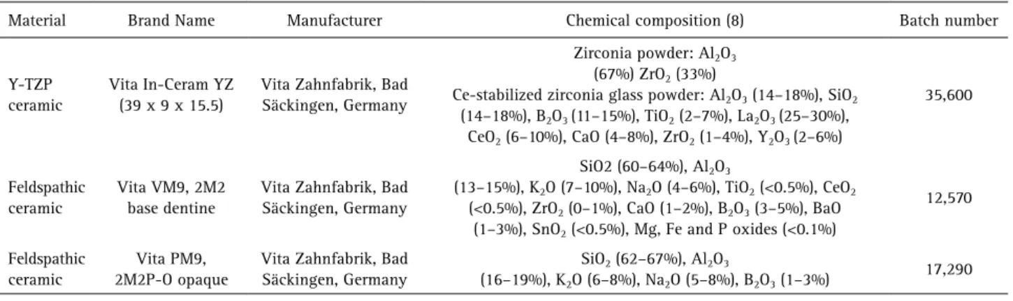
placed concentrically on 3 hardened steel balls with a 2.5 mm diameter, positioned 120° apart on a support circle with a 10 mm diameter. The load was applied with a flat punch with a 1.4 mm diameter at the center of the specimen. The biaxial flexural strength calculus was performed according to ISO 6872 (19).
Apparent Fracture Toughness Test for Monolayers
The apparent fracture toughness was measured by the Vickers hardness indentation technique using an indentation tester (Micromet 2100; Buehler UK Ltd) following the ASTM C 1421-99 standard (20). Three indentations (9.8 N, 30 s) were performed in each specimen fractured in a biaxial flexural strength test. To standardize the slow crack growth started by the tension area after loading, the crack length measurement was performed 30 s after the indentation (21).
Microstructural Analyses of Monolayers
XRD (X-ray diffraction) analyses in the VM9 powder, sintered VM9, PM9 press pellet, and pressed PM9 were performed in a diffractometer (XRD 6000, Shimadzu, Tokyo, Japan) with Cu Kα X-ray scanning from 10 to 80° 2θ degrees with a step size of 0.05° and 3 s step interval.
For the SEM (Scanning Electron Microscopy) analyses (n=3) of VM9, PM9 and PM9 press pellet specimens, the specimens were polished and etched with 2% HF (hydrofluoric acid) for 15 s and sputter-coated with gold– palladium. The specimens were examined in a scanning electron microscope (JEOL – JSM 6510, Jeol, Tokyo, Japan), operating at 10 kV. Three scanning electron micrographs from each group were obtained at a magnification of 500×, 1000× and 2000×. The Image J software (National Institutes of Health, Bethesda, MD, USA) was used to analyze the crystalline phase concentration of VM9, PM9 press pellet and pressed PM9. The SEM images of 1000x magnification (n=9) were used to measure the crystalline area, three times per image. The mean of the measurements was used for statistical analysis.
Preparation of Bilayers
Sixty pre-sintered zirconia (Vita In-Ceram YZ, Vita Zahnfabrik) were finished using 600-grit size SiC papers in a polishing machine, sintered in the oven (Vita Zyrcomat, Vita Zahnfabrik) at 1530 °C for 2 h with 1.5 h of rising time and cooling until 200 °C, according to the manufacturer´s recommendation. After sintering, the discs were 12 mm diameter and 1 mm thick.
The zirconia discs to be veneered with VM9 were cleaned by a cleaning firing procedure according to the manufacturer’s instructions. The powder and liquid of Vita VM9 were mixed to obtain a thin aqueous mixture
(washbake layer), which was applied very thinly with a brush on the cleaned zirconia framework. The firing of the washbake layer was carried out following the manufacturer’s recommendations. The discs were placed inside an oversized polyether mold with the washbake layer upward. The porcelain was applied into the mold as previously described, and the sintering was conducted (first dentine firing) and followed by a second dentine firing for correcting the final shape 2.0 mm thick (1 mm of zirconia and 1 mm of veneer porcelain) and 12 mm diameter.
To prepare the bilayered zirconia specimens with the pressed Vita PM9 porcelain, the zirconia disc was positioned inside a metallic matrix (12 mm diameter and 2 mm thick) and was waxed up, obtaining 1 mm of zirconia layer and 1 mm of wax layer. The investing, pressing, cooling and divesting procedures were performed as described previously. The bilayered disc was cut out from the press sprue by a diamond disc.
The finishing and polishing procedures were similar to the monolayered groups, until a 2-mm-thick layer (1 mm zirconia and 1 mm veneer porcelain) and 12 mm diameter were achieved.
Cooling Protocols
After polishing, the bilayered specimens (VM9 and PM9) were submitted to a self-glaze firing, according to the manufacturer´s instructions. The specimens were divided into 2 sub-groups: 1) Fast cooling: immediate removal of the specimens after the end of firing (900 °C), leaving them on the bench until they achieve the room temperature and 2) Slow cooling: after the end of firing, the oven was switched off and kept closed with the specimens inside, until dropping to room temperature.
Biaxial Flexural Strength for Bilayers
The biaxial flexural strength test of the bilayered specimens followed the same configuration used for the monolayered specimens. The porcelain side was positioned downward under tensile stress (Fig. 1).
The following equation was used to calculate the biaxial flexural strength, according to Lin et al. (22):
Where σ is the biaxial flexural strength, M is the maximum bending moment, and ta and tbare the thicknesses
of zirconia and porcelain layers, respectively. Ea e Eb are
527
Conventional versus pressed porcelain for zirconia
following equation:
M value was obtained by:
Where W is the load, ν is the Poisson’s ratio of bilayer, A is the radius of the circle of support points, and R is the equivalent radius of loading calculated by:
Where B is the radius of the tip of the piston and d is the thickness of specimens.
Mode of Failure Analysis
After the test, the type of fracture was observed by visual analysis, classified as only porcelain fracture or complete fracture (zirconia and porcelain fracture). The zirconia/porcelain interface was analyzed by SEM in three fractured specimens randomly selected from each group. Qualitative fractographic analysis was performed.
Apparent Fracture Toughness Test for Bilayers
The porcelain apparent fracture toughness in bilayer specimens was measured by indentation technique, as described for the monolayers’ test (n=15).
Residual Stress Measurements
To calculate the residual stress on the porcelain surface of the bilayer specimens after slow and fast cooling
requires measuring fracture toughness before and after the ultimate firing (glaze). Ten specimens of VM9/zirconia and 10 specimens of PM9/zirconia were made according to previous description. After polishing, the apparent fracture toughness was measured by the Vickers hardness indentation technique (21), as described before. For each specimen, three indentations (9,8 N; 30 s) were performed to calculate the fracture toughness (n=5). The bilayer specimens were submitted to a self-glaze firing, according to the manufacturer´s instructions, cooled by slow or fast cooling method and final fracture toughness was measured after glazing (n=5). Residual stress (MPa) was calculated according to the following equation (23):
Where, KIc is the fracture toughness measured before glaze cooling, KIc is the fracture toughness measured after glaze cooling (slow or fast), and c is the half size of the median crack measured before the glaze cooling.
Statistical Analysis
The statistical analysis for monolayers (biaxial flexural strength and apparent fracture toughness) was performed by Student-t test. One-way ANOVA was used for analyzing the crystalline phase concentration. Two-way ANOVA (porcelain and cooling method) followed by Tukey’s Honestly Significant Difference (HSD) post hoc test were used to analyze the biaxial flexural strength and apparent fracture toughness of the bilayered specimens (α=0.05). The used software was SPSS 19.0 (Statistical Package for Statistical Science Inc, Chicago, IL, USA).
Results
The Student’s t tests performed for monolayer specimens revealed that the biaxial flexural strength of the pressed
D. Longhini et al.
feldspathic porcelain Vita PM9 exhibited significantly higher values (105.20±10.91MPa) than the conventional VitaVM9 (81.76±7.72MPa) (p<0.01). Regarding fracture toughness, no significant difference (p=0.41) was observed between Vita VM9 (1.59±0.1 MPa.m1/2) and Vita PM9 (1.62±0.08 MPa.m1/2).
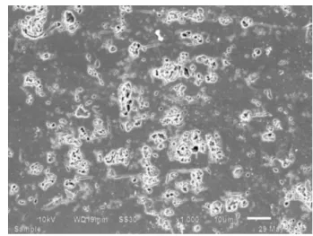
The XRD showed that both porcelains have leucite in composition as a crystalline phase before or after sintering (VM9) or pressing (PM9). SEM analysis showed a similar microstructure for VM9 (Fig. 2), pressed PM9 (Fig. 3) or pressed pellet (Fig. 4). The crystalline phase concentration was: PM9 pellet = 19.5±3.2 %; PM9 pressed = 18.4±2.1 %; VM9 = 21.5±2.7 %, and one-way ANOVA was not significant (F2,24 = 2.9541, p=0.07).
The means of the flexural strength (σ, MPa) and standard deviations (SD) of the bilayers biaxial flexural strength, cooled at slow or fast rate were: VM9/Slow = 167.15±30.53; VM9/Fast = 186.83±51.45; PM9/Slow =
234.89±49.19; PM9/Fast = 232.24±40.55. The two-way ANOVA was performed for the biaxial flexural strength (MPa) of the bilayered specimens for the porcelain material and the cooling method variables. This analysis indicated significance only for the porcelain material variable (p<0.01) better for the bilayer Vita PM9/zirconia.
With regards to fracture characteristics, 4 specimens showed complete fractures (13.3%) and 26 showed only porcelain fractures (86.6%) in VM9/zirconia group, but for the PM9/zirconia group, 10 discs failed in complete fractures (33.3%) and 20 showed only porcelain fractures (66.6%). A fractographic analysis of VM9 bilayer and PM9 bilayer showed that the fracture originated from defects in the porcelain layer. Cracks started from the porcelain surface and from the zirconia/porcelain interface, which may propagate each other to extend through the specimen (Figs. 5 and 6).
There was no statistically significant difference among the groups in apparent fracture toughness of porcelain in the bilayers (Slow/VM9 = 0.99 MPa.m1/2, Fast/VM9 = 1.06 MPa.m1/2, Slow/PM9 = 1.10 MPa.m1/2 and Fast/PM9 = 1.12 MPa.m1/2). All the specimens showed compressive residual stress (MPa): Slow/VM9 = -5.19 MPa; Fast/VM9 = -16.5 MPa; Slow/PM9 = -11.62 MPa; Fast/PM9 = -13.03 MPa.
Discussion
Besides the similarity in composition between the studied porcelains, as revealed in the XRD analysis and crystalline phase concentration, the higher values of biaxial flexural strength for the monolayer pressed porcelain PM9 compared to the conventional layered VM9 porcelain may be due to the processing technique. In this study, both conventional and pressed porcelain were similar in composition and microstructure. The XRD analysis detected leucite contents, and the SEM images revealed comparable Figure 2. SEM images at 1000× magnification of Vita VM9.
529
Conventional versus pressed porcelain for zirconia
Figure 6. SEM images at 75× magnification of broken PM9 bilayer showing fracture originating at interface from defects in the porcelain layer.
microstructures (Figs. 2-4) and crystal concentration. An explanation may be that there is a possible leucite crystal growth in the PM9 after the pressing cycle, but it was not confirmed since the crystal concentration before and after firing was not different.
The better flexural strength of the pressed porcelains as PM9 may be related to highly controlled manufacturing of the ingots. The prefabricated ingots are melted and overpressed into a previously invested mold cavity in a controlled procedure(7,16), which can achieve a more homogeneous structure, avoiding large pore formation (15). Conversely, in hand-layer technique, the powder and liquid are hand mixed and modeled with a brush, which makes it more susceptible to internal porosity concentration. In addition, it is easy to accumulate other technical errors due to the multiple stages of uncontrolled factors (8). Heintze and Rousson (18) found less frequent veneer chipping rates in pressed technique compared to the conventional one, either for zirconia or metal-ceramic fixed dental prostheses. However, better results were also found for conventional porcelain in some studies(7,15), both attributing it to the lack of dental laboratory experience with the press technique. On the other hand, Lima et al. (8) did not show difference between the VM9 and PM9 porcelains in four-point flexural strength after fatigue.
The PM9 porcelain showed a higher performance in the bilayer specimens test. Again, this may be related to the more controlled technique. Moreover, some studies showed that pressed porcelains have better wetting with the zirconia substructure, due to the pressure and vacuum during injection (16,22). Kim et al. (17), in failure analysis, observed that in the conventional porcelain group, the fracture was always found in the porcelain body, while the pressed porcelain group showed mainly the complete
fracture of the zirconia/porcelain set. They attributed the low resistance of the conventional porcelain to technical failures and structural weakness caused by internal porosities and the higher resistance of pressed porcelain to its stronger adhesion to zirconia (17). In the present study, there was more incidence of zirconia/porcelain combined fracture in the group with PM9 compared to the VM9 group. The fractographic qualitative analysis shows that for both PM9 and VM9 bilayer specimens, the fracture started from internal defect close to the interface and from the surface. The study of Guazzato et al. (24) explained that different fracture modes could occur depending on the setting of biaxial flexural strength testing. It is influenced by the material at the bottom and physical properties such as elastic modulus of the bilayer specimen materials. When the porcelain is on the bottom, as it was in the present study, the fracture likely starts from a major defect at the surface to propagate to the inside (24). In the conducted study, defects at the interface may originate a secondary crack initiation.
With regard to the cooling rate in the firing cycle, an increasing number of authors propose slow cooling (2,10,13), aiming to reduce the thermal gradient by a homogeneous cooling of internal and external porcelain surface below Tg (13). This would avoid excessive development of high magnitude transient stresses (13). However, some studies observed better results for fast cooling (4,14). Almeida-Junior et al. (14) suggested that compressive stress is developed at the fast cooled veneering porcelain, which could improve the resistance of the surface layer but may develop high tension stress at the zirconia/ porcelain interface (14). However, better results of bond strength were observed by Göstemeyer et al. (4) associated with compressive stress in the veneering porcelain, which
D. Longhini et al.
would protect the adhesion interface (4). In this study, all groups showed compressive residual stress on surface after both slow and fast glaze cooling. This may be explained by the thermal gradients generated after cooling, since the Y-TZP conducts substantially less heat than veneer porcelain (10). Due to the tempering effect, the residual stresses distribution change from the surface to the bulk, with compressive stresses at the surface and compensating tensile stress in the bulk (3,11). Residual stress may be related to cooling rate, material thickness, and thermal properties such as thermal expansion coefficient, conductivity and diffusivity (11). Higher values of compressive residual stress were found in fast cooling methods, particularly for VM9. This result may be related to differences between pressed technique (PM9) and conventional technique (VM9) or because the higher mismatch of the thermal coefficient expansion between zirconia infrastructure (10.5 ppm/oK) and VM9 (8.8-9.2 ppm/oK), rather than that for PM9 (9-9.5 ppm/oK).
It is important to highlight that in the clinical routine the crowns are finished before glazing, different from several studies, where the specimens are usually finished after the cooling method (4,14). Taking into account that compressive stresses in a zirconia/porcelain bilayer after the sintering are locked on the surface of veneer, the compression zone on the surface may be relieved when finishing is performed (25). In the present study, specimens were polished prior the final glaze firing. Nevertheless, no significant difference was found for the fast and slow cooling rates.
Fracture toughness is related to the mechanical resistance of the material to the catastrophic failure, defined as the critical stress intensity level at which a given flaw starts growing (21). In contrast to the flexural strength results, the statistical results for apparent fracture toughness were not significant either for monolayer or bilayer specimens. However, for monolayer specimens the values were higher (1.59 to 1.62 MPa.m1/2) than those for the bilayers (0.99 to 1.12 MPa.m1/2). Although this is not the main objective of the study, the lower fracture toughness of porcelain in bilayer specimens may be associated to residual stresses and may make the porcelain weaker and more susceptible to chipping. According to Pang et al., more chipping rates in zirconia/porcelain restorations may be associated to greater residual stresses and low fracture toughness of the veneering porcelain (6). With regard to the cooling rate for the bilayers, again, it was not able to interfere in the fracture toughness.
As a limitation, it may be reminded that the study was performed in flat specimens (discs), which can be difficult for the clinical conditions. Moreover, the mechanical properties were evaluated by means of static testing.
Further studies about all-ceramic zirconia/porcelain crowns or bridges under fatigue could help in understanding the intraoral behavior of these systems.
In conclusion, pressed porcelain may have better mechanical behavior because it has higher flexural strength than the conventional hand layered porcelain, either in monolayer or bilayer structures, despite the similarity in apparent fracture toughness, composition and microstructure. The cooling rates did not impair the biaxial flexural strength and apparent fracture toughness in bilayer specimens, but fast cooling increased compressive residual stress on porcelain surface, particularly for the conventional VM9.
Resumo
Este estudo teve como objetivo caracterizar uma porcelana convencional e uma prensada indicadas para infraestrutura de zircônia em termos de resistência à flexão biaxial (RFB), tenacidade à fratura aparente (TF) e composição microestrutural, e investigar o efeito da taxa de resfriamento do ciclo de glaze na RFB de espécimes bicamada de zircônia/porcelana. Espécimes monocamada de porcelana convencional Vita VM9 e porcelana prensada Vita PM9 (n=15) (12 mm de diâmetro x 1,2 mm de espessura) foram preparados para o ensaio de RFB (MPa). Tenacidade à fratura aparente (MPa.m½) foi mensurada pela técnica da indentação (n=15). O teste de t-Student foi aplicado para as análises estatísticas. Microscopia eletrônica de varredura e difração de raios-X foram utilizadas para análise microestrutural da porcelana. Para o ensaio de RFB nos bicamadas, discos de zircônia (12 mm de diâmetro x 1 mm de espessura) (Vita In-Ceram YZ) foram recobertos com as duas porcelanas (1 mm de espessura). Após a simulação da queima de glaze, os espécimes foram submetidos ao método de resfriamento rápido ou lento (n=15). A tenacidade à fratura aparente (MPa.m½) foi mensurado na superfície da porcelana dos bicamadas (n=15) e o estresse residual foi calculado. Foi utilizado ANOVA a dois fatores (porcelana e método de resfriamento) para análise estatística dos espécimes bicamada (α=0,05). Monocamadas de Vita PM9 exibiram valores de resistência à flexão biaxial significativamente maiores (p<0,01), mas não houve diferença estatística (p=0,41) em relação à TF entre as porcelanas. Para os espécimes bicamada, ANOVA a dois fatores para RFB foi significante apenas para a variável porcelana (p<0,01), com superioridade para Vita PM9/zircônia. ANOVA a dois fatores para TF dos espécimes bicamadas não apresentou significância para nenhuma variável. Todos os grupos apresentaram estresse residual de compressão. Porcelanas prensadas parecem ser mecanicamente mais efetivas para recobrimento de zircônia.
References
1. Sailer I, Feher A, Filser F, Gauckler LJ, Luthy H, Hammerle CH. Five year clinical results of zirconia frameworks for posterior fixed partial dentures. Int J Prosthodont 2007;20:383-388.
2. Choi JE, Waddell JN, Swain MV. Pressed ceramics onto zirconia. Part 2: indentation fracture and influence of cooling rate on residual stresses. Dent Mater 2011;27:1111-1118.
3. Swain MV. Unstable cracking (chipping) of veneering porcelain on all-ceramic dental crowns and fixed partial dentures. Acta Biomater 2009;5:1668-1677.
4. Göstemeyer G, Jendras M, Dittmer MP, Bach F, Stiesch M, Kohorst P. Influence of cooling rate on zirconia/veneer interfacial adhesion. Acta Biomater 2010;6:4532-4538.
5. Pjetursson BE, Sailer I, Makarov NA, Zwahlen M, Thoma DS. All-ceramic or metal-ceramic tooth-supported fixed dental prostheses (FDPs)? A systematic review of the survival and complication rates. Part II: Multiple-unit FDPs. Dent Mater 2015;31;624-639.
531
Conventional versus pressed porcelain for zirconia
retrieved zirconia-ceramic and metal-ceramic fixed dental prostheses. Dent Mater 2015;31:1198-1206.
7. Guess PC, Bonfante EA, Silva NR, Coelho PG, Thompson VP. Effect of core design and veneering technique on damage and reliability of Y-TZP-supported crowns. Dent Mater 2013;29:307-316.
8. Lima JM, Souza AC, Anami LC, Bottino MA, Melo RM, Souza RO. Effects of thickness, processing technique, and cooling rate protocol on the flexural strength of a bilayer ceramic system. Dent Mater 2013;29:1063-1072.
9. Sailer I, Makarov NA, Thoma DS, Zwahlen M, Pjetursson BE. All-ceramic or metal-ceramic tooth-supported fixed dental prostheses (FDPs)? A systematic review of the survival and complication rates. Part I: Single crowns (SCs). Dent Mater 2015;31:603-623.
10. Tan JP, Sederstrom D, Polansky JR, McLaren EA, White SN. The use of slow heating and slow cooling regimens to strengthen porcelain fused to zirconia. J Prosthet Dent 2012;107:163-169.
11. Mainjot AK, Schajer GS, Vanheusden AJ, Sadoun MJ. Influence of zirconia framework thickness on residual stress profile in veneering ceramic: measurement by hole-drilling. Dent Mater 2012;28:378–384. 12. DeHoff PH, Barrett AA, Lee RB, Anusavice KJ. Thermal compatibility of
dental ceramic systems using cylindrical and spherical geometries. Dent Mater 2008;24:744-752.
13. Benetti P, Kelly JR, Della Bona A. Analysis of thermal distributions in veneered zirconia and metal restorations during firing. Dent Mater 2013;29:1166-1172.
14. Almeida AA Jr, Longhini D, Domingues NB, Santos C, Adabo GL. Effects of extreme cooling methods on mechanical properties and shear bond strength of bilayered porcelain/3Y-TZP specimens. J Dent 2013;41:356-362.
15. Nadja N, Andreas B, Caroline S, Christoph H, Irena S. A randomized controlled clinical trial of 3-unit posterior zirconia-ceramic fixed dental prostheses (FDP) with layered or pressed veneerin ceramics: 3-year results. J Dent 2015;43:1365-1370.
16. Aboushelib MN, Kleverlaan CJ, Feilzer AJ. Microtensile bond strength of different components of core veneered all-ceramic restorations. Part II: Zirconia veneering ceramics. Dent Mater 2006;22:857-863. 17. Kim JH, Lee SJ, Park JS, Ryu JJ. Fracture load of monolithic CAD/CAM
lithium disilicate ceramic crowns and veneered zirconia crowns as a posterior implant restoration. Implant Dent 2013;22:66-70. 18. Heintze SD, Rousson V. Survival of zirconia- and metal-supported fixed
dental prostheses: a systematic review. Int J Prosthodont 2010;23:493-502.
19. International Organization for Standardization. ISO 6872. Dentistry - ceramic materials. Geneva: ISO; 2008.
20. ASTM Standard C1421-99. Standard test method for determination of fracture toughness of advanced ceramics at ambient temperature. West Conshocken, PA:ASTM;1999.
21. Scherrer SS, Denry IL, Wiskott HW. Comparison of three fracture toughness testing techniques using a dental glass and a dental ceramic. Dent Mater 1998;14:246-255.
22. Lin WS, Ercoli C, Feng C, Morton D. The effect of core material, veneering porcelain, and fabrication technique on the biaxial flexural strength and Weibull analysis of selected dental ceramics. J Prosthodont 2012;21:353-362.
23. Cesar PF, Gonzaga CC, Miranda WG, Yoshimura HN. Effect of ion exchange on hardness and fracture toughness of dental porcelains. J Biomed Mater Res B Appl Biomater 2007;83:538-545.
24. Guazzato M, Proos K, Sara G, Swain MV. Strength, reliability, and mode of fracture of bilayered porcelain/core ceramics. Int J Prosthodont 2004;17:142-149.
25. Guazzato M, Quach L, Albakry M, Swain MV. Influence of surface and heat treatments on the flexural strength of Y-TZP dental ceramic. J Dent 2005;33:9-18.