Article
J. Braz. Chem. Soc., Vol. 22, No. 5, 884-890, 2011. Printed in Brazil - ©2011 Sociedade Brasileira de Química 0103 - 5053 $6.00+0.00
A
*e-mail: [email protected]
Studying the Distribution of Selenium in Buffalo and Cow’s Milk Whey
João B. Pereira Juniorand Kelly G. Fernandes*
Grupo de Espectrometria Analítica Aplicada, Faculdade de Química, Instituto de Ciências Exatas e Naturais, Universidade Federal do Pará, 66075-110 Belém-PA, Brazil
Neste estudo, a cromatograia por exclusão de tamanho (SEC) com detecção online ultravioleta (UV), espectrometria de absorção atômica em forno de graite (GF AAS) e a espectrometria de massa por tempo de voo com dessorção/ionização de matriz assistida por laser (MALDI-TOF-MS) foram usadas para estudar a associação de selênio com macromoléculas e compostos presentes no soro de leite de búfala e bovino. Os peris de SEC-UV obtidos para as amostras de soro de leite de búfala e soro de leite bovino indicaram a presença de espécies de alta e baixa massa molecular. A combinação das informações obtidas com SEC-UV, GF AAS e MALDI-TOF-MS para as frações < 10 kDa conirmou a associação de selênio com espécies de baixa massa molecular.
In this study, size exclusion chromatography (SEC) coupled with an online ultraviolet detection (UV) and graphite furnace atomic absorption spectrometry (GF AAS) and matrix-assisted laser desorption ionisation time-of-light mass spectrometry (MALDI-TOF-MS) were used to study the association of selenium with macromolecules and compounds present in buffalo and cow’s milk whey. SEC-UV proiles obtained from buffalo and cow’s milk whey samples indicate the presence of high molecular weight (HMW) and low molecular weight (LMW) species in both samples studied. The combined information obtained with SEC-UV, GF AAS and MALDI-TOF-MS for fractions < 10 kDa conirmed the association of selenium with LMW species.
Keywords: buffalo and cow’s milk whey, SEC offline GF AAS, SEC online UV, MALDI-TOF-MS
Introduction
Buffalo (Bubalus bubalis) milk is a food highly nutritious and beneicial to the human health due to the biological value of its constituents. It has pronounced differences when compared to cow’s milk, due to the presence of greater percentages of constituents such as fat, protein, lactose, total solids and some minerals.1 Buffalo
milk is used in northern Brazil for making different dairy products, including butter, various cheeses and yogurt.2
However, studies concerning the chemical composition of buffalo milk are limited compared with cow’s milk ones.
Milk proteins are classiied according to their structural and physicochemical properties, and consist of the following: casein, whey proteins, fat globule membrane proteins, enzymes and growth factors. However, from a nutritional and industrial perspective, casein and whey proteins are more widely applicable and have signiicant economic value. The total protein concentration and ratio
of casein and whey protein fractions vary considerably in milk from different species.α-lactalbumin, bovine serum albumin, immunoglobulin, lactoferrin and lysozyme are whey proteins that provide health beneits. β-lactoglobulin, which is not found in human milk,3 is the most abundant
protein in cow’s milk whey and is associated with allergies, particularly in children.
The elemental composition of food plays important biological roles. However, the bioavailability and toxicity of these elements strongly depend on the chemical form in which they occur in biological systems.4 Therefore,
speciation studies are necessary in order to obtain more information about the elemental composition of foods.
The nutritional bioavailability and toxicity of selenium depend on its concentration and the chemical form in which it is ingested.6-8 In fact, studies have shown that selenium in
the form of seleno-amino acids is more easily absorbed by the body than inorganic forms.9 The scientiic community
has shown increasing interest in the determination of the elemental composition of milk, because of its important role in human health.10,11 The concentration of selenium
in milk has been reported to vary by geographical region, and depends on factors such as selenium levels in soil and water, as well as in local plants and food.12
Studies have shown that only a small proportion of selenium (< 3%) is associated with the lipid fraction.13
However, reported values for the amount of selenium in casein and whey milk fractions are conlicting. In some studies, selenium was found mainly associated with the casein fraction, whereas in others, more selenium was found in whey fractions.13,14 The reason for these divergent
results is not clear.
Although the chemical composition of buffalo milk has been previously reported,15,16 to date, no studies have been
conducted on the determination and speciation of selenium in buffalo milk whey.
Recently, selenium fractionation, distribution and speciation in foods such as nuts, cow’s milk and infant formulas have been reported.12,16-21 In addition, several
works in the literature have described elemental speciation in milk samples by size exclusion chromatography (SEC),22 in combination with speciic element detectors,
such as electrothermal atomic absorption spectrometry (ET AAS),18,23 inductively coupled plasma optical emission
spectrometry (ICP OES)24,25 and inductively coupled plasma
mass spectrometry (ICP-MS).4,16,17 Of these techniques, the
most appropriate are ET AAS and ICP-MS, due to their low detection limits for trace elements. However, investigations coupling online liquid chromatography with an ET AAS detector are limited compared to those using ICP-MS. Thus, current published procedures for speciation are based on chromatographic separation and fraction collection, followed by fraction quantiication ofline by ET AAS. Despite this disadvantage, procedures based on ET AAS detection have been extensively used for speciation studies.26,27
In this study, SEC coupled with online UV and GF AAS detection was used to evaluate selenium binding to high molecular weight (HMW) and low molecular weight (LMW) species present in buffalo and cow’s milk whey. In addition, matrix-assisted laser desorption ionisation time-of-light mass spectrometry (MALDI-TOF-MS) was used to characterize organic moieties associated with Se containing LMW fractions present in buffalo and cow’s milk whey.
Experimental
Instrumentation
A ProStar 210 Liquid Chromatographic System (Varian, Mulgrave, Australia) equipped with a BioSep-SEC S 3000 column (300 × 7.8 mm id) (Phenomenex, Torrance, CA, USA) and a Rheodyne (Model 7125, Cotati, CA, USA) sample injection valve itted with a 20 µL loop was used in the SEC chromatographic separation of milk whey proteins. Selenium determinations in SEC separated fractions were carried out using a Varian Model SpectrAA 220 atomic absorption spectrometer (Mulgrave, Victoria, Australia) equipped with a graphite furnace atomiser (a GTA 100 autosampler) and a deuterium lamp background corrector (Varian). A selenium hollow cathode lamp was employed as a radiation source, operating at 10 mA. Absorbance signals were measured using the 196.0 nm line at a spectral resolution of 0.2 nm. For GF AAS, argon (99.998% purity) (Linde, Pará, Brazil) was used as gas purge (3.0 L min-1) during
all steps of the graphite furnace heating program, except atomisation. Pyrolytic coated graphite tubes (Varian) were used throughout. All signals were measured as integrated absorbance.
Milk samples were separated into fat, milk whey and casein micelles, using a centrifuge (Sigma 2K15, Germany). The mobile phase was degassed using a model USC 1400 ultrasonic bath, (Unique, São Paulo-SP, Brazil). Following SEC separation, eluted fractions were concentrated by lyophilisation using a Model L 101 Lyophiliser (Liotop, São Carlos-SP, Brazil). Molecular mass analysis was conducted using MALDI-TOF-MS (Model Axima CRF, Shimadzu, Kyoto, Japan).
Reagents and materials
All reagents used were analytical grade. All dilutions were made using distilled-deionised water (resistivity 18.2 MΩ cm) obtained from an ELGA water puriication system (Elgastat, Buckinghamshire, England). Nitric acid (Quimex, São Paulo-SP, Brazil) was puriied by distillation in a quartz distiller (Quimis) and used to prepare the aqueous reference solutions.
Tris(hydroxymethyl)-aminomethane (Sigma, St. Louis, USA) and sodium dodecyl sulfate (Sigma, Tokyo, Japan) were used in the preparation of the mobile phase. Hydrochloric acid (Quimex) was used to adjust the pH of the mobile phase.
The following protein standards were used to calibrate the size exclusion column: bovine serum albumin (67 kDa), β-lactoglobulin (18.4 kDa) and selenomethionine (0.196 kDa) (Sigma, St. Louis, USA). Ferritin (440 kDa) was used to obtain the column void volume.
A standard stock solution containing 1000 mg L-1
selenium (Sigma) was used. A solution of 1000 mg L-1
palladium (Sigma) in 1% v/v nitric acid (99.999% purity) (Aldrich, Milwaukee, WI, USA) was used as a selenium chemical modiier.
Millex-SR 0.45 µm ilters (Millipore, Bedford, MA, USA) were used to ilter milk whey samples. C18 Zip TipTM
micropipette tips (Millipore) were used to purify fractions for MALDI-TOF-MS analysis.
Milk samples
The two types of milk whey samples studied, buffalo and cow’s milk, were obtained from a farm in the Marajó Island (Pará State, Brazil) and from the Institute of Animal Health and Production of the Universidade Federal Rural da Amazônia (Pará State, Brazil), respectively. Once collected, milk samples were conditioned in polyethylene lasks and stored in a freezer at −20 °C.
Sample preparation: centrifugation
Milk samples were fractionated into components by centrifugation at 5000 rpm during 60 min at 4 °C. Milk whey samples were then removed using a micropipette, fat and casein micelles were discarded. Milk whey samples were passed through a ilter (0.45 µm), the iltrate was diluted (1 + 1 v/v) with the mobile phase, and 20 µL of this dilution was injected onto the SEC-UV system.
SEC-UV conditions
The mobile phase used in this study was prepared by dissolving 0.5% m/v SDS in 2.5 mmol L-1 Tris in deionised
water and adjusting the pH to pH 7.4 with hydrochloric acid. The chromatographic conditions are summarised in Table 1.
The SEC column was calibrated using 2 mg mL-1
of each protein standard (albumin, β-lactoglobulin and selenomethionine); dissolved in 0.5% m/v SDS in 2.5 mmol L-1 Tris-HCl (pH 7.4). Chromatographic proiles
were monitored using a UV detector at a wavelength of 295 nm.
Selenium determination by GF AAS
C o l u m n f r a c t i o n s w e r e m a n u a l l y c o l l e c t e d (0.6 mL min-1) in polypropylene containers and then
lyophilised. These concentrated SEC column fractions were then diluted with 200 µL of 0.028 mol L-1 nitric acid.
Selenium content in the column fractions was determined by GF AAS using palladium (5 µg) as chemical modiier. The concentration of selenium in the buffer blank solvent (mobile phase) and milk whey samples was also investigated. Calibrations were conducted using selenium standards (5–15 µg L-1) in a 0.028 mol L-1 nitric acid
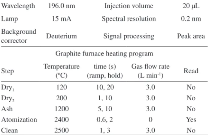
medium. The temperature program for the atomiser for an injection volume of 20 µL is shown in Table 2.
Molecular mass characterization by MALDI-TOF-MS
SEC-UV elution peaks were collected in polypropylene containers. Fractions were then concentrated by lyophilisation. These concentrated fractions were dissolved in 0.05% v/v TFA and 50% v/v acetonitrile and then desalted using C18 Zip TipTM micropipette tips. An aliquot
of 0.5 µL of each sample was mixed with 0.5 µL of sinapinic acid (10 mg mL-1) MALDI matrix. MS spectra were
acquired in linear mode using an acceleration voltage of 20 kV and a vacuum pressure of 7.0 × 10⁻⁶ bar. Laser pulses
were generated by a nitrogen laser (337 nm, 10 pulses per seconds). External calibration of the instrument was conducted using a bovine serum albumin (67 kDa) standard.
Table 1. Optimized chromatographic conditions for SEC
Column BioSep-SEC-S 3000 (300 × 7.8 mm i.d.)
Sample volume 20 µL
Mobile phase 0.5% (m/v) SDS in 2.5 mmol L-1 Tris-HCl (pH 7.4)
Flow rate 0.6 mL min-1
UV detection 295 nm
Separation range 0.196–67 kDa
Table 2. Instrument conditions for the determination of selenium in
milk whey
Wavelength 196.0 nm Injection volume 20 µL
Lamp 15 mA Spectral resolution 0.2 nm
Background
corrector Deuterium Signal processing Peak area
Graphite furnace heating program
Step Temperature (ºC)
time (s) (ramp, hold)
Gas low rate (L min-1) Read
Dry1 120 10, 20 3.0 No
Dry2 200 1, 10 3.0 No
Ash 1200 5, 10 3.0 No
Atomization 2400 0.6, 2 0 Yes
Results and Discussion
UV proiles and MALDI-TOF mass spectra
Experimental SEC conditions, such as mobile phase (SDS in Tris-HCl at pH 7.4 and phosphate buffer solution at pH 6.8), mobile phase low rate (0.2-1.0 mL min-1),
retention time and wavelength (280 and 295 nm) were optimised, and optimal SEC conditions are shown in Table 1. The following equation was obtained from column calibration with albumin (Kav = 0.015), β-lactoglobulin
(Kav = 0.075) and selenomethionine (Kav = 0.562):
log (MW) = −4.6569 Kav + 4.899 (r2 = 1.0), where MW and
Kav are the molecular weight (Da) and partition coeficient,
respectively. The partition coeficient, Kav, was calculated
using the formula, Kav= (Velution− V0) / (Vtotal− V0), where
V0 is column void volume (5.92 mL), and the total column
volume, Vtotal, is 30 mL. The eluent from the SEC column
was passed through a UV detector cell and the elution proiles of the milk whey sample fractions were recorded. Before chromatographic separation of each sample, a blank was injected in order to control for the cleanliness of the chromatographic system. SEC proiles of the two types of milk whey are shown in Figure 1a (buffalo milk whey) and Figure 1b (cow’s milk whey).
As it can be seen in Figure 1a, three main peaks are distinguishable in the buffalo milk whey sample. Peaks I (Kav = 0.008), II (Kav = 0.319) and III (Kav = 0.518)
eluted at volumes corresponding to apparent molecular weights (MW) of 72.3, 2.6 and 0.3 kDa, respectively. Peak I (72.3 kDa apparent MW) contains high molecular weight (HMW) macromolecules, and may correspond to large protein species or complexes such as serum albumin (67 kDa) and lactoferrin (87.0 kDa). It has been reported that milk whey fractions with MWs between 40 and 180 kDa may contain serum albumin and lactoferrin (Peak I).4 Peak II (2.6 kDa) and peak III (0.3 kDa)
represent low molecular weight (LMW) components of buffalo milk whey.
The SEC elution profile obtained for the cow’s milk whey sample was completely different from the chromatographic proile observed for the buffalo milk whey sample. Figure 1b shows that four peaks were detected in cow’s milk whey: peak I (52.8 kDa, Kav = 0.038); peak II
(34.6 kDa, Kav = 0.078); peak III (1.7 kDa, Kav = 0.358) and
peak IV (0.47 kDa, Kav= 0.480). Peak I may contain serum
albumin and lactoferrin or other HMW compounds, and peaks II, III and IV may contain β-lactoglobulin (dimeric form)4 (peak II) and LMW species (peaks III and IV). Cow’s
milk whey fractions contained signiicantly less LMW compounds and an increase in HMW species compared to milk whey buffalo fractions.
According to Martino et al.,4 non-protein compounds
as such lactose (0.360 kDa), orotic acid (0.156 kDa) and inorganic mineral salts have been found in milk whey by UV detector at a wavelength of 280 nm. LMW species < 0.196 kDa were not detected in both samples (λ = 295 nm). This can be due to the wavelength used or non-presence of these compounds in the samples studied. The chromatographic run time was extended to 100 min to ensure the elution of all fractions. However, no peaks were observed at retention times higher than 30 min.
Figure 2 shows the MALDI-TOF mass spectra obtained from cow’s milk whey and buffalo milk whey fractions with apparent molecular weights < 10 kDa. Two molecular ions were observed at m/z values of 2.6 and 0.73 kDa for the buffalo milk whey fraction (Figure 2a). The 2.6 kDa molecular ion is similar to the value obtained during calibration of the SEC column (see Figure 1a). In contrast, MALDI-TOF mass spectra from cow’s milk whey fractions (Figure 2b) contained three ine structures corresponding to m/z of 2.4, 2.8 and 5.1 kDa. A similar molecular weight of 2.8 kDa was previously reported in the literature in a comparative study of raw cow’s milk whey vs. human milk whey by Martino et al.4 LMW species < 0.58 kDa were
not found in both samples studied. Because of instrument
Figure 1. SEC elution proiles obtained using an online UV detector
problems, it was not possible to obtain results for molecular weights > 10 kDa.
Total concentration and distribution of selenium
Total selenium contents in buffalo and cow’s milk whey were measured in samples diluted 1 + 19 v/v with 0.028 mol L-1 nitric acid and using analytical calibration
solutions prepared in water medium. No matrix effects were observed in milk whey samples. The total concentrations of selenium determined for buffalo and cow’s milk whey were 254.4 and 250.8 µg L-1, respectively. These results
compare well with the sum of selenium concentrations measured for each of the SEC column elution fractions from buffalo (258.7 µg L-1) and cow’s milk whey (204.2 µg L-1),
following SEC separation. The relatively high selenium concentration values obtained may be related to the geographical location of the farms and local food sources.28
Selenium recoveries obtained for buffalo and cow’s milk whey following SEC were 101.7 and 81.4%, respectively.
Selenium levels in the solvent buffer blank control (mobile phase) were lower than the detection limit. The detection limit (3 × σ/b) and quantiication limit (10 × σ/b), where σ
is the standard deviation and b the angular coeficient of the calibration graph, were 2.08 and 6.97 µg L-1, respectively
(n = 10). The characteristic mass (m0)was 55.2 pg for a
20 µL sample aliquot.
Graphite furnace AAS selenium determination results for each SEC fraction for buffalo and cow’s milk whey are plotted in Figure 3. Relative standard deviations were
≤ 2.0% (n = 3).
Figure 3a demonstrates that in buffalo milk whey, selenium was detected co-eluting with both HMW and LMW fractions. In particular, high selenium levels were detected in peak II (39.1% in the 2.6 kDa fraction) and peak III (41.0% at 0.3 kDa). The co-elutions of selenium with peaks II and III conirm the association of selenium with chemical species of MW 2.6 and 0.3 kDa. In contrast, as it can be seen in Figure 2b, in the cow’s milk whey sample, selenium predominantly co-elutes with two HMW peaks
Figure 3. Selenium co-elution proiles detected by GF AAS for (a) buffalo milk whey and (b) cow’s milk whey SEC fractions.
Acknowledgments
The authors are grateful to Fundação de Amparo à Pesquisa do Estado do Pará (FAPESPA), Conselho Nacional de Desenvolvimento Cientíico e Tecnológico (CNPq) and Coordenação de Aperfeiçoamento de Pessoal de Nível Superior for research funding and fellowships. K. G. F. and J. B. P. J. are also thankful to Dr. Geraldo Narciso da Rocha Filho (FAQUI/UFPA, Pará) for donation of the HPLC, Dr. Dulcidéia da Conceição Palheta (UFPA, Pará) for use of the GF AAS, Dr. Arthur Luiz da Costa da Silva and MSc Alessandra Ciprandi (ICB/UFPA) for analysis by MALDI-TOF-MS.
References
1. Nader Filho, A.; Schoken-Iturrino, R. P.; Rossi Júnior, O. D.; Cavagliano, C. P. G.; Rev. ILCT1984, 39, 25.
2. Figueiredo, E. L.; Lourenço Junior, J. B.; Toro, M. J. U.; Rev.
Bras. Tecn. Agroindustrial2010, 4, 19.
3. Sgarbieri, V. C.; Braz. J. Food Technol.2005, 8, 43.
4. Martino, F. A. R.; Sánchez, M. L. F.; Medel, A. S.; J. Anal. At. Spectrom. 2002, 17, 1271.
5. Lun, J.; Holmgren, A.; J. Biol. Chem. 2009, 284, 723. 6. Shibata, Y.; Morita, M.; Fuwa, K.; Adv. Biophys. 1992, 28, 31. 7. Fishbein, L.; Metals and their Compounds in the Environment,
Occurrence, Analysis and Biological Relevance; Merian, E.,
ed., VCH: Weinheim, 1991, pp. 1153.
8. Cámara, C.; Cobo, M. G.; Palacios, M. A.; Muñoz, R.; Donard, O. F. X.; Quevaullier, P., Maier, E.A., Griepink, B., eds. In
Quality Assurance for Environmental Analysis, Elsevier:
Amsterdam, 1995, pp. 237-264.
9. Volderheide, A.; Wrobel, K.; Kannamkumarath, S.; B’Hymer, C.; Montes-Bayón, M.; Ponte de Leon, C.; Caruso, J.; J. Agric.
Food Chem.2002, 50, 5722.
10. Rodríguez Rodríguez, E. M.; Alaejos, M. S.; Romero, C. D.;
J. Agric. Food Chem. 1999, 47, 1520.
11. Prohaska, T.; Köllensperger, G.; Krachler, M.; De Winne, K.; Stingeder, G.; Moens, L.; J. Anal. At. Spectrom. 2000, 15, 335.
12. Muñiz-Naveiro, O.; Domínguez-González, R.; Bermejo-Barrera, A.; Bermejo-Bermejo-Barrera, P.; Cocho, J. A.; Fraga, J. M.;
Talanta 2007, 71, 1587.
13. Debski, B.; Piccinao, M. F.; Milner, J. A.; J. Nutr. 1987, 117, 1091.
14. Muñiz-Naveiro, O.; Domínguez-González, R.; Bermejo-Barrera, A.; Bermejo-Bermejo-Barrera, P.; Cocho, J. A.; Fraga, J. M.; Bermejo-Barrera, P.; Anal. Bioanal. Chem. 2005, 381, 1145. 15. Verruma, M. R.; Salgado, J. M.; Sci. Agric. 1994, 51, 131. 16. Pereira Junior, J. B.; Fernandes, K. G.; Müller, R. C. S.;
Nóbrega, J. A.; Palheta, D. C.; Quim. Nova2009, 32, 2333.
at 52.8 kDa (23.5%) and 34.6 kDa (43.5%). Although co-elution with two other, smaller, LMW peaks (4.1-19.2%) was also detected, the highest levels of selenium were found associated with HMW macromolecules. The distribution of selenium in the buffalo and cow’s milk whey samples measured in this study is signiicantly different, most likely due to differences in milk composition.
Conclusion
17. Brätter, P.; Blasco, I. N.; de Brätter, V. E. N.; Raab, A.; Analyst
1998, 123, 821.
18. Martino, F. A. R.; Sánchez, M. L. F.; Medel, A. S.; Anal. Chim. Acta2001, 442, 191.
19. Bermejo, P.; Peña, E.; Domínguez, R.; Bermejo, A.; Fraga, J. M.; Cocho, J. A.; Talanta2000, 50, 1211.
20. Kannamkumarath, S. S.; Wrobel, K.; Wuilloud, R. G.; Talanta
2005, 66, 153.
21. Gonçalves, A. M.; Fernandes, K. G.; Ramos, L. A.; Cavalheiro, E. T. G.; Nóbrega, J. A.; J. Braz. Chem. Soc.2009, 20, 760. 22. Naozuka, J.; Marana, S. R.; Oliveira, P. V.; J. Food Comp. Anal.
2010, 23, 78.
23. Leon, C. A.P.; Montes-Bayón, M.; Caruso, J. A.; J. Chromatogr. A.2002, 974, 1.
24. Bermejo, P.; Barciela, J.; Peña, E.; Domínguez, R.; Bermejo, A.; Fraga, J. M.; Cocho, J. A.; J. Anal. At. Spectrom.2001, 16, 188.
25. Bocca, B.; Alimonti, A.; Coni, E.; Di Pasquale, M.; Giglio, L.; Bocca, A. P.; Caroli, S.; Talanta2000, 53, 295.
26. de Brätter, V. E. N.; Recknagel, S.; Gawlik, D.; Fresenius J. Anal. Chem. 1995, 353, 137.
27. Cornelis, R.; Caruso, J. A.; Crews, H.; Heumann, K.; Handbook
of Elemental Speciation, John Wiley: Chichester, 2005. v. 2
28. Gierus, M.; Ciência Rural2007, 37, 1212.
Submitted: April 18, 2010