https://doi.org/10.1590/0004-282X20170160
REVIEW
Hereditary spastic paraplegia from 1880 to
2017: an historical review
Paraplegia espástica hereditária de 1880 a 2017: uma revisão histórica
Ingrid Faber
1, Eduardo Rafael Pereira
2, Alberto R. M. Martinez
1, Marcondes França Jr
1, Hélio Afonso Ghizoni Teive
2Hereditary spastic paraplegia (HSP) is a group of
neu-rodegenerative monogenic disorders characterized by
dysfunction of the longer tracts of the spinal cord: the
corticospinal tract and, to a lesser extent, the posterior
columns
1, 2, 3, 4. his culminates in the key clinical aspects of
HSP: spasticity of the lower limbs, followed by a – usually less
severe – weakness, urinary urgency and decreased vibration
sense
1, 2, 3, 4. he HSP may segregate as autosomal dominant
(AD-HSP), autosomal recessive (AR-HSP), X-linked or
mito-chondrial traits
1, 2, 3, 4. On clinical grounds, one may present
with a pure form of the disease, which is more frequent in
families with an autosomal dominant inheritance pattern.
On the other hand, a complex or complicated clinical
pic-ture, characterized by the presence of other neurological or
non-neurological dysfunctions, is more frequently found in
AR-HSP or X-linked HSP
1, 2, 3, 4. hese features encompass, for
example, ataxia, parkinsonism, peripheral neuropathy,
cog-nitive dysfunction, cataracts, and icthyosis
1, 2, 3, 4, 5, 6, 7.
he diagnosis of an inherited spastic paraplegia is based
essentially on the clinical picture, together with the
investi-gation of other, potentially afected, members of the kindred.
Exclusion of acquired myelopathies is fundamental in the
absence of family history. Some conditions that can mimic
HSP are: human T-lymphotropic virus-related myelopathy,
primary progressive multiple sclerosis, vitamin B12 deiciency,
copper deiciency, spinal cord tumors or malformations and
degenerative disorders of the spine
4, 5, 6, 7. he presence of a
family history or consanguinity strengthens the
possibil-ity of a hereditary condition; its absence, on the contrary,
should not dissuade the clinician from the diagnosis
1, 2, 3, 4, 5, 6, 7.
An apparently isolated case can be related to the occurrence
of mildly afected family members, incomplete penetrance,
false paternity or de novo mutations, for example.
In the last three decades, advances in molecular
genet-ics have revealed that HSPs constitute a large and
heteroge-neous group of diseases
4, 5. he knowledge of the key
histori-cal marks of HSPs may assist neurologists in understanding
why a common ground of spasticity and weakness of the
lower limbs has evolved into more than 80 diferent genetic
subtypes. hese frequently overlap clinically and/or
geneti-cally with other degenerative disorders such as ataxias,
leu-kodystrophies, spinal muscular atrophy, amyotrophic lateral
sclerosis (ALS) and peripheral neuropathies
7. Historically,
the diferentiation between HSPs and inherited ataxias
has been the most challenging. he most common ataxia
worldwide, SCA3, as well as other autosomal dominant
1Universidade Estadual de Campinas, Faculdade de Ciências Médicas, Departamento de Neurologia, Campinas SP, Brasil;
2Universidade Federal do Paraná, Hospital de Clínicas, Departamento de Medicina Interna, Serviço de Neurologia, Setor de Distúrbios do Movimento, Curitiba
PR, Brasil.
Correspondence: Ingrid Faber;Rua Tessália Vieira de Camargo, 126; 13083-887 Campinas SP, Brasil; E-mail: ingridfaber81@gmail. com
Conflict of interest: There is no conflict of interest to declare.
Received 03 July 2017; Received in final form 21 August 2017; Accepted 24 August 2017.
ABSTRACT
The authors have constructed a brief timeline of major clinical research related to hereditary spastic paraplegia (HSP). This timeline
summarizes the evolution of HSP research, from the first clinical descriptions by Adolf von Strümpell in 1880 to the present day, with the
transformation of these diseases into a rapidly-growing and heterogeneous group of neurogenetic diseases.
Keywords:
hereditary spastic paraplegia;timeline; review.
RESUMO
Os autores constroem uma breve linha do tempo com as principais pesquisas clinicas relacionadas as paraplegias espásticas hereditárias.
Desde a descrição clínica inicial em 1880, feita por Adolf von Strümpell, até os dias atuais com a transformação dessas doenças em um
grupo de doenças neurogenéticas com grande variabilidade na apresentação fenotípica e genotípica.
ataxias (examples: SCA1, SCA2, SCA6, SCA7), are frequently
accompanied by pyramidal signs, and spastic
parapare-sis can be the inaugural symptom
8, 9, 10. In regard to the AR
group, absent relexes are a hallmark of its most frequent
subtype, Friedreich’s ataxia. he late form of Friedreich’s
ataxia, beginning at age 25 or older, is alternatively
asso-ciated with retained relexes, and may present initially as
spastic paraparesis
11, 12. he key to diferentiating
compli-cated HSP from ataxias with associated corticospinal tract
dysfunction is to focus on the patient’s main disabilities.
As a rule, individuals with HSP are overtly spastic and have
mild or no complaints relating to the upper limbs. Whereas,
in the ataxia group, there is no pronounced dissociation
between upper and lower limbs. Although useful on clinical
grounds, more recently-discovered genes are increasingly
challenging the neurologist’s abilities to diferentiate these
conditions. An ever-growing number of genetic subtypes
can present either with pure ataxia or pure spastic
para-paresis, and a discordant phenotype can occur within the
same family. Some examples of this overlap are: HSP-SPG7,
autosomal recessive spastic ataxia of Charlevoix-Saguenay,
HSP-SPG35, HSP-SPG46, cerebrotendinous xanthomatosis,
Alexander’s disease and spastic ataxias (SPAX 1 to 5)
13, 14, 15, 16.
Considering the complexity associated with the study
of HSP, the present article is devoted to taking the reader
through the most import discoveries in the ield, from its
original description in 1880, through to the challenges faced
today. We have focused on the outstanding works regarding
clinical, neuropathology, neuroimaging, neurophysiology,
and genetics of these rare motor neuron disorders.
Hereditary spastic paraplegia – first descriptions
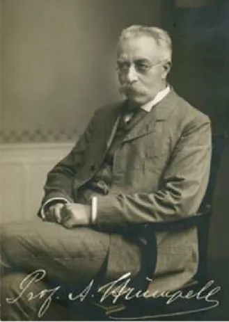
In 1880, the Baltic-German (region known today as
Estonia and Latvia) neurologist Ernst Adolf Gustav Gottfried
von Strümpell (Figure 1) published the irst case series of
patients with HSP. Strümpell reported on two siblings with
a probable AD-HSP whose symptoms manifested at 37 and
56 years of age. Clinically, these patients showed a pure form
of spastic paraplegia. After the death of one of the siblings,
neuropathology showed degeneration of the lateral
cortico-spinal tract, fasciculus gracilis and spinocerebellar tract
17, 18.
Wilhelm Erb was probably the irst to describe a condition
dominated by limb spasticity. Although controversy
per-sists, most authors today consider Erb’s works to be the
irst description of primary lateral sclerosis. A case
descrip-tion by Otto Adolph Seeligmüller, in 1876, has long been
dis-puted as the irst description of HSP. Nevertheless, a critical
analysis of Seeligmüller’s publication highlighted that the
patient studied had a clinical picture dominated by
muscu-lar atrophy and bulbar paralysis, features not suggestive of
HSP itself. In 1888, Maurice Lorrain, a French neurologist,
published a more detailed contribution to the anatomical
and clinical study of HSPs; hence HSP also being known as
Strümpell-Lorrain disease
19, 20.
Clinical phenotype
Rhein
21was the irst, in 1916, to drive attention to the
exqui-site clinical heterogeneity some families with HSP displayed. His
observations were accompanied by many case series describing
pleomorphic clinical pictures encompassing: retinal
degenera-tion
22, 23, dementia
24, extrapyramidal symptoms
25, mental
handi-cap
26, hand amyotrophy
27and other features. he term Strümpell’s
familial spastic paraplegia was reserved for families that
dis-played only spastic paraparesis, a condition that was thought to
be much rarer
28. Currently, it is recognized that pure forms are
more prevalent than complicated ones, their relatively benign
clinical course, not afecting life expectancy, greatly contributed
to their underdiagnosis at that time. Most epidemiological
stud-ies were based on postmortem or hospitalization records
29.
Anita Harding, a professor of neurology at the University
of London, was an important pioneer in the ield of molecular
neurogenetics. In the 1980s, she published a series of
ground-breaking works addressing HSP. In 1981, she presented what
was, at the time, the largest investigation into pure HSP, with
22 families studied
30. Eighteen presented with AD-HSP, three
were AR-HSP and, in the remaining kindred, the inheritance
was uncertain due to the paucity of individuals afected. his
work has consolidated the knowledge that pure subtypes of
the disease are almost always dominantly inherited
30. Harding
also shed light on the importance of examining irst degree
(Extracted from http://www.200.uk-erlangen.de/de/geschichte/20-koepfe-der-erlangeruniversitaetsmedizin/struempell/, April 08th, 2017).
relatives who considered themselves asymptomatic, as that
practice unraveled AD-HSP in ive of the 22 kindreds in this
work
30. She also reinforced the knowledge that spasticity, and
not weakness, is the main source of disability in this scenario,
an aspect that remains useful in diferentiating HSP from other
myelopathies
5, 31. Harding’s major contribution to the ield of
HSP was published in
he Lancet
journal in 1983
32. Entitled
“Classiication of the hereditary ataxias and paraplegias”, this
paper established an accurate diferentiation between ataxias
and HSP, providing a workable base for the etiological
inves-tigations that would come
32. he now current clinical
classi-ication of HSP into pure and complicated forms was
pro-posed. Harding deined that, besides the spastic paraparesis,
additional mild signs such as vibration and segmental
posi-tion sense deicits, slight distal amyotrophy and sphincter
dys-function were also conceivably present in the pure phenotype.
Before this work, the literature had been confusing with regard
to this topic. Pure HSP was subdivided into two groups
accord-ing to age of onset, before or after 35 years of age. he
compli-cated forms were recognized to be rarer and genetically
het-erogeneous. his work also provided reinement of the HSP
phenotype, emphasizing the variability of disease
progres-sion, even within the same family. Sadly, Harding’s outstanding
career ended prematurely at the age 42 when she succumbed
to a cancer
33. Professor Victor Dubowitz wrote, on the
occa-sion of her obituary: “the neuromuscular world and, indeed,
world neurology has lost one of its most colorful, most
produc-tive, and best-loved characters”
33.
In 2006, the German Network for Hereditary Spastic
Paraplegia presented the spastic paraplegia rating scale,
developed to quantify the disease progression clinically.
Validated measures of disease severity are essential to
under-standing the disease’s natural history. Additionally, by their
ability to measure treatment impact, they are decisive in the
development of future clinical trials
34.
Structural characterization: neuropathology
and neuroimaging
While the clinical descriptions of HSP lourished during
the 1980s, the pathological comprehension of the disease saw
lourishing moments 100 years before. We return to Adolf von
Strümpell, the irst to identify the disease clinically, who was
also the irst to describe its pathological hallmarks six years
later, in 1886
35. An unrelated case was also documented by him
in 1904
36. Both pathological specimens showed degeneration
of the corticospinal tract in the spinal cord with a
distal-prox-imal gradient. Since then, the disease has been understood
as a distal axonopathy of the longest large myelinated ibers
of the spinal tract. In 1952, Schwarz
37gave an extensive review
of the pathologic literature, highlighting the contributions of
Newmark (1904, 1906, 1911)
38, Jakob
39and Kahlstorf
40, that
cul-minated with the diferentiation of Strümpell’s disease from
other conditions such as cerebellar ataxias and motor
neu-ron disorders, establishing that, at irst, lesions are restricted
to the spinal cord and afecting the corticospinal and posterior
tracts
41. In 2004, DeLuca and colleagues quantitatively
exam-ined the neuronal population of six HSP patients, the
larg-est pathological report of the disease
42. Marked symmetrical
reduction in area and axonal density of the corticospinal tract
in the lower regions of the spinal cord was found. On the other
hand, sensory tracts demonstrated a signiicant reduction in
area and axonal density only in the upper regions of the spinal
cord. his supported the concept that the extent of axonal loss
in HSP is tract-speciic
42.
In the past decade, neuroimaging techniques have emerged
as powerful tools to investigate structural abnormalities in
a wide variety of neurological disorders in vivo
. In this ield,
Agosta et al.
43, were able to investigate a larger cohort. hey
demonstrated that microstructural brain abnormalities are
rather difuse and not restricted to motor pathways, when one
analyzes multiple subtypes of HSP. Lindig
44and Rezende
45, both
in 2015, made substantial contributions to the delineation of the
extent of neurodegeneration speciically associated with
HSP-SPG4. Both authors showed correlations between white matter
tract disruption and disease severity, indicating that difusion
tensor imaging measures could be used as biomarkers. Rezende
and colleagues also demonstrated the absence of cortical
thin-ning as a hallmark of this pure subtype of HSP
44, 45. Regarding
HSP-SPG11, both França et al.
46and Pan et al.
47revealed
wide-spread white matter microstructural disruption. Pan et al. also
demonstrated a distal-proximal gradient, whereas França et al.
identiied major deep grey matter volumetric loss
46, 47.
Neurophysiology
At the same time that Anita Harding made her
contribu-tions to the HSP classiication, Schady and colleagues
48pub-lished their neurophysiological indings of central
motorcon-duction studies by transcranial magnetic stimulation in HSP.
he abnormalities found were worse in the lower than in
the upper limbs, conirming neurological examination
ind-ings and strengthening the hypothesis made from observing
pathological specimens in a length-dependent dysfunction
48.
Advances in genetics
After the accurate clinical classiication developed by
Harding
32, the research involving HSP evolved from
anec-dotal case descriptions to the systematic study of families
alike. Numerous HSP genetic studies have been published
since the 1980s. Several genetic subtypes of HSP have been
described and numbered sequentially, based on the order of
the gene discovery (SPG1, SPG2, SPG3, etc. )
1.
In 1994, Jouet et al.
49, reported that three disorders;
X-linked spastic paraplegia, MASA syndrome (mental
retar-dation, aphasia, shuling gait and adducted thumbs) and
X-linked hydrocephalus were, in fact, allelic conditions, all
resulting from mutations in the gene for neural cell
adhe-sion molecule (
L1CAM
or
SPG1
)
49. his was the irst gene
glycoprotein involved in neuronal migration and
diferentia-tion. Also in 1994, Hazan et al., identiied the locus implicated
in the majority of cases of dominantly inherited HSP. he
SPAST
or SPG4 gene was cloned in 1999 by the same group
and responds to up to 60% of AD-HSP
50. his gene encodes
spastin, an AAA protein family member related to
microtu-bule severing and endoplasmatic reticulum morphogenesis,
key factors in the process of intra-axonal transport
50.
In 1998, Griiths and colleagues were the irsts to create an
animal model of HSP
51. hey developed a mouse knockdown of
proteolipoprotein (causing HSP-SPG2), unraveling the proteins
critical role in maintaining the integrity of myelinated axons.
Afected mice developed a clear spastic gait, related to iber
swelling and impaired axonal transport
51. his model opened
up a new window of understanding of the mechanisms through
which corticospinal axons degenerate in HSP. During the same
year, the irst gene for autosomal recessive HSP was identiied
52,
when the HSP-SPG7 gene was cloned. Paraplegin, encoded by
the SPG7 gene, localizes to the mitochondria, and ragged red
muscle ibers (a typical inding in mitochondrial disease), and
was readily detected in this work. Key indings suggestive of this
phenotype encompass ataxia, ptosis, progressive external
oph-thalmoplegia and optic atrophy. he latter can also occur in
iso-lation as an autosomal dominant trait
52.
In 2007,
SPG11
was identiied as the gene most commonly
related to spastic paraplegia with mental impairment and
thin corpus callosum. Stevanin, led by Brice and other
col-leagues, identiied loss of function mutations afecting this
gene in 11 families
53. Since then, HSP-SPG11 has been
rec-ognized as the most common AR-HSP. Surprisingly,
SPG11
mutations can also primarily present with other phenotypes,
such as parkinsonism or ALS
54.
he next-generation sequencing techniques,
encom-passing gene panels, whole exome sequencing and whole
genome sequencing have initiated a new era of knowledge
in the ield. he possibility of sequencing dozens, or
hun-dreds, of genes simultaneously, has enormously increased
the number of genes known and has expanded both the
phenotype and genotype of HSP
54, 55.
A brilliant example of the new possibilities opened up
by this technology is the work done by Gaia Novarino et al.,
an Italian physician devoted to unraveling the molecular
pathways of many neurological disorders
55. Novarino’s work,
published in Science in 2014, presented the discovery, and
functional validation of, as many as 18 new HSP-related
genes. Her investigations have also contributed to linking
HSP-related genes to other degenerative disorders, especially
ALS, Alzheimer’s and Parkinson’s diseases. Today, more than
80 diferent loci related to hereditary spastic paraplegias are
recognized, with more than 50 genes already identiied
56.
Brazilian contributions
Brazilian researchers have made substantial contributions
to the ield of HSP. Clinically, Teive et al.
57described the irst
cases of HSP with thin corpus callosum in Brazil in 2001, at a
time when very few families worldwide had been identiied,
other than in Japan
57. In 2007, a multinational group of
research-ers, including the Brazilian biologist Zatz, identiied mutations
causing HSP-SPG8
58. In 2014, França et al.
59addressed the SPG4
molecular epidemiology, conirming it as the most common
HSP in Brazil
59. In 2015, Melo et al.
60described the
over-expres-sion of the
KLC2
gene as the cause of spastic paraplegia with
optic atrophy and neuropathy. his discovery was unique in
the ield, as it was the irst HSP-related mutation to be
docu-mented in an untranslated region
60.
Final remarks
his article presents a comprehensive historical
time-line of the scientiic discoveries related to HSP (Figure2).
Ground zero was the irst description of HSP made by Adolf
von Strüpell in 1880, which outlined the clinical picture, the
inheritance pattern and, later, the neuropathological
ind-ings of “pure” HSP
17. DeLuca later reinforced the
pathologi-cal discoveries of the 19
thcentury in the largest pathological
series of HSP ever reported
42. he period from the early 20
thcentury to the late 1970s was characterized almost
exclu-sively by case descriptions of single individuals or families
with spastic paraplegia as a common ground. It was only
Figure 2. A brief timeline of clinical research related to HSP.
1880
Stümpell: the
first report
Strümpell:
“dying back”
1979
The complicated
phenotypes
Anita Harding:
the clinical
classification
1991
Schady:
central motor
conduction
Jouet:
L1CAM gene
1999
Hazan:
SPG4 gene
DeLuca:
detailed
pathology
2006
Shule: German
Network for HSP
Rating Scale
Gaia Novarino:
14 new genes
2017
80 loci
molecular
after Harding’s studies in 1981 and 1983, that HSP acquired
a more accurate clinical case deinition and a classiication
based on clinical presentation and inheritance pattern
30,32.
his classiication, still useful today, heralded a period of
systematic study of HSP. In 1991, Schady et al. were the irsts
to report abnormal neurophysiological indings in patients
with HSP
48. Jouet et al., in 1996, inaugurated the molecular
era with his discovery of the irst HSP gene
49. Griiths et al.,
in 1998,
51developed the irst animal model of the disease,
unfolding a new technique of studying HSP biological
path-ways. As many as 50 leading world scientists have identiied
HSP-related genes to -date, with most of them having been
discovered in the last ive years, since the advent of the
next-generation sequencing techniques. Functional studies of
the proteins encoded by these genes are now opening
pos-sibilities in the understanding of the biological processes,
not only of HSP, but also of other degenerative disorders,
such as ALS, peripheral neuropathies and spinal muscular
atrophy
55, 56. he understanding of the molecular pathways
of HSP, together with the establishment of disease
biomark-ers, will hopefully lead to a better, and more personalized,
treatment for HSP patients.
References
1. Fink JK. Hereditary spastic paraplegia: clinical principles and genetic advances. Semin Neurol.
2014;34(3):293-305. https://doi.org/10.1055/s-0034-1386767
2. Klebe S, Stevanin G, Depienne C. Clinical and genetic heterogeneity in hereditary spastic paraplegias: from SPG1 to SPG72 and still counting. Rev Neurol(Paris).
2015;171 (6-7):505-30. https://doi.org/10.1016/j.neurol.2015.02.017
3. Tesson C, Koht J, Stevanin G. Delving into the complexity of hereditary spastic paraplegias: how unexpected phenotypes and inheritance modes are revolutionizing their nosology. Hum Genet. 2015;134(6):511-38. https://doi.org/10.1007/s00439-015-1536-7
4. Lo Giudice T, Lombardi F, Santorelli FM, Kawarai T, Orlacchio A. Hereditary spastic paraplegia: clinical-genetic characteristics and evolving molecular mechanisms. Exp Neurol. 2014;261:518-39. https://doi.org/10.1016/j.expneurol.2014.06.011
5. Faber I,Servelhere KR, Martinez ARM, D’Abreu A, Lopes-Cendes I, França MC Jr. Clinical features and management of hereditary spastic paraplegia. ArqNeuropsiquiatr. 2014;72(3):219-26. https://doi.org/10.1590/0004-282X20130248
6. Souza PV, de Rezende Pinto WB, de Rezende Batistella GN, Bortholin T, Oliveira AS. Hereditary spastic paraplegia: Clinical and genetic hallmarks. Cerebellum 2017;16 (2):525-51. https://doi.org/10.1007/s12311-016-0803-z
7. Müller vom Hagen J, Karle KN, Schüle R, Krägeloh-Mann I, Schöls L. Leukodystrophies underlying cryptic spastic paraparesis: frequency and phenotype in 76 patients. Eur J Neurol.. 2014;21(7):983-8. https://doi.org/10.1111/ene.12423
8. Teive HA, Iwamoto FM, Camargo CH, Lopes-Cendes I,
Werneck LC. Machado-Joseph disease versus hereditary spastic paraplegia: case report. ArqNeuropsiquiatr. 2001;59(3B):809-11. https://doi.org/10.1590/S0004-282X2001000500030
9. Pedroso JL, Souza PV, Pinto WB, Braga-Neto P, Albuquerque MV, Saraiva-Pereira ML et al. SCA1 patients may present as hereditary spastic paraplegia and must be included in spastic-ataxias group. Parkinsonism RelatDisord. 2015;21(10):1243-6. https://doi.org/10.1016/j.parkreldis.2015.07.015
10. Synofzik M, Schüle R. Overcoming the divide between ataxias and spastic paraplegias: shared phenotypes, genes, and pathways. MovDisord. 2017;32(3):332-45. https://doi.org/10.1002/mds.26944
11. Martinez AR, Moro A, Abrahao A, Faber I, Borges CR, Rezende TJ et al. Non neurological involvement in late-onset Friedreich ataxia (LOFA): exploring the phenotypes. Cerebellum. 2017;16 (1):253-56. https://doi.org/10.1007/s12311-015-0755-8
12. Abrahão A, Pedroso JL, Braga-Neto P, Bor SengShu E,
Carvalho PA, Barsottini OG. Milestones in Friedreich ataxia: more than a century and still learning. Neurogenetics. 2015;16:151-60. https://doi.org/10.1007/s10048-015-0439-z
13. Bot ST, Willemsen MA, Vermeer S, Kremer HP, Warrenburg BP. Reviewing the genetic causes of spastic-ataxias. Neurology. 2012;79(14):1507-14. https://doi.org/10.1212/WNL.0b013e31826d5fb0
14. Pedroso JL, Raskin S, Barsottini OGP, Oliveira ASB. Adult onset Alexander disease presenting with progressive spastic paraplegia. Parkinsonism RelatDisord.
2013;20(2):241-2. https://doi.org/10.1016/j.parkreldis.2013.10.014
15. Rezende SA, Fernandes M, Munhoz RP, Raskin S, Schelp AO, Knaap MS et al. Cerebellar ataxia as the irst manifestation of Alexander’s disease. ArqNeuropsiquiatr. 2012;70(4):309-10. https://doi.org/10.1590/S0004-282X2012000400018
16. Saute JA, Giugliani R, Merkens LS, Chiang JP, DeBarber AE, Souza CF. Look carefully to the heels! A potentially treatable cause of spastic paraplegia.J Inherit Metab Dis. 2015;38(2):363-4. https://doi.org/10.1007/s10545-014-9745-0
17. Strümpell A.BeitragezurPathologie des Rückenmarks. Arch PsychiatrNervenkr. 1880;10(3):676-717.
https://doi.org/10.1007/BF02224539
18. Kolle K, editor. GroßeNervena¨rzte, vol 3. Stuttgart: Thieme; 1963. Adolf Struümpell (1853–1925), p. 184-190.
19. Seeligmuller AS. Sklerose der Seintenstrange des RückenmarksbeivierKindernderselbenFamilie. Dtsch MedWochenschr.1876;2:185-6.
20. Engmann B, Wagner A, Steinberg H. Adolf von Strümpell: a key neglected protagonist of neurology. J Neurol. 2012;259(10):2211-20. https://doi.org/10.1007/s00415-012-6486-6
21. Rhein, JHW. Family spastic paralysis. J NervMent Dis. 1916;44(2):115-44,224-42.
22. Jequier M, Streiff EB. Paraplégie, dystrophiesquelettique et dégénérescencetapéto-rétiniennefamiliales. Archiv Julius Klaus-Stift ungVererbungsforschSozialanthropolRassenhyg. 1947;22(3-4):129-67.
23. Kjellin K. Familial spastic paraplegia with amyotrophy, oligophrenia and central retinal degeneration. Arch Neurol. 1959;1(2):133-40. https://doi.org/10.1001/archneur.1959.03840020007002
24. Van BogaertL..Studies on familial spastic paraplegia; classical forms with massive optic atrophy in certain members of the family of van L. and genetic considerations on spastic familial paraplegia. ActaNeurolPsychiatr Belg. 1952;52(12):795-807. French.
25. Dick AP, Stevenson CJ. Hereditary spastic paraplegia: report of a family with associated extrapyramidal signs. Lancet. 1953;1(6767):921-3. https://doi.org/10.1016/S0140-6736(53)92061-3
26. Johnston, AW, McKusick VA. A sex-linked recessive form of spastic paraplegia. Am J Hum Genet. 1962;14:83-94.
28. Behan WM, Maia M Strümpell’s familial spastic paraplegia: genetics and neuropathology. J NeurolNeurosurg Psychiatry. 1974;37(1):8-20. https://doi.org/10.1136/jnnp.37.1.8
29. Bell J.On hereditary ataxia and spastic paraplegia. In: Lewis T, Bulloch W. Treasury of human inheritance. London: Cambridge University Press; 1939. Vol. 4, p. 141-281.
30. Harding AE. Hereditary “pure” spastic paraplegia: a clinical and genetic study of 22 families. J NeurolNeurosurg Psychiatry. 1981;44(10):871-83. https://doi.org/10.1136/jnnp.44.10.871
31. Salinas S, Proukakis C, Crosby A, Warner TT. Hereditary spastic paraplegia: clinical features and pathogenetic mechanisms. Lancet Neurol. 2008;7(12):1127-38. https://doi.org/10.1016/S1474-4422(08)70258-8
32. Harding AE. Classiication of the hereditary ataxias and paraplegias. Lancet 1983;1(8334):1151-5. https://doi.org/10.1016/S0140-6736(83)92879-9
33. Dubowitz V. Anita Harding obituary. NeuromuscDisord.
1995;5(6):519-20. https://doi.org/10.1016/0960-8966(95)90017-9
34. Schüle R, Holland-Letz T, Klimpe S, Kassubek J, Klopstock T, Mall V et al. The Spastic Paraplegia Rating Scale (SPRS): a reliable and valid measure of disease severity. Neurology. 2006;67(3):430-4. https://doi.org/10.1212/01.wnl.0000228242.53336.90
35. Strümpell A.Uebereinebestimmte Form der primarenkombinierten Systemerkrankung des Rückenmarks. Arch PsychiatrNervenkr. 1886;17(1):227-38. https://doi.org/10.1007/BF02172796
36. Strümpell A.Die primareSeitenstrangsklerose (spastischeSpinalparalyse). Deutsche ZtschrNervenh. 1904;27(3-4):291-339. https://doi.org/10.1007/BF01667115
37. Schwarz GA. Hereditary (familial) spastic paraplegia. Arch Neurol Psychiatry. 1952;68(5):655-62.
https://doi.org/10.1001/archneurpsyc.1952.02320230081010
38. Newmark L.KlinischerBerichtüber den siebenten Fall von spastischerParaplegie in einerFamilie und Ergebnis der drittenAutopsieausderselbenFamilie. Deutsche ZtschrNervenh. 1911;42(5-6):419-31. https://doi.org/10.1007/BF01629416
39. Jakob C. Sobre un caso de paraplegia espasmódica familiar progresivaconexamenhistopatologico completo. RevSocMed Arg. 1909;17:665-703.
40. Kahlstorf A. Klinischer undhistopathologischer Beitragzurhereditarenspastischen Spinalparalyse. Z GesamteNeurolPsychiatr. 1937;159(1):774-80. https://doi.org/10.1007/BF02870699
41. Fink JK. Advances in the hereditary spastic
paraplegias. Exp Neurol. 2003;184 (suppl 1):S106-10. https://doi.org/10.1016/j.expneurol.2003.08.005
42. Deluca GC, Ebers GC, Esiri MM. The extent of axonal loss in the long tracts in hereditary spastic paraplegia. NeuropatholApplNeurobiol. 2004;30(6):576-84. https://doi.org/10.1111/j.1365-2990.2004.00587.x
43. Agosta F, Scarlato M, Spinelli EG, Canu E, Benedetti S, Bassi MT et al. Hereditary spastic paraplegia: beyond clinical
phenotypes toward a uniied pattern of central nervous system damage. Radiology. 2015;276(1):207-18. https://doi.org/10.1148/radiol.14141715
44. Lindig T, Bender B, Hauser TK, Mang S, Schweikardt D, Klose U et al. Gray and white matter alterations in hereditary spastic paraplegia type SPG4 and clinicalcorrelations. J Neurol. 2015;262(8):1961-71. https://doi.org/10.1007/s00415-015-7791-7
45. Rezende TJ, de Albuquerque M, Lamas GM, Martinez AR, Campos BM, Casseb RF et al. Multimodal MRI-based study in patients with SPG4 mutations. PLoS One. 2015;10(2):e0117666. https://doi.org/10.1371/journal.pone.0117666
46. França MC Jr, Yasuda CL, Pereira FR, D’Abreu A, Lopes-Ramos CM, Rosa MVet al. White and grey matter abnormalities in patients with SPG11 mutations. J NeurolNeurosurg Psychiatry. 2012;83(8):828-33. https://doi.org/10.1136/jnnp-2011-300129
47. Pan MK, Huang SC, Lo YC, Yang CC, Cheng TW,
Yang CC et al. Microstructural integrity of cerebral iber tracts in hereditary spastic paraparesis with SPG11 mutation. AJNR Am J Neuroradiol. 2013;34(5):990-6. https://doi.org/10.3174/ajnr.A3330
48. Schady W, Dick JPR, Crampton SA. Central motor conduction studies in hereditary spastic paraplegia. J NeurolNeurosurg Psych. 1991;54(9):775-9. https://doi.org/10.1136/jnnp.54.9.775
49. Jouet M, Rosenthal A, Armstrong G, MacFarlane J, Stevenson R, Paterson J et al. X-linked spastic paraplegia (SPG1), MASA syndrome and X-linked hydrocephalus result from mutations in the L1 gene. Nat Genet. 1994;7(3):402-7. https://doi.org/10.1038/ng0794-402
50. Hazan J, Fonknechten N, Mavel D, Paternotte C, Samson D, Artiguenave F et al. Spastin, a new AAA protein, is altered in the most frequent form of autosomal dominant spastic paraplegia. Nat Genet. 1999;23(3):296-303. https://doi.org/10.1038/15472
51. Grifiths I, Klugmann M, Anderson T, Yool D, Thomson C,
Schwab MH et al. Axonal swellings and degeneration in mice lacking the major proteolipid of myelin. Science. 1998;280(5369):1610-3. https://doi.org/10.1126/science.280.5369.1610
52. Casari G, De Fusco M, Ciarmatori S, Zeviani M, Mora M, Fernandez P et al. Spastic paraplegia and OXPHOS impairment caused by mutations in paraplegin, a nuclear-encoded mitochondrial metalloprotease. Cell. 1998;93(6):973-83. https://doi.org/10.1016/S0092-8674(00)81203-9
53. Stevanin G, Santorelli FM, Azzedine H, Coutinho P, Chomilier J, Denora PS et al. Mutations in SPG11, encoding spatacsin, are a major cause of spastic paraplegia with thin corpus callosum. Nat Genet. 2007;39(3):366-72. https://doi.org/10.1038/ng1980
54. Kara E, Tucci A, Manzoni C, Lynch DS, Elpidorou M,
Bettencourt C et al. Genetic and phenotypic characterization of complex hereditary spastic paraplegia. Brain. 2016;139(7):1904-18. https://doi.org/10.1093/brain/aww111
55. Novarino G, Fenstermaker AG, Zaki MS, Hofree M, Silhavy JL, Heiberg AD et al. Exome sequencing links corticospinal motor neuron disease to common neurodegenerative disorders. Science. 2014;343(6170):506-11. https://doi.org/10.1126/science.1247363
56. Schüle R, Wiethoff S, Martus P, Karle KN, Otto S, Klebe S et al. Hereditary spastic paraplegia: clinicogenetic lessons from 608 patients. Ann Neurol. 2016;79(4):646-58. https://doi.org/10.1002/ana.24611
57. Teive HA, Iwamoto FM, Della Coletta MV, Camargo CH, Bezerra RD, Minguetti G et al. Hereditary spastic paraplegia associated with thin corpus callosum. ArqNeuropsiquiatr. 2001;59(3B):790-2. https://doi.org/10.1590/S0004-282X2001000500025
58. Valdmanis PN, Meijer IA, Reynolds A, Lei A, MacLeod P, Schlesinger D et al. Mutations in the KIAA0196 gene at the SPG8 locus cause hereditary spastic paraplegia. Am J Hum Genet. 2007;80(1):152-61. https://doi.org/10.1086/510782
59. França MC Jr, Dogini DB, D’Abreu A, Teive HA, Munhoz RP, Raskin S et al. SPG4-related hereditary spastic paraplegia: frequency and mutation spectrum in Brazil. Clin Genet. 2014;86(2):194-6. https://doi.org/10.1111/cge.12252