DOI: 10.1590/0004-282X20130205
ARTICLE
Regional cerebral blood flow and cognitive
function in patients with
obsessive-compulsive disorder
Fluxo sanguíneo regional cerebral e função cognitiva em pacientes com
transtorno obsessivo compulsivo
Huirong Guo1, Ning Zhao2, Zheng Li1, Binhua Zhu1, He Cui1, Youhui Li1*
1Department of Psychiatry, The First Affiliated Hospital of Zhengzhou University, Zhengzhou; 2Department of Neurology, Henan Provincial People’s Hospital.
Correspondence: Youhui Li; Department of Psychiatry; The First Affiliated Hospital of Zhengzhou University; Zhengzhou 450052 – China; E-mail: [email protected]
Conflict of interest: There is no conflict of interest to declare.
Support: The present research was funded by The Science and Technology Research Funded Projects of Henan Provincial Health Bureau (200703056). Received 22 April 2013; Received in final form 17 July 2013; Accepted 24 July 2013.
ABSTRACT
Objective: To explore the relationship between regional cerebral blood flow (CBF) and cognitive function in obsessive-compulsive disorder (OCD). Method: Single-photon emission computed tomography (SPECT) was performed for 139 OCD patients and 139 controls, and the radioactivity rate (RAR) was calculated. Cognitive function was assessed by the Wisconsin Card Sorting Test (WCST). Results: The RARs of the prefrontal, anterior temporal, and right occipital lobes were higher in patients than controls. For the WCST, correct and classification numbers were significantly lower, and errors and persistent errors were significantly higher in OCD patients. Right prefrontal lobe RAR was negatively correlated with correct numbers, right anterior temporal lobe RAR was positively correlated with errors, and the RARs of the right prefrontal lobe and left thalamus were positively correlated with persistent errors. Conclusion: OCD patients showed higher CBF in the pre-frontal and anterior temporal lobes, suggesting that these areas may be related with cognitive impairment.
Keywords: obsessive-compulsive disorder, regional cerebral blood flow, cognitive function. RESUMO
Objetivo: Estudar a relação entre a perfusão sanguínea regional cerebral e as funções cognitivas em pacientes com transtorno obsessivo compulsivo (TOC). Método: Foram estudados 139 pacientes com TOC e 139 controles. As imagens do fluxo sanguíneo cerebral foram feitas por single photon emission computed tomography e o radioactivity rate (RAR) foi calculado usando como referência a radioatividade do córtex cerebelar. As funções cognitivas foram avaliadas através do Wisconsin Card Sorting Test (WCST). Resultados: O RAR do lobo pré-frontal, do lobo temporal anterior e do lobo occipital direito foram mais elevados nos pacientes do que nos controles (P<0.05). Quanto ao WCST, os escores de números corretos (P<0.01) e de classificação (P<0.05) foram menores no grupo TOC do que nos controles; entretanto, os escores de erros e de erros persistentes nos controles foram mais elevados do que nos pacientes (P<0.01). O RAR do lobo pré-frontal direito estava relacionado de modo negativo com os números corretos; o RAR do lobo temporal anterior apresentava correlação positiva com os erros; o RAR do lobo pré-frontal direito e do tálamo esquerdo apresentavam correlação positiva com erros persistentes. Conclusão: Pacientes com TOC apresentaram perfusão sanguínea cerebral mais intensa no lobo pré-frontal e no lobo temporal anterior. O lobo pré-frontal direito, o lobo temporal e o tálamo esquerdo podem estar relacionados com alterações cognitivas.
Palavras-chave: transtorno obsessivo-compulsivo, fluxo sanguíneo regional cerebral, função cognitiva.
Obsessive-compulsive disorder (OCD) is an intractable psychiatric condition. OCD morbidity is 2-3%, which severely afects patients’ quality of life1. In order to investigate the
biological basis underlying changes in cerebral function in
METHODS
Subjects
OCD subjects were chosen from patients who were treated at the Psychiatric Department of the First Ailiated Hospital of Zhengzhou University. All of the subjects were untreated and in their irst episode, with Yale-Brown Obsessive Compulsive Scale (Y-BOCS) ≥16 and symptoms in agreement with the diagnostic criteria of the 10th
revi-sion of the International Statistical Classiication of Diseases and Related Health Problems (ICD-10). Patients with other mental disorders, somatic disease, pregnancy, or lactation
and patients who once had taken medicine for cerebrovas
-cular diseases or psychological disorders, hormone drugs, or contraceptive pills in the preceding month were excluded. A total of 139 patients were included, (86 male and 53 female), with ages ranging from 17-49 (26.4±7.8). he score range of the subjects tested with Yale-Brown Obsessional Scale was 16-39 (29.32±8.75). he scores of the subjects examined with Hamilton Depression Scale (HAMD) ranged from 9-19 (15.33±3.11). he scores of Hamilton Anxiety Scale (HAMA)
were between 2 and 13 (7.97±5.02). A total of 139 control sub
-jects were selected from among First Ailiated Hospital of Zhengzhou University employees and Zhengzhou University undergraduate students, including 86 males and 53 females
ranging from 19-49 years old (27.8±7.0). here were no sta
-tistically signiicant diferences between the two groups with regard to gender or age (P>0.05). All the subjects were in -formed about the research and agreed to participate.
SPECT imaging
he subjects took oral potassium chlorate (400 mg) to
block 99mTcO4- intake by the choroid plexus. hirty minutes
later, the subjects were injected with 9.25×10⁹ Bq of 99mTc
-ethycysteine dimer (ECD) via the median cubital vein. After 1 hour, the subjects were scanned with a dual head SPECT (GE Millennium Hawkeye VG) equipped with parallel hole, low-energy, high-resolution collimators. Matrices (64×64) were captured, stepping 6 degree, and a total of 60 frames were collected with 35 s per frame. A Butterworth ilter was used for rescanning, and the cross-section was paralleled to the orbitomeatal line. All of the subjects were examined between 10:00 and 11:00 A.M.. he ratio between the mean radioactivity rate (RAR) in each brain region and that in the cerebellum was calculated by overlaying two or three regions of interest (ROIs) into a dataset and taking the mean RAR of the bila teral cerebella as a reference value.
Cognitive function assessment
We employed the revised version of the WCST comprised
of 4 stimulus cards and 48 response cards2. he cards were
printed with designs with diferent numbers ( from 1 to 4), colors (red, yellow, green, and blue) and shapes (rectangular,
round, ive-star, and triangular) that can be classiied by co-lor, shape, or number. If the subjects correctly classiied the cards by color six consecutive times, they converted to the following classiication (e.g., shape), and then did the next classiication (e.g., number). After the three kinds of classi -ication, the classiications were repeated once more. If the subjects accomplished classiication correctly six times in total or used up all 48 cards without successful completion, the examination was ended. he observation indices inclu-ded the total numbers of correct, error, preservative errors, non-maintained errors, and inished classiication.
Statistics
All data are expressed as mean±standard deviation (SD) and were assessed with SPSS13.0 statistical software (SPSS Inc., Chicago, IL, USA). Statistical diferences were calculated
for brain RAR values and WCST results using the Student’s t
test, followed by a Pearson correlation analysis to determine the relationship between the two groups. P<0.05 was consi-dered statistically signiicant.
RESULTS
SPECT results
he RAR of the bilateral prefrontal lobes (P<0.01), ante -rior temporal lobe (P<0.01), and right occipital lobe (P<0.05)
were signiicantly higher in patients with OCD than in con
-trol subjects (Table 1).
WCST results
As shown in Table 2, for the WCST, scores of correct num
-bers (P<0.01) and classiication num-bers (P<0.05) in OCD pa -tients were lower than those of the control group, and scores of errors and persistent errors were higher in patients than in controls (P<0.01).
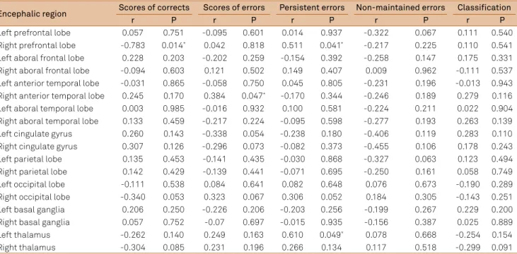
Relationship between RAR and cognitive function in OCD patients
As shown in Table 3, the RAR value of the right prefrontal lobe was negatively correlated with the correct number score (r=-0.783, P=0.014), the right anterior temporal lobe RAR was positively correlated with the error score (r=0.384, P=0.047), and the RARs of the right prefrontal lobe and left thalamus were positively correlated with the persistent error score (r=0.511, P=0.041; r=0.610, P=0.049).
DISCUSSION
Functional brain imaging of patients with OCD has be
-come a research topic of interest in recent years. Trivedi3
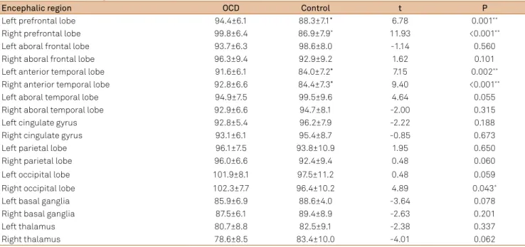
Table 1. RARs by brain region.
Encephalic region OCD Control t P
Left prefrontal lobe 94.4±6.1 88.3±7.1* 6.78 0.001**
Right prefrontal lobe 99.8±6.4 86.9±7.9* 11.93 <0.001**
Left aboral frontal lobe 93.7±6.3 98.6±8.0 -1.14 0.560
Right aboral frontal lobe 96.3±9.4 92.9±9.2 1.62 0.101
Left anterior temporal lobe 91.6±6.1 84.0±7.2* 7.15 0.002**
Right anterior temporal lobe 92.8±6.6 84.4±7.3* 9.40 <0.001**
Left aboral temporal lobe 94.9±7.5 99.5±9.6 4.64 0.055
Right aboral temporal lobe 92.9±6.6 94.7±8.1 -2.00 0.315
Left cingulate gyrus 92.8±5.4 96.2±7.9 -2.22 0.188
Right cingulate gyrus 93.1±6.1 95.4±8.7 -0.85 0.673
Left parietal lobe 96.1±7.5 93.8±10.9 1.95 0.650
Right parietal lobe 96.0±6.6 92.4±9.4 0.48 0.060
Left occipital lobe 101.9±8.1 97.5±11.2 0.48 0.059
Right occipital lobe 102.3±7.7 96.4±10.2 4.89 0.043*
Left basal ganglia 85.9±6.9 88.6±4.0 -3.64 0.078
Right basal ganglia 87.5±6.1 89.4±8.9 -2.63 0.201
Left thalamus 80.7±8.8 82.5±9.1 -2.38 0.337
Right thalamus 78.6±8.5 83.4±10.0 -4.01 0.062
Values are mean±SD *P<0.05 **P<0.01. OCD: obsessive-compulsive disorder; RAR: radioactivity rate.
Table 2. WCST scores.
Item OCD Control t P
Scores of corrects 33.24±5.39 38.15±4.64 -3.657 0.006**
Scores of errors 13.21±6.89 7.53±5.97 5.881 0.000**
Persistent errors 6.79±4.89 2.06±1.29 4.873 0.001**
Non-maintained errors
6.49±2.74 5.59±4.41 1.713 0.139
Complete Classification
4.42±1.62 5.72±0.94 -2.595 0.012*
Values are mean±SD *P<0.05 **P<0.01. WCST: Wisconsin Card Sorting Test;
OCD: obsessive-compulsive disorder.
temporal lobes may contribute to the onset of OCD, which
does not support the previous view of neural circuit dysfunc
-tion. he frontal lobe is involved in abstract thinking, and the prefrontal lobe is associated with goal-directed action, wor-king memory, and executive function. he temporal lobe is responsible for memory, perception, and emotion. Clinically, patients with OCD are characterized by thinking-trans fer dysfunction, excessive focus on details, and morbid hy
per-mnesia about unnecessary fears, which are relevant to dys
-function of the frontal and temporal lobes.
Many patients with mental disorders have cognitive func tional dysfunction. Previous studies demonstrated that patients with OCD exhibit extensive cognitive functional dysfunction including the domains of attention, memory,
abs tract thinking, and spatial memory6,7. Research has shown
that OCD patients with depression have obviously decreased executive function8. In this study, cognitive functional testing revealed that the performance of patients with OCD for the
total number of correct responses, total error number, pre
-servative error number, and number of categories was much worse than that of the control group, which demonstrates
that attention and executive function were impaired in pa
-tients with OCD. he reason may be that pa-tients with OCD could not concentrate on a task, and when they made mis-takes, patients with OCD tended to pay excessive attention to the wrong rules, which had an unfavorable efect on ma-king judgments of correct rules. hus, they needed to spend more time changing their ways of solving problems and checking whether an answer was correct in the next round. herefore, our results are in accordance with those reported by Savage and colleagues9.
circuit of the prefrontal lobe-cingulate gyrus-basal ganglia re -gion, with increased blood perfusion in this region. However,
other studies4 indicated that functional changes in patients
with OCD only involved individual brain areas. Alptekin and
colleagues4 performed SPECT and described signiicantly in
-creased perfusion of the bilateral prefrontal lobe cortex in
patients with OCD. Adler5 found that the front of the
here have been some reports about the relationship bet-ween brain function abnormalities and cognitive deicits. In
1997, Lucey et al.10 assessed controls and subjects with OCD
(19 in each group) with the WCST and SPECT and discovered a positive correlation between obsessive thinking and error number and a negative correlation between the number of categories achieved and perfusion of the inferior prefrontal
gyrus and left caudate nucleus. Lacerda et al.11 performed a
similar study and identiied a positive correlation between perfusion of the frontal lobe and anterior cingulate gyrus and non-persistent errors. In addition, their results revealed that perfusion of the right thalamus was negatively correlated with the number of preservative errors, which was positively correlated with symptom severity. In the present study, we analyzed the relationship between WCST results and SPECT imaging in subjects with OCD. he results demonstrated that perfusion of the right prefrontal lobe was negatively corre -lated with the categories-achieved number, perfusion of the
right anterior temporal lobe was positively correlated to er-ror number, and perfusion of the right prefrontal lobe and left thalamus were positively correlated with preservative errors. hese indings suggest that dysfunction of the frontal and temporal lobes and thalamus may play a key role in cognitive impairment in patients with OCD. Considering that there are various clinical phenotypes of OCD, the inclusion of more subjects would increase the signiicance of our results.
In conclusion, OCD patients exhibited higher CBF, mainly in the prefrontal and anterior temporal lobes and the left thalamus, indicating that these regions may play important roles in cognitive impairment in OCD.
Acknowledgements
he authors would like to thank Prof. Zeping Xiao at Shanghai Mental Health Center and Prof. Wei Gu at he Second Xiangya Hospital of Central South University for their valuable help and assisting in revising the manuscript.
1. Chamberlain SR, Blackwell AD, Fineberg NA, et al. The neuropsychology of OCD: the importance of failures in cognitive and behavioural inhibition as candidate endophenotypic makers. Neurosci Biobehav Rev 2005;29:399-419.
2. Zhang LX, Yang YC, et al. A study of cognitive function in OCD patients. Chinese J Psychiatry 2005;38:23-26.
3. Trivedi MH. Functional neuroanatomy of obsessive-compulsive disorder. J Clin Psychiatry 1996;57:26-28.
References
4. Alptekin K, Degirmenci B, Kivircik B, et al. Tc-99m HMPAO brain perfusion SPECT in drug-free obsessive-compulsive patients without depression. Psychiatry Res 2001;107:51-56.
5. Adler CM, McDonough-Ryan P, Sax KW, Holland SK, Arndt S, Strakowski SM. fMRI of neuronal activation with symptom provocation in unmedicated patients with obsessive-compulsive disorder. J Psychiatr Res 2000;34:317-324.
Table 3. Pearson correlation analysis between RAR values and WCST performance.
Encephalic region Scores of corrects Scores of errors Persistent errors Non-maintained errors Classification
r P r P r P r P r P
Left prefrontal lobe 0.057 0.751 -0.095 0.601 0.014 0.937 -0.322 0.067 0.111 0.540
Right prefrontal lobe -0.783 0.014* 0.042 0.818 0.511 0.041* -0.217 0.225 0.110 0.541
Left aboral frontal lobe 0.228 0.203 -0.202 0.259 -0.154 0.392 -0.258 0.147 0.175 0.331
Right aboral frontal lobe -0.094 0.603 0.121 0.502 0.149 0.407 0.009 0.962 -0.111 0.537
Left anterior temporal lobe -0.031 0.865 -0.058 0.750 0.045 0.805 -0.231 0.196 -0.013 0.943
Right anterior temporal lobe 0.245 0.170 0.384 0.047* -0.170 0.344 -0.246 0.189 0.279 0.116
Left aboral temporal lobe 0.003 0.985 -0.016 0.932 0.100 0.581 -0.224 0.211 0.022 0.904
Right aboral temporal lobe 0.133 0.459 -0.217 0.224 -0.095 0.598 -0.277 0.193 0.263 0.139
Left cingulate gyrus 0.260 0.143 -0.338 0.054 -0.238 0.180 -0.406 0.119 0.283 0.110
Right cingulate gyrus 0.307 0.126 -0.296 0.073 -0.082 0.373 -0.455 0.106 0.178 0.243
Left parietal lobe 0.135 0.453 -0.141 0.435 -0.030 0.868 -0.327 0.063 0.123 0.494
Right parietal lobe 0.142 0.429 -0.139 0.441 -0.071 0.695 -0.250 0.161 0.058 0.749
Left occipital lobe -0.111 0.538 0.084 0.641 0.082 0.648 0.076 0.673 -0.190 0.289
Right occipital lobe -0.340 0.053 0.323 0.067 0.306 0.052 0.184 0.305 -0.143 0.251
Left basal ganglia 0.206 0.250 -0.226 0.206 -0.203 0.256 -0.199 0.267 0.229 0.200
Right basal ganglia 0.057 0.752 -0.07 0.697 -0.015 0.935 -0.156 0.387 0.025 0.889
Left thalamus -0.262 0.140 0.249 0.163 0.610 0.049* 0.078 0.668 -0.254 0.154
Right thalamus -0.304 0.085 0.231 0.196 0.266 0.134 0.117 0.518 -0.299 0.091
6. Clayton IC, Richard JC, Edwards CJ. Selective attention in obsessive-compulsive disorder. J Abnorm Psychol 1999;108:171-175.
7. Okasha A, Rafaat M, Mahallawy N, et al. Cognitive dysfunction in obsessive-compulsive disorder. Acta Psychiatr Scand 2000;101:281-285.
8. Aycicegi A, Dinn WM, Harris CL, Erkmen H. Neuropsychological function in obsessive compulsive disorder: effects of comorbid conditions on task performance. Eur Psychiatry 2003;18:241-248.
9. Savage CR, Baer L, Keuthen NJ, Brown HD, Rauch SL, Jenike MA. Organizational strategies mediate nonverbal memory impairment in obsessive-compulsive disorder. Biol Psychiatry 1999;5:905-916. 10. Lucey JV, Burness CE, Costa DC, et al. WCST errors and cerebral blood
flow in OCD. Br. J Med Psychol 1997;70:403-411.