Arq Neuropsiquiatr 2008;66(1):88-89
88
InfantIle glIosarcoma
José Roberto Tude Melo
1, André Luiz Pitanga Bastos de Souza
2,
Rodolfo Casimiro Reis
2, Marco Antônio Cardoso de Almeida
3glIosarcoma InfantIl
Complexo Hospitalar Universitário Professor Edgard Santos da Universidade Federal da Bahia (HUPES-UFBA) e Hospital São Rafael (HSR), Salvador BA, Brazil: 1 Neurocirurgião, Doutorando em Medicina pela UFBA; 2Acadêmico da Faculdade de Medicina da UFBA; 3Chefe do Serviço de Anatomia
Patológica do HSR.
Received 9 August 2007, received in inal form 4 October 2007. Accepted 29 November 2007.
Dr. José Roberto Tude Melo – HUPES/UFBA - Rua Augusto Viana sn / 2° andar - 40110-060 Salvador BA - Brasil. E-mail: [email protected]
Since the irst reports published by Stroebe in 1895, more than 100 cases of gliosarcoma have been reported,
eight of which were in children1. Gliosarcoma accounts for
1.8 to 8% of all glioblastomas1. It is classiied by the World Health Organization (WHO) as a grade IV tumor and is
composed of malignant glial and mesenchymal tissue2-4.
This tumor affects supratentorial regions, particularly the peripheral region of the temporal lobe, normally near the
falx cerebriand diploe. It usually affects the adult
pop-ulation in the ifth to sixth decades of life, males being more frequently affected than females (male/female ra-tio 1.8:1)1,2,5. Sixty percent of patients experience paresis
and 55% report headaches6. At computed tomography
(CT), the appearance of the tumor is extremely variable; however, the imaging presentation is generally of a het-erogeneous hyperdense lesion with intense peritumoral edema and necrotic areas. There have been some reports of cases of hypodense images in which the histological di-agnosis was gliosarcoma. In children, the tumor may ap-pear as an extensive cystic mass. It is important to differ-entiate gliosarcoma from meningioma in which there is more homogeneity with contrast enhancement and less
edema1,7. Magnetic resonance imaging (MRI) shows a
hy-perintense lesion with irregular contrast enhancement
at T1. T2 sequence appears as an isointense lesion7.
His-tologically, the tumor is composed of areas of mesen-chymal and glial differentiation. The glial component is composed of pleomorphic astrocytic cells that are immu-nopositive to glial ibrillary acidic protein (GFAP), prolif-erative endothelium and occasional sites of necrosis. The sarcomatous component is comprised of uniform spin-dle cells arranged in fascicles between reticulin ibers. Vimentin and S-100 are used as markers of connective
tissue8. The pathogenesis of gliosarcoma is still
contro-versial and some authors sustain the idea of a sarcoma-tous transformation of the endothelial cells of glioblas-tomas, while others afirm that the sarcomatous compo-nent originates from vascular smooth muscle cells,
ibro-blasts, pericytes and undifferentiated mesenchymal cells. However, the most acceptable theory, based on genetic studies, is the dedifferentiation of glial cells into
mesen-chymal ones8,9. Prognosis is similar to that of most
glio-blastomas, with a mean survival time of 6 to 14.8 months following diagnosis, although one case has been report-ed in the literature in which the patient had a 22-year
in-terval without recurrences8. The mean interval between
recurrences ranges from 53.5 to 62 weeks9. The
predomi-nance of the sarcomatous component is associated with
a better prognosis and a greater recurrence-free interval9.
Some authors claim that metastases are rare, while others report that they occur in up to one third of cases, princi-pally in the lungs, pleura, lymph nodes, bone marrow, liv-er, spinal cord, kidney and peripancreatic areas6,7. The role of chemo- and radiotherapy in the treatment of gliosar-coma remains to be deined, although some authors be-lieve that these adjuvant forms of therapy are important, particularly in the case of children1,8.
We describe an infantile case of gliosarcoma after the authorization of the parents.
Arq Neuropsiquiatr 2008;66(1)
89
Infantile gliosarcoma Melo et al.
case
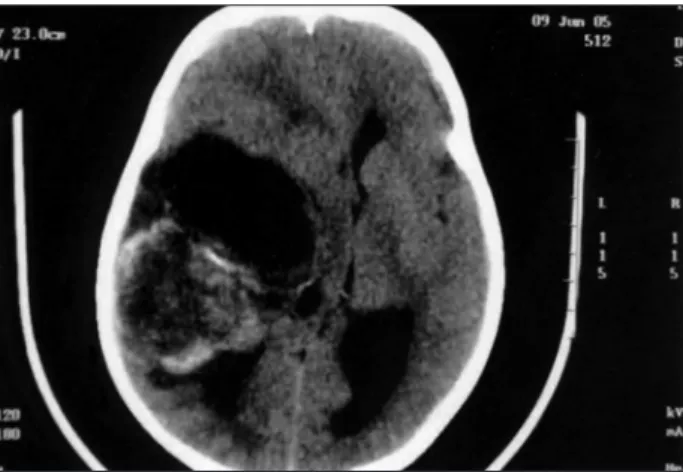
A 4 year-old-boy was taken to hospital with complaints of a rapidly worsening headache, generalized seizures and left hemi-paresis, principally in the face and arm. Computed tomography (CT) showed a bulk tumor in the right temporal lobe, with ir-regular contrast enhancement (Fig 1). A radical resection of the neoplasia was performed, and the histopathological diagnosis was gliosarcoma (Fig 2). Adjuvant chemo- and radiotherapy was recommended but was not adhered to by the patient’s guard-ians due to the poor socioeconomic conditions of the family. The child was brought back to the hospital three months later at which time his level of consciousness had decreased and he was suffering from seizures. He died ive days after the second neurosurgical intervention.
DIscussIon
There are few reports in the literature of gliosarcoma in children. Of the 145 cases of infantile neoplasia that
have undergone surgery in our institute, this has been the only case of gliosarcoma.
No discrepancy was found between the clinical and imaging presentations, in disagreement with previous re-ports in the literature. Imaging scans provided us with a heterogenous temporal image with irregular enhancement under endovenous contrast, which was in agreement with descriptions published in previous reports.
There was a 3-month overall survival in this case, prin-cipally due to the discontinuation of adjuvant therapy as a result of the family’s socioeconomic limitations. However, it is well-known that infantile gliosarcoma sometimes
re-sponds to adjuvant chemo- or radiotherapy10,11.
references
1. Okami N, Kawamata T, Kubo O. Yamane F, Kawamura H , Hori T. In-fantile gliosarcoma: a case and a review of the literature. Child’s Nerv Syst 2002;18:351-355.
2. Ohgaki H, Biernat W, Reis R, Hegi M, Kleihues P. Gliosarcoma. In Klei-hues P, Cavenee WK (eds). Pathology and genetics of tumors of the ner-vous system. Lyon: IARC Press 2000:42-44.
3. Goldstein SJ, Young B, Markesberry WR. Congenital malignant gliosar-coma. Am J Neuroradiol 1981;2:475-476.
4. Parekh HC, O’Donovan DG, Sharma RR, Keogh AJ. Primary cerebral gliosarcoma: report of 17 cases. Br J Neurosurg 1995;9:171-178. 5. Meis JM, Martz KL, Nelson JS. Mixed glioblastoma multiforme and
sarcoma: a clinicopathologic study of 26 Radiation Therapy Oncology Group cases. Cancer 1991;67:2342-2349.
6. Witwer BP, Salamat MS, Resnick DK. Gliosarcoma metastatic to the cer-vical spinal cord: case report and review of the literature. Surg Neurol 2000;54:373-379.
7. Moreira RK, Koppe D, Zignani J, et al. Gliossarcoma de tronco cerebral em paciente pediátrico: relato de caso. Radiol Bras 2004;37:61-63. 8. Winkler PA, Büttner A, Tomezzoli A, Weis S. Histologically repeatedly
conirmed gliosarcoma with long survival: review of the literature and
report of a case. Acta Neurochir (Wien) 2000;142:91-95.
9. Salvati M, Caroli1 E, Raco A. Gliosarcomas: analysis of 11 cases do two subtypes exist? J Neuro Oncol 2005;74:59-63.
10. Ono N, Nakamura M, Inoue HK, Tamura M, Murata M. Congenital gliosarcoma; so-called sarcoglioma. Childs Nerv Syst 1990;6:416-420. 11. Radkowski MA, Naidich TP, Tomita T, Byrd SE, McLone DG.
Neona-tal brain tumors: CT and MR indings. J Comput Assist Tomogr 1988;
12:10-20.