Ar
ti
cle
J. Braz. Chem. Soc., Vol. 21, No. 1, 169-178, 2010. Printed in Brazil - ©2010 Sociedade Brasileira de Química 0103 - 5053 $6.00+0.00
*e-mail: [email protected]
#Present address: Instituto de Química, Universidade Federal do Rio de Janeiro, 22640-000 Rio de Janeiro-RJ, Brazil
Novel 2-(R-phenyl)amino-3-(2-methylpropenyl)-[1,4]-naphthoquinones: Synthesis,
Characterization, Electrochemical Behavior and Antitumor Activity
Acácio I. Francisco,a Annelise Casellato,a,# Amanda P. Neves,a J. Walkimar de M. Carneiro,a
Maria D. Vargas,*,a Lorenzo do C. Visentin,b Alviclér Magalhães,c Celso A. Câmara,d Claudia Pessoa,e
Letícia V. Costa-Lotufo,e José D. B. Marinho Filhoe and Manoel O. de Moraese
aInstituto de Química, Universidade Federal Fluminense, 24020-141 Niterói-RJ, Brazil bInstituto de Química, Universidade Federal do Rio de Janeiro, 22640-000 Rio de Janeiro-RJ, Brazil
cInstituto de Química, Universidade Estadual de Campinas, 13083-970 Campinas-SP, Brazil dInstituto de Química, Universidade Federal Rural de Pernambuco, 52171-900 Recife-PE, Brazil eDepartamento de Fisiologia e Farmacologia, Universidade Federal do Ceará, 60430-270 Fortaleza-CE, Brazil
Novas 2-(R-fenil)amino-3-(2-metilpropenil)-[1,4]-naftoquinonas (R = H, 4-OMe, 4-Ferrocenil, 4-Me, 3-Me, 4-I, 3-I, 4-CN, 3-CN, 4-NO2 e 3-NO2) derivadas do nor-lapachol
[2-hidroxi-3-(2-metilpropenil)-1,4-naftoquinona] foram obtidas em bons rendimentos. A estrutura dos compostos foi proposta com base em estudos de difração de raios-X (R = OMe, 2b), dados de RMN de 1H
e 13Ce cálculos teóricos utilizando o funcional B3LYP e a base 6-311+G(2d,p). Os potenciais de
meia-onda das aminonaftoquinonas e o deslocamento químico do hidrogênio da cadeia 3-propenil dos compostos 2a-k mostraram boa correlação com as constantes de Hammett dos substituintes presentes no anel fenileno. A avaliação da citotoxicidade evidenciou atividade antitumoral promissora para o substrato metóxi-nor-lapachol 1 e o derivado 4-ferrocenil 2c.
Novel 2-(R-phenyl)amino-3-(2-methyl-propenyl)-[1,4]-naphthoquinones (R = H, 4-OMe, 4-Ferrocenyl, 4-Me, 3-Me, 4-I, 3-I, 4-CN, 3-CN, 4-NO2 and 3-NO2) derived from nor-lapachol
[2-hydroxy-3-(2-methylpropenyl)-1,4-naphthoquinone] were obtained in good yields. Their structures were proposed on the basis of a single crystal X-ray diffraction study (R = OMe, 2b),
1H and 13C NMR studies and calculations using the B3LYP functional and the 6-311+G(2d,p)
basis set. The half-wave potentials of the aminonaphthoquinones and 1H NMR chemical shifts of
the 3-propenyl hydrogen in 2a-k show good correlation with the substituent Hammett constants on the phenylamino ring. The antitumor assays showed promising activity for substrate
methoxy-nor-lapachol 1 and the 4-ferrocenyl derivative 2c.
Keywords: Nor-lapachol, arylamine, aminonaphthoquinone, electrochemistry, B3LYP,
antitumor activity
Introduction
Naphthoquinones are widely distributed in nature and some of these molecules have an important role in the biochemistry of microbial energy production, by means of photosynthesis and respiratory chain.1 Compounds
containing the quinone group are known for exhibiting antitumor,2 trypanocide,3 moluscicide,4 fungicide5 and
antimalarial6 activities. The presence of an amino group
in quinones has led to interesting biologically active compounds.7-12
Biological activity of quinones is often related to their electrochemical behavior.13 The ability to accept
one or two electrons to form the corresponding radical anion (Q•−) or dianion (Q2−) species is believed to induce
formation of reactive oxygen species, responsible for the oxidative stress in cells.14 The electron-accepting capacity
of naphthoquinones may be tuned by carbonyl position changes (1,2- x 1,4-naphthoquinones)15 or different
Novel 2-(R-phenyl)amino-3-(2-methylpropenyl)-[1,4]-naphthoquinones J. Braz. Chem. Soc.
170
this type of molecules has proven to be useful.16,17 We
reported recently18 the synthesis of a series of
2-arylamino-1,4-naphthoquinones from 2-methoxy-1,4-naphthoquinone and arylamines in the presence of MgCl2•6H
2O and
p-toluenesulfonic acid as catalysts. The reactions of both electron-donor and electron-attracting substituted anilines having given good yields of the respective products, we decided to investigate the analogous reactions of the methoxy-derivatives of nor-lapachol [2-hydroxy-3-(2-methyl-propenyl)-1,4-naphthoquinone]4 and lapachol
[2-hydroxy-3-(3-methyl-2-butenyl)-1,4-naphthoquinone].19
Nor-lapachol is obtained from lapachol by the Hooker oxidation20 and has been used as a substrate for the
synthesis of several active compounds.9-12 The incorporation
of polyamines to this quinone, for example, has led to signiicant increase in the DNA topoisomerase II-α inhibition, compared to the original naphthoquinone.13,14
Furthermore, arylamino derivatives of nor-α and nor-β lapachones present potent antitumor11 and trypanocide
activities.9
Herein is the first report on the synthesis and characterization of the novel 2-arylamine derivatives of nor-lapachol 2a-k, including
2-(4-ferrocenyl-phenyl)amino-3-(2-methylpropenyl)-1,4-naphthoquinone 2c (Figure 1) and
cytotoxic screening against the several cancer cell lines (SF-295, HCT-8, MDAMB-435 and HL-60). Because correlations between electrochemical potentials and the inhibitory activity of naphthoquinones on Epstein-Barr virus early antigen activation21 and their cytotoxicity22 has
been reported, we also investigated the redox properties of these compounds by cyclic voltammetry.
Results and Discussion
Syntheses
The compounds 2a-k (Figure 2) were synthesized from
2-methoxy-3-(2-methylpropenyl)-[1,4]-naphthoquinone with various aromatic amines, in the presence of the catalysts 4-toluenesulfonic acid and MgCl2•6H
2O in
methanol under relux.18 The products are stable in the solid
state and in solution. Compounds 2a, 2b were obtained
in a pure state, whereas 2c-k were puriied by column
chromatography using a mixture of ethyl acetate / hexane (1:5) as eluent. They were obtained in yields ranging from 84 to 73% and formulated on the basis of analytical and spectroscopic data (see Experimental).
The 1H NMR and infrared spectra of compounds 2a-k
are consistent with their composition and structure. The
1H NMR spectra exhibit signals in the d 7.5-8.2 ppm
region as double dublets and triple dublets, attributed to the four naphthoquinone aromatic hydrogens H5-H8. Attributions were made on the basis of 1H x 1H-COSY
experiments, J values and multiplicity. All expected resonances were observed in the 13C NMR spectra of
compounds 2a-k. The carbonyl peaks appear around d183 and 180, and those attributed to C2 bound to the nitrogen at about d 145. The other chemical shifts are compatible with the structures proposed for these compounds. We observed that the chemical shift of H18 (Figure 2) in the
1H NMR spectra of 2a-k is directly inluenced by the
nature of the R substituent group in the phenylamino ring [5.80 (4-OMe) < dH(18) < 6.08 (4-NO2)].
X-ray structure / Theoretical calculations
The structure of 2-(4-methoxy-phenyl)amino-3-(2-methylpropenyl)-1,4-naphthoquinone 2b was determined
by a single crystal X-ray diffraction study (Figure 3). The average C-C, C-O, C=O and C-N bond lengths are in good agreement with the literature.36The naphthoquinone ring
system of 2b is approximately planar, the dihedral angle
between the naphthoquinone plane and the arylamine phenyl ring being 47.3°(1). The torsion angles around the fragments involved in the H-bond are: C(2)-N(1)-C(11)-C(12), −158.94(17)o, C(1)-C(2)-N(1)-C(11)
−154.47(16)o and N(1)-C(2)-C(1)-O(2) −3.2(2)°. The
planar unsaturated side chain is twisted about 54° with respect to the ring system. This is the irst reported structure of an amine derivative of nor-lapachol.
The packing of 2b involves molecules that interact
through classical and non-classical hydrogen bonds, forming a 1D ininite network along the crystallographic [100] direction (Figures 4 and 5). The carbonyl O1i atom O O OMe + NH2 R O O NH cat1 / cat2
MeOH, reflux
R
1
Figure 1. Synthesis of the novel arylamines derived from methoxy-nor
-lapachol 1.
O
O
NH 2a2b (R=H) (R=4-OMe) 2c (R=4-Fc) 2d (R=4-Me) 2e (R=3-Me) 2f (R=4-I)
2g (R=3-I) 2h (R=4-CN) 2i (R=3-CN) 2j (R=4-NO2)
2k (R=3-NO2)
R 1 3 2 4 5 6 7 8 9
10 H(18) 19 11 12 13 14 15 16 17 20 21
Francisco et al. 171 Vol. 21, No. 1, 2010
makes a classical and a non-classical hydrogen bonds with H1 to the N1 atom (amino group) and with H12 to the C12 atom (C11-C16 phenyl ring) of a neighboring molecule, forming a six-membered ring [symmetry code: i = x-1, y, z]. In addition the other C=O group interacts via O2ii with H5 to the C5 forming now a ten-membered ring. For more details of the crystal structure, see Supplementary material.
Starting from the experimental structure of 2b
(Figure 3), the geometries of 2a-2e and 2h-2i were fully
optimized with the B3LYP/6-31G(d) method.37 Energies
and molecular properties were obtained from a single-point calculation on the optimized geometries using the 6-311+G(2d,p) basis set38 and the B3LYP functional.39 To
conirm that the most stable conformation in the gas-phase is similar to that found in the solid state the geometry of an alternative conformation for 2b, with the 2-methylpropenyl
group bonded to position 3 of the naphthoquinone ring rotated by 180o was also optimized. This alternative
conformation is 0.4 kcal mol-1 less stable than the solid
state conformation. The barrier for conversion between the two conformers calculated at the 6-31G(d) level is 5.5 kcal mol-1.
Calculations of the absolute 1H NMR chemical shifts
using the GIAO approach40 confirmed that
electron-attracting groups yield higher dH(18) values than
electron-donor groups. Calculations including solvent (chloroform) show essentially the same behavior. Interestingly, the
dH(18) value for the alternative conformation with the 2-methylpropenyl side chain rotated by 180o is shifted
highield by 1.67 ppm, compared to the same hydrogen in the most stable conformation. The fact that the experimental values are intermediate between the calculated values for the two conformations suggests that these conformations are in equilibrium in solution, although the variable temperature 1H NMR spectra of 2a do not show broadening
of H18 down to −90 oC in CD 2Cl2.
Figure 3. View of the ORTEP plot for 2b with labeled atoms and 50% probability ellipsoids.
Novel 2-(R-phenyl)amino-3-(2-methylpropenyl)-[1,4]-naphthoquinones J. Braz. Chem. Soc.
172
UV-Vis spectra
The UV-Vis spectra of the compounds obtained in CHCl3 show two absorption bands. PBE1PBE/6-311+G(2d,p) calculations indicate that the band in the 275-290 nm region can be attributed to the aromatic and quinone π-π∗
transitions and the low-energy band in the visible region between 456 and 512 nm is attributed predominantly to π phenyl-π* naphthoquinone transitions. Electron-donor
substituents blue shift the latter band, whereas electron-attracting groups red shift it.
Cyclic voltammetry
The redox behavior of compounds 2a-k was evaluated
by cyclic voltammetry (CV) at room temperature in acetonitrile/Bu4NPF6 (0.1 mol L-1). The CVs were obtained
in the potential range from +1.3 to –2.1V vs FcH/FcH+ as
internal standard (Table 1). Two quasi-reversible pairs of waves were observed for compounds 2a-i in the negative
region of the CV, which are attributed to the one-electron transfer to the naphthoquinone moiety. The redox potentials of the naphthoquinone unit are directly inluenced by the substituents in the phenylamino ring: electron-donor groups present lower E1/2 when compared to electron-releasing groups. The complexity of the CV observed for 2j indicated
that the nitro group is also electroactive in the cathodic region studied41 and because the reduction potentials for the
nitro and the quinone moieties are similar, we were unable to assess the voltammetric parameters for this derivative. The data indicate that derivative 2c, R= ferrocenyl (Fc,
E1/2 = −1.23 V) exhibit electronic properties similar to
compound 2b. This was also observed for the Fc-arylamine
derivative of lawsone.18
Good correlation of the E1/2(1) potentials with the σp and σp− Hammett constants42 was obtained (Figures 6 and 7,
respectively) except for the Fc group (Figure 7) for which the low σp− value (−0.03)43 has been correlated to low
resonance contribution. The linear correlation coeficients for both plots suggest that all naphthoquinones of this series are reduced by the same mechanism. E1/2(1) potentials do not show linear correlation with σm values.
Antitumor assays
The antitumor screening of compounds 2a-k was
carried out against three cancer cell lines: SF-295 (central nervous system), HCT-8 (colon), MDAMB-435 (breast) through an MTT assay35 and the results, summarized in the
Figure 5. View of the self-assembly 1D by classical and non-classical hydrogen bonds along the [100] crystallography direction. [Symmetry codes:
(i) x−1, y, z; (ii) x+1, y, z].
Table 1. Voltammetric data (V) for 2a-k vs FcH/FcH+ as internal standard
Compound E1/2 (1) E1/2 (2) E1/2 (1) – E1/2 (2)
2a −1.19 −1.54 0.35
2b −1.23 −1.59 0.36
2c −1.23 -
-2d −1.20 −1.58 0.38
2e −1.23 −1.50 0.27
2f −1.17 −1.57 0.40
2g −1.16 −1.48 0.32
2h −1.10 −1.47 0.37
2i −1.15 −1.53 0.38
2j - -
Francisco et al. 173 Vol. 21, No. 1, 2010
Supplementary information, show that the Fc-derivative
2c and the methoxy-substrate 1 presented significant
proliferation inhibition against MDA-MB435, higher than the positive control doxorubicin (DOX). In a second set of experiments, four cell lines were used for IC50 determination of previously selected compounds (1 and 2c). Only
methoxy-nor-lapachol 1 was highly active against MDA-MB435
and moderately active against HL-60 and HCT-8 cell lines (Table 2). The loss of activity of the ferrocenyl derivative 2c
may be due to decomposition during dilution and defreezing
of the solution, since this compound is slightly unstable in solution in the presence of oxygen.
Experimental
Materials and methods
Reagents and solvents were used without further puriication. Microanalyses were performed using a Perkin-Elmer CHN 2400 micro analyser at the Central Analítica, Instituto de Química, USP-São Paulo, Brazil. Melting points were obtained with a Mel-Temp II, Laboratory Devices-USA apparatus and are uncorrected. IR spectra (KBr pellets) were recorded on a FT-IR Spectrum One (Perkin Elmer) spectrophotometer. 1H and 13C NMR spectra were recorded
with a Varian Unit Plus 300 MHz spectrometer in CDCl3; coupling constants are reported in Hertz (Hz) and chemical shifts in parts per million (ppm) relative to internal standard Me4Si. The hydrogen signals were attributed through coupling constant values and 1H × 1H - COSY experiments. Electronic
spectra were taken on a Diode Array 8452A (Hewlett Packard-HP) spectrophotometer using spectroscopic grade solvents (Tedia Brazil) in 10-3 and 10-4 mol L-1 solutions.
Cyclic voltammograms were obtained on an Epsilon-BAS potentiostat-galvanostat from 1 × 10⁻³ mol L-1 solutions
in chloroform containing 0.1 mol L-1 of TBABF 4 as
supporting electrolyte, at room temperature and under argon atmosphere. A standard three component system was used: a carbon-glassy working electrode, a platinum wire auxiliary electrode, and an Ag/AgCl reference electrode for organic media. Ferrocene was used as an internal standard (E1/2 0.40 V vs NHE). Density functional calculations were carried out using the Gaussian03W molecular orbital package.23 Geometries were fully optimized using the
B3LYP functional24 with the standard 6-31G(d) basis set25
Solvent effects (chloroform) were estimated by single-point calculations on the gas-phase optimized geometries by mean of the continuum solvation model using the conductor-like polarisable continuum model26 (CPCM) at the same level.
NMR absolute chemical shifts were calculated using the GIAO (Gauge Independent Atomic Orbital) method with the B3LYP/6-311+G(2d,p) approach on the B3LYP/6-31G(d) optimized geometry. The electronic spectra were calculated using the TD (Time Dependent) methodology available in Gaussian. The PBE1PBE functional together with the 6-311+G(2d,p) basis set was employed.
Synthesis of the compounds 2a-k
Compounds 2a-k were synthesized by the same
procedure we reported recently for the synthesis of
Figure 6. Correlation between E1/2(1) and σp.
Figure 7. Correlation between E1/2(1) and σp−.
Table 2. Cytotoxic activity expressed by IC50 in μg mL-1 of compounds
1, 2c and doxorubicin (DOX), with the respective conidence intervals
Compound SF295 HCT-8 MDA-MB435 HL-60
2c > 5 > 5 > 5 > 5
1 > 5 2.53
(1.38-4.66) (0.14-0.64)0.30 (0.59-2.32)1.17
DOX 0.42
Novel 2-(R-phenyl)amino-3-(2-methylpropenyl)-[1,4]-naphthoquinones J. Braz. Chem. Soc.
174
2-arylamine-1,4-naphthoquinones derived from 2-hydroxy-1,4-naphthoquinone and were obtained in yields that varied from 71% (2c) to 84% (2h).18
[2-methoxy-3-(2-methylpropenyl)-[1,4]-naphthoquinone] (155 mg, 0.64 mmol) 1 in MeOH (6.00 mL) was heated under
relux in the presence of 4-toluenesulfonic acid (17.2 mg, 0.1 mmol) and MgCl2•6H
2O (20.3 mg, 0.1 mmol) as
catalysts for 10 min. to dissolve most of 1. After addition
of the respective arylamine (0.96 mmol), the reactions were monitored by TLC (1:9 ethyl acetate/hexane) and were stopped when 1 was no more observed. The
resulting solids were iltered, washed with water and cold MeOH and dried under vacuum. The melting points and elemental analysis data are indicative of pure compounds. In contrast, the analogous reactions of methoxylapachol under the same conditions yielded the corresponding 1-aza-1,2-dihydro-5,10-anthraquinones in low yields described previously.19
2 ( P h e n y l ) a m i n o 3 ( 2 m e t h y l p r o p e n y l ) 1 , 4 -naphthoquinone(2a)
Yield: 78%. mp 131-133 oC. Anal. Calc. for C
20H17NO2:
C 79.14; H 5.47; N 4.67%; found: C 79.19; H 5.65; N 4.62%. 1H NMR (300 MHz, CDCl
3): d 8.13 (ddd, 1H,
J 6.51, J 1.51, J 0.49 Hz), 8.10 (ddd, 1H, J 6.51, J 1.51, J 0.49 Hz), 7.73 (td, 1H, J 6.51, J 6.51, J 1.51 Hz), 7.65 (td, 1H, J 6.51, J 6.51, J 1.51 Hz), 7.23 (br t, 1H, J 7.52 Hz), 7.07 (tt, 1H, J 7.52, J 2.05, J 1.13 Hz), 7.91 (m, 1H), 6.89 (dt, 1H, J 7.93, J 2.05, J 1.34 Hz), 5.87 (m, 1H), 1.38 (d, 3H, J 1.54 Hz), 1.24 (d, 3H, J 1.32 Hz). 13C NMR (75
MHz, CDCl3): d 188.2, 184.3, 140.2, 139.5, 137.9, 134.8, 133.6, 132.6, 130.8, 127.9, 126.7, 124.2, 120.1, 118.9, 117.0, 100.2, 25.6, 20.6. IR (KBr) νmax/cm-1: 3278, 3102,
3051, 2902, 1670, 1590, 1567.UV-Vis (CHCl3) λmax/nm: 290 (log ε 4.15), 463 (3.37).
2-(4-Methoxy-phenyl)amino-3-(2-methylpropenyl)-1,4-naphthoquinone(2b)
Yield: 80%. mp 167-169 oC. Anal. Calc. for C
21H19NO3:
C 75.66; H 5.74; N 4.20%; found: C 75.56; H 5.69; N 4.22%. 1H NMR (300 MHz, CDCl
3): d8.10 (m, 2H), 7.72
(td, 1H, J 7.49, J 7.49, J 1.54 Hz), 7.63 (td, 1H, J 7.70, J 1.54 Hz), 5.80 (m, 1H), 6.85 (br d, 2H, J 9.25 Hz), 6.77 (br d, 2H, J 9.25 Hz), 3.80 (s, 3H), 1.40 (d, 3H, J 1.54 Hz), 1.25 (d, 3H, J 1.32 Hz). 13C NMR (75 MHz, CDCl
3):
183.7, 182.9, 156.5, 140.4, 138.8, 134.4, 133.0, 132.1, 130.8, 130.4, 126.0, 124.5, 118.4, 115.4, 114.7, 112.9, 55.4, 25.3, 20.2. IR (KBr) νmax/cm-1: 3288, 3100, 3031,
2889, 1668, 1600, 1577. UV-Vis (CHCl3) λmax/nm: 289 (log ε 4.20), 511 (3.45).
2-(4-Ferrocenyl-phenyl)amino-3-(2-methylpropenyl)-1,4-naphthoquinone(2c)
Yield: 71%. mp 125-126 oC. Anal. Calc. for
C30H25FeNO2: C 73.93; H 5.17; N 2.87%; found: C 73.84; H 5.11; N 2.81%. 1H NMR (300 MHz, CDCl
3): d8.16 (ddd,
1H, J 7.40, J 1.57, J 0.51 Hz), 8.13 (ddd, 1H, J 7.40, J 1.57, J 0.51 Hz), 7.79 (td, 1H, J 7.40, J 7.40, J 1.57 Hz), 7.69 (td, 1H, J 7.40, J 7.40, J 0.51 Hz), 7.55 (dd, 2H, J 6.12, J 2.09 Hz), 7.17 (dd, 2H, J 6.12, J 2.09 Hz), 5.82 (m, 1H), 4.61 (dd, 2H, J 2.37, J 1.98 Hz), 4.31 (dd, 2H, J 2.37, J 1.98 Hz), 4.04 (s, 5H), 1.35 (d, 3H, J 1.61 Hz), 1.24 (d, 3H, J 1.47 Hz). 13C NMR (75 MHz, CDCl
3): 184.1, 181.8,
155.8, 139.4, 138.1, 131.7, 133.5, 131.3, 130.2, 129.6, 125.8, 124.3, 118.2, 114.9, 114.5, 112.3, 69.2, 68.7, 66.3, 24.9, 19.8. IR (KBr) νmax/cm-1: 3281, 3098, 2880, 1670,
1599, 1571, 1108, 994. UV-Vis (CHCl3) λmax/nm: 294 (log ε 4.29), 510 (3.64).
2-(4-Methyl-phenyl)amino-3-(2-methylpropenyl)-1,4-naphthoquinone(2d)
Yield: 81%. mp 141-142 oC. Anal. Calc. for C
21H19NO2:
C 79.47; H 6.03; N 4.41%; found: C 79.43; H 6.01; N 4.38%.1H NMR (300 MHz, CDCl
3): d8.13 (m, 2H), 7.74
(td, 1H, J 7.57, J 1.47 Hz), 7.66 (td, 1H, J 7.57, J 1.47 Hz), 7.05 (br d, 2H, J 8.06 Hz), 6.80 (br d, 2H, J 8.06 Hz), 5.85 (m, 1H), 2.34 (s, 3H), 1.40 (d, 3H J 1.47 Hz), 1.26 (d, 3H, J 1.22 Hz). 13C NMR (75 MHz, CDCl
3): 182.1, 181.3,
157.4, 141.4, 137.5, 133.9, 132.9, 132.0, 131.0, 130.9, 125.6, 123.9, 119.1, 114.3, 114.1, 112.5, 49.3, 25.1, 20.8. IR (KBr) νmax/cm-1: 3272, 3101, 3045, 2900, 1668, 1587,
1565.UV-Vis (CHCl3) λmax/nm: 286 (log ε 4.00), 505 (3.25).
2-(3-Methyl-phenyl)amino-3-(2-methylpropenyl)-1,4-naphthoquinone(2e)
Yield: 84%. mp 151-152 oC. Anal. Calc. for C
21H19NO2:
C 79.47; H 6.03; N 4.41%; found: C 80.01; H 6.03; N 4.53%.
1H NMR (300 MHz, CDCl
3): d8.12 (m, 2H); 7.74 (td, 1H,
J 7.48, J 7.48, J 1.51 Hz), 7.66 (td, 1H, J 7.48, J 7.48, J 1.51 Hz), 7.70 (m, 2H); 7.06 (br d, 2H, J 8.30 Hz); 5.84 (m, 1H); 6.80 (br d, 2H, J 8.30 Hz); 2.34 (s, 3H); 1.40 (d, 1H, J 1.22 Hz); 1.26 (d, 1H, J 1.33 Hz. 13C NMR (75 MHz,
CDCl3): 181.9, 181.2, 157.3, 141.5, 137.2, 133.8, 133.5, 132.4, 131.9, 131.0, 130.5, 129.5, 125.3, 123.4, 118.9, 114.0, 113.8, 112.1, 50.3, 24.5, 20.3. IR (KBr) νmax/cm-1:
3277, 3106, 3049, 2906, 1669, 1588, 1570.UV-Vis (CHCl3) λmax/nm: 287 (log ε 4.15), 507 (3.31).
2-(4-Iodo-phenyl)amino-3-(2-methylpropenyl)-1,4-naphthoquinone (2f)
Yield: 73%. mp 111-112 oC. Anal. Calc. for C
20H16INO2:
Francisco et al. 175 Vol. 21, No. 1, 2010
1H NMR (300 MHz, CDCl
3): d8.13 (dd, 1H, J 7.52, J 1.58
Hz), 8.10 (dd, 1H, J 7.52, J 1.39 Hz), 7.74 (td, 1H, J 7.52, J 7.52, J 1.39 Hz), 7.66 (td, 1H, J 7.52, J 7.52, J 1.58 Hz), 7.54 (br d, 2H, J 8.71 Hz), 6.65 (br d, 2H, J 8.71 Hz), 5.90 (m, 1H), 1.47 (d, 3H, J 1.53 Hz), 1.24 (d, 3H, J 1.33 Hz).
13C NMR (75 MHz, CDCl
3): 184.3, 183.0, 140.3, 139.8,
137.7, 136.8, 134.9, 133.4, 132.8, 130.7, 126.5, 124.6, 118.9, 117.7, 100.3, 87.2, 25.7, 20.8. IR (KBr) νmax/cm-1:
3327, 3065, 2994, 1664, 1594, 1566. UV-Vis (CHCl3) λmax/nm: 290 (log ε 4.01), 460 (3.47).
2-(3-Iodo-phenyl)amino-3-(2-methylpropenyl)-1,4-naphthoquinone(2g)
Yield: 75%. mp 117-118 oC. Anal Calc. for C
20H16INO2:
C 55.96; H 3.76; N 3.26%; found: C 55.92; H 3.74; N 3.23%. 1H NMR (300 MHz, CDCl
3): d8.11 (dd, 1H, J 7.61,
J 1.64 Hz), 8.08 (dd, 1H, J 7.61, J 1.45 Hz), 7.72 (td, 1H, J 7.61, J 1.45 Hz), 7.63 (td, 1H, J 7.61, J 1.64 Hz), 7.40 (m, 1H), 6.90 (m, 3H), 5.89 (m, 1H), 1.44 (d, 3H, J 1.49 Hz), 1.23 (d, 3H, J 1.28 Hz). 13C NMR (75 MHz, CDCl
3):
184.1, 182.9, 140.5, 140.1, 137.3, 136.1, 134.6, 133.1, 132.4, 130.1, 126.9, 126.3, 124.3, 123.2, 118.1, 116.9, 100.1, 87.1, 25.2, 20.5. IR (KBr) νmax/cm-1: 3331, 3069,
2998, 1664, 1595, 1568.UV-Vis (CHCl3) λmax/nm: 284 (log ε 4.30), 456 (3.40).
2-(4-Cyano-phenyl)amino-3-(2-methylpropenyl)-1,4-naphthoquinone(2h)
Yield: 79%. mp 161-163 oC. Anal. Calc. for C
21H16N2O2:
C 76.81; H 4.91; N 8.53%; found: C 76.79; H 4.87; N 8.50%.
1H NMR (300 MHz, CDCl
3): d 8.14 (dd, 1H, J 1.59, J 7.46
Hz), 8.12 (dd, 1H, J 1.49, J 7.46 Hz), 7.77 (td, 1H, J 7.46, J 7.46, J 1.49 Hz), 7.70 (td, 1H, J 7.46 Hz, J 7.46, J 1.59 Hz), 7.52 (br d, 2H, J 8.58 Hz), 6.90 (br d, 2H, J 8.58 Hz), 6.03 (m, 1H), 1.50 (d, 3H, J 1.48 Hz), 1.24 (d, 3H, J 1.27 Hz). 13C
NMR (75 MHz, CDCl3): 184.4, 182.8, 141.9, 141.4, 138.6, 135.1, 133.2, 132.0, 130.7, 127.0, 126.7, 121.7, 120.3, 119.2, 118.8, 106.1, 100.3, 25.9, 21.0. IR (KBr) νmax/cm-1: 3282,
3079, 2972, 2927, 2218, 1664, 1594, 1567.UV-Vis (CHCl3) λmax/nm: 290 (log ε 4.19), 471 (3.48).
2-(3-Cyano-phenyl)amino-3-(2-methylpropenyl)-1,4-naphthoquinone(2i)
Yield: 82%. mp 173-174 oC. Anal Calc. for C
21H16N2O2:
C 76.81; H 4.91; N 8.53%; found: C 76.77; H 5.01; N 8.55%. 1H NMR (300 MHz, CDCl
3): d8.15 (ddd, 1H, J
5.25 Hz, J 1.60 Hz, J 0.46 Hz), 8.12 (ddd, 1H, J 5.25 Hz, J 1.60 Hz, J 0.46 Hz), 7.76 (td, 1H, J 5.25, J 5.25, J 1.60 Hz), 7.69 (td, 1H, J 5.25, J 5.25, J 1.60 Hz), 7.34 (m, 2H), 7.11 (m, 2H), 5.95 (m, 1H), 1.47 (d, 3H, J 1.37 Hz), 1.25 (d, 3H, J 1.14 Hz). 13C NMR (75 MHz, CDCl
3): 183.7,
182.4, 140.8, 138.7, 138.2, 134.7, 132.8, 132.7, 130.2, 128.3, 126.7, 126.5, 126.2, 126.0, 124.9, 118.4, 118.1, 111.5, 25.3, 20.5). IR (KBr) νmax/cm-1: 3281, 3081, 2970,
2925, 2218, 1661, 1591, 1564. UV-Vis (CHCl3) λmax/nm: 284 (log ε 4.02), 475 (3.19).
2-(4-Nitro-phenyl)amino-3-(2-methylpropenyl)-1,4-naphthoquinone(2j)
Yield: 84%. mp 205-207 oC. Anal. Calc. for C
20H16N2O4:
C 68.96; H 4.63; N 8.04%; found: C 68.86; H 4.55; N 8.03%. 1H NMR (300 MHz, CDCl
3): d 8.14 (m, 4H), 7.87
(br s, 1H), 7.78 (td, 1H J 7.59, J 7.59, J 1.47 Hz), 7.71 (td, 1H, J 7.59, J 1.57 Hz), 6.91 (br d, 2H, J 9.08 Hz), 6.08 (m, 1H), 1.27 (d, 3H, J 1.17 Hz), 1.53 (d, 3H, J 1.43 Hz).
13C NMR (75 MHz, CDCl
3): 184.0, 182.3, 143.4, 142.4,
141.4, 138.0, 134.7, 132.7, 132.9, 130.7, 126.6, 123.6, 120.4, 120.8, 118.3, 99.8, 25.6, 20.7. IR (KBr) νmax/cm-1:
3283, 3084, 2974, 2911, 1593, 1663, 1500, 1332. UV-Vis (CHCl3) λmax/nm: 281 (log ε 4.37), 469 (3.63).
2-(3-Nitro-phenyl)amino-3-(2-methylpropenyl)-1,4-naphthoquinone(2k)
Yield: 83%. mp 210-211 oC. Anal. Calc. for C
20H16N2O4:
C 68.96; H 4.63; N 8.04%; found: C 68.49; H 4.66; N 8.01%. 1H NMR (300 MHz, CDCl
3): d8.15 (ddd, 1H, J
7.57, J 1.46, J 0.49 Hz), 8.13 (ddd, 1H, J 7.57, J 1.46, J 0.49 Hz), 7.91 (ddd, 1H, J 8.06, J 1.93, J 0.98 Hz), 7.82 (br s, 1H), 7.77 (td, 1H, J 7.57, J 7.57, J 1.46 Hz), 7.69 (td, 1H, J 7.57, J 1.46 Hz), 7.67 (br s, 1H), 7.40 (br t, 1H, J 8.06 Hz), 7.21 (dd, 1H, J 8.06, J 1.95 Hz), 6.01 (m, 1H), 1.43 (d, 3H, J 1.46 Hz), 1.24 (d, 3H, J 0.98 Hz). 13C NMR
(75 MHz, CDCl3): 183.8, 182.4, 147.3, 141.1, 138.6, 134.7, 132.8, 132.7, 130.2, 128.1, 127.1, 126.5, 126.2, 118.7, 118.4, 117.9, 116.3, 25.4, 20.6). IR (KBr) νmax/cm-1: 3279,
3080, 2970, 2908, 1597, 1661, 1498, 1330.UV-Vis (CHCl3) λmax/nm: 275 (log ε 4.39), 470 (3.59).
X-ray crystallography
The X-ray diffraction data for 2b were collected at
295 K from a Enraf-Nonius Kappa-CCD27 diffractometer
with graphite monochromatized Mo Kα radiation. The cell parameters were obtained and reined using PHICHI28 and
EvalCCD29 programs. Intensities for (1) were corrected by
Lorentz polarization and absorption with the SADABS30
program. The structure was solved by SHELXS-97 Direct Methods,29 and reined with SHELXL-97,32 contained
within the WinGX-32 crystallography program.33 The
Novel 2-(R-phenyl)amino-3-(2-methylpropenyl)-[1,4]-naphthoquinones J. Braz. Chem. Soc.
176
their respective C atoms, with Uiso(H) = 1.2Ueq(Csp2). H
atoms bonded to C atoms in the methyl group were located geometrically and with the C-H distances ixed at 0.96Å for Csp3 and with U
iso(H) = 1.5Ueq(Csp3). The positional
parameters of atom H1 bonded to N1 was obtained from a Fourier difference map and reined freely with an isotropic displacement parameter; the distance for N1-H1 is 0.87(2). X-ray data are listed in Table 3 and ORTEP-334 for Windows
was used to draw the Figures.
Antitumor assays
The compounds (1-5 mg mL-1) were tested for
cytotoxic activity against four cancer cell lines: SF-295 (Central Nervous System), HCT-8 (colon), MDAMB-435 (breast) and HL-60 (human leukemia). All cell lines were maintained in RPMI 1640 medium supplemented with 10% fetal bovine serum, 2 mmol L-1 glutamine, 100 U mL-1
penicillin, and 100 µg mL-1 streptomycin at 37 oC with 5%
CO2. Each compound was dissolved in DMSO and diluted with water to obtain a concentration of 1 mg mL-1. They
were incubated with the cells for 72 h. The negative control received the same amount of DMSO (0.5% in the highest concentration). Doxorubicin (0.1-0.58 µg mL-1) was used
as a positive control. The cell viability was determined by
reduction of the yellow dye 3-(4,5-dimethyl-2-thiazol)-2,5-phenyl-2H-tetrazolium bromide (MTT) to a blue formazan product as described by Mosmann.35
Conclusions
The eleven novel aminonaphthoquinones 2a-k, obtained
from methoxy-nor-lapachol and various arylamines, were synthesized in good yields and showed interesting electrochemical behavior due to the nature of the substituents in the phenylamino ring, presenting a good correlation with Hammett parameter, which conirms that the reaction with electron-donor or electron-attracting groups follow a single mechanism. Unfortunately, because the arylamine derivatives of nor-lapachol were not active against the tested tumor cells, correlation between structure, electrochemical data and antitumor activity could not be attempted.
Supplementary Information
Supplementary data associated with this paper are available free of charge at http://jbcs.sbq.org.br, as a PDF ile and contain the results of the theoretical calculations, crystallographic data, NMR spectra (1H and APT), cyclic
voltammograms and antitumor assays of compounds 2a-k.
Crystallographic data for the structural analysis of the three complexes have been deposited with the Cambridge Crystallographic Data Center, CCDC 734112. Copies of this information may be obtained free of charge from The Director, CCDC, 12 Union Road, Cambridge, CB2 1EZ, UK (fax: +44 1233336 033; e-mail: [email protected]).
Acknowledgments
The authors thank Prof. A. V. Pinto and Dr. M. C. F. R. Pinto for providing nor-lapachol and the Brazilian agencies Conselho Nacional de Desenvolvimento Científico e Tecnológico (CNPq) and Fundação de Amparo à Pesquisa do Estado do Rio de Janeiro (FAPERJ) for inancial support. Pronex-FAPERJ (grant number E-26/171.512/2006) is acknowledged. J. W. D. Carneiro, M. D. Vargas, A. P. Neves and C. A. Camara are recipients of CNPq research fellowships. A. I. Francisco and A. Casellato were beneited with CAPES fellowships. We also thank the X-ray diffraction laboratory (LDRX) of Universidade Federal Fluminense for data collection.
References
1. Goodwin, T. W.; Mercer, E. I.; Introduction to Plant Biochemistry; Pergamon Press: New York, 1972.
Table 3. Crystal data and structure reinement for compound 2b
Formula C21H19O3N
Formula weight 333.37
Crystal system, space group Triclinic, P-1 Crystal size / mm 0.30 × 0.20 × 0.15 Unit cell dimensions
a, b ,c /Å α, β, γ /o
a =7.8709(16) b = 9.3748(19) c = 12.117(2) α = 86.11(3)° β = 81.60(3)° γ = 78.26(3)°
Volume / Å3 865.3(3)
Z, Calculated density / (g cm-3) 2 / 1.279
T / K 295
Absorption coeficient /mm-1 0.086
θ range 3.99 to 25.00
F(000) 352
Relections collected / unique 10324 / 3023
Rint 0.0358
Max. and min. transmission 0.9873 and 0.9748 Data/ restraints / parameters 3023 / 0 / 232
S 1.027
Francisco et al. 177 Vol. 21, No. 1, 2010
2. Fry, F. H.; Jacob, C.; Curr. Pharm. Design2006, 12, 4479; Asche, C.; Mini-Rev. Med. Chem.2005, 5, 449; Hassani, M.;
Cai, W.; Holley, D. C.; Lineswala, J. P.; Maharjan, B. R.; Ebrahimian, G. R.; Seradj, H.; Stocksdale, M. G.; Mohammadi, F.; Marvin, C. C.; Gerdes, J. M.; Beall, H. D.; Behforouz, M.;
J. Med. Chem.2005, 48, 7733; Lee, J. H.; Cheong, J. H.; Park,
Y. M.; Choi, Y. H.; Pharmacol. Res.2005, 51, 553; Liu, K. K. C.; Li, J.; Sakya, S.; Mini-Rev. Med. Chem.2004, 4, 1105;
Kongkathip, N; Siripong, P.; Sangma, C.; Luangkamin, S.; Niyomdecha, M.; Pattanapa, S.; Piyaviriyalgul, S.; Kongsaeree, P.; Bioorg. Med. Chem.2003, 11, 3179; da Silva, A. J. M.; Netto, C. D.; Pacienza-Lima, W.; Torres-Santos, E. C.; Rossi-Bergmann, B.; Maurel, S.; Valentin, A.; Costa, P. R. R.; J. Braz. Chem. Soc.2009, 20, 176.
3. Pinto, C. N.; Dantas, A. P.; De Moura, K. C. G.; Emery, F. S.; Polequevitch, P. F.; Pinto, M. D.; De Castro, S. L.; Pinto, A. V.; Arzneim. Forsch./Drug Res. 2000, 50, 1120; Goulart, M. O. F.; Zani, C. L.; Tonholo, J.; Freitas, L. R.; de Abreu, F. C.; Oliveira, A. B.; Raslan, D. S.; Starling, S.; Chiari, E.; Bioorg. Med. Chem. Lett. 1997, 7, 2043.
4. Barbosa, T. P.; Camara, C. A.; Silva, T. M. S.; Martins, R. M.; Pinto, A. C.; Vargas, M. D.; Bioorg. Med. Chem.2005, 13, 6464;
dos Santos, A. F.; Ferraz, P. A. L.; de Abreu, F. C.; Chiari, E.; Goulart, M. O. F.; Sant’Ana, A. E. G.; Planta Med.2001, 67,
92; Dos Santos, A. F.; Ferraz, P. A. L.; Pinto, A. V.; Pinto, M.C. F. R.; Goulart, M. O. F.; Sant’Ana, A. E. G.; Int. J. Parasitol.
2000, 30, 1199.
5. Gafner, S.; Wolfender, J.-L.; Nianga, M.; Stoeckli- Evans, H.; Hostettmann, K.; Phytochemistry1996, 42, 1315.
6. Dos Santos, E. V. M.; Carneiro, J. W. de M.; Ferreira, V. F.;
Bioorg. Med. Chem.2004, 12, 87.
7. Stefanska, J. B.; Dzieduszycka, M.; Morteli, S.; Antonini, I.; Borowski, E.; J. Org. Chem.1993, 58, 1568; Lin, T. –S.; Xu, S.-P.; Zhu, L.-Y.; Cosby, L.; Sartonelli, A.; J. Med. Chem.1989,
32, 1467; Konoshima, T.; Kozuka, M.; Koyami, J.; Okatani, J.; Tagahara, K.; Tokuda, H.; J. Nat. Prod.1989, 52, 987;
Ossowski, T.; Goulart, M. O. F.; de Abreu, F. C.; Sant’Ana, A. E. G.; Miranda, P. R. B.; Costa, C. O.; Liwo, A.; Falkowski, P.; Zarzeczanska, D.; J. Braz. Chem. Soc. 2008, 19, 175; Neves, A. P.; Barbosa, C. C.; Greco, S. J.; Vargas, M. D.; Visentin, L. C.; Pinheiro, C. B.; Mangrich, A. S.; Barbosa, J. P.; da Costa, G. L.; J. Braz. Chem. Soc. 2009, 20, 712.
8. Lin, T.-S; Xu, S.-P; Zhu, L.-Y; Divo, A.; Sartonelli, A.; J. Med. Chem.1991, 34, 1634.
9. da Silva Junior, E. N.; de Souza, M. C. B. V.; Fernandes, M. C.; Menna-Barreto, R. F. S.; Pinto, M. do C. F. R.; Lopes, F. de A.; de Simone, C. A.; Andrade, C. K. Z.; Pinto, A. V.; Ferreira, V. F.; de Castro, S. L.; Bioorg. Med. Chem.2008, 16, 5030; da
Silva Junior, E. N.; de Moura, M. A. B. F.; Pinto, A. V.; Pinto, M. C. F. R.; de Souza, M. C. B. V.; Araujo, A. J.; Pessoa, C.; Costa-Lotufo, L. V.; Montenegro, R. C.; de Moraes, M. O.;
Ferreira, V. F.; Goulart, M. O. F.; J. Braz. Chem. Soc.2009,
20, 635; Tapia, R. A.; Cantuarias, L.; Cuellar, M.; Villena, J.; J. Braz. Chem. Soc.2009, 20, 999.
10. da Silva Junior, E. N.; Menna-Barreto, R. F. S.; Pinto, M. do C. F. R.; Silva, R. S. F.; Teixeira, D. V.; de Souza, M. C. B. V.; de Simone, C. A.; de Castro, S. L.; Ferreira, V. F.; Pinto, A. V.;
Eur. J. Med. Chem. 2008, 43, 1774; da Silva Junior, E. N.; de Souza, M. C. B. V.; Pinto, A. V.; Pinto, M. do C. F. R.; Goulart, M. O. F.; Barros, F. W. A.; Pessoa, C.; Costa-Lotufo, L. V.; Montenegro, R. C.; de Moraes, M. O.; Ferreira, V. F.; Bioorg. Med. Chem.2007, 15, 7035.
11. Cunha, A. S.; Lima, E. L. S.; Pinto, A. C.; Esteves-Souza, A.; Echevarria, A.; Camara, C. A.; Vargas, M. D.; Torres, J. C.;
J. Braz. Chem. Soc. 2006, 17, 439; Esteves-Souza, A.;
Figueiredo, D. V.; Esteves, A.; Câmara, C. A.; Vargas, M. D.; Pinto, A. C.; Echevarria, A.; Braz. J. Med. Biol. Res.2007, 30,
1399; Cunha, A. S.; Vargas, M. D.; Gattass, C. R.; Pinto, A. C.; Camara, C. A.; Esteves, A. S.; Lima, E. L. S.; Oncol. Rep.
2008, 20, 225.
12. Silva, R. S. F.; Costa, E. M.; Trindade, U. L. T.; Teixeira, D. V.; Pinto, M. de C. F. R.; Santos, G. L.; Malta, V. R. S.; de Simone, C. A.; Pinto, A. V.; de Castro, S. L.; Eur. J. Med. Chem.2006,
41, 526.
13. Kovacic, P.; Becvar, L. E.; Curr. Pharm. Des. 2000, 6, 143;
Monks, T. J.; Hanzlik; P.; Cohen, G. M.; Ross, D.; Graham, D. G.; Toxicol. Appl.Pharmacol.1992,112, 2.
14. Ferraz, P. A. L.; de Abreu, F. C.; Pinto, A. V.; Glezer, V.; Tonholo, J. Goulart, M. O. F.; J. Electroanal. Chem. 2001, 507,
275; Kumagai, Y.; Tsurutani, Y.; Shinyashiki, M.; Takeda, S. H.; Nakai, Y.; Yoskikawa, T.; Shimojo, N.; Environ. Toxicol. Pharmacol. 1997, 3, 245.
15. Fragoso, T. P.; Carneiro J. W. D.; Vargas, M. D.; J. Mol. Model.,
submitted.
16. González, F. J.; Aceves, J. M.; Miranda, R.; González, I.;
J. Electroanal. Chem.1991, 310, 293.
17. Ferraz, P. A. L.; Abreu, F. C.; Pinto, A. V.; Glezer, V.; Tonholo, J.; Goulart, M. O. F.; J. Electroanal. Chem.2001, 507, 275. 18. Francisco, A. I.; Vargas, M. D.; Carneiro, J. W. de M.;
Lanznaster, M.; Torres, J. C.; Camara, C. A.; Pinto, A. C.;
J. Mol. Struct.2008, 891, 228.
19. Camara, C. A.; Pinto, A. C.; Rosa; M. A.; Vargas, M. D.;
Tetrahedron2001, 57, 9569.
20. Fieser, L. F.; Fieser, M.; J. Am. Chem. Soc.1948, 70, 3215. 21. Koyama, J.; Morita, I.; Kobayashi, N.; Osakai, T.; Cancer Lett.
2004, 212, 1.
22. Pan, A-S.; Gonzalez, H.; Mol. Pharmacol. 1990, 37, 966.
Novel 2-(R-phenyl)amino-3-(2-methylpropenyl)-[1,4]-naphthoquinones J. Braz. Chem. Soc.
178
K.; Fukua, R.; Hasegawa, J.; Ishida, M.; Nakajim, T.; Honda, Y.; Kitao, O.; Nakai, H.; Klene, M.; Li, X.; Knox, J. E.; Hratchian, H. P.; Cross, J. B.; Adamo, C.; Jaramillo, J.; Gomperts, R.; Stratmann, R. E.; Yazyev, O.; Austin, A. J.; Cammi, R.; Pomelli, C.; Ochteski, J. W.; Ayala, P. Y.; Morokuma, K.; Voth, G. A.; Salvador, P.; Dannenberg, J. J.; Zakrzewski, V. G.; Dapprich, S.; Daniels, A. D.; Strain, M. C.; Faras, O.; Malick, D. K.; Rabu, A. D.; Raghavachari, K.; Foresman, J. B.; Ortiz, J. V.; Cui, Q.; Baboul, A. G.; Clifford, S.; Cioslowski, J.; Stefanov, B. B.; Liu, G.; Liashenko, A.; Piskorz, P.; Komaromi, I.; Martin, R. L.; Fox, D. J.; Keith, T.; Al-Laham, M. A.; Peng, C.; Nanayakkra, A.; Challacombe, M.; Gill, P. M. W.; Johnson, B.; Chen, W.; Wong, M. W.; Gonzalez, C.; Pople, J. A.; Gaussian 03, Revision B.02,
Gaussian, Inc.: Pittsburg P. A., 2003. 24. Beck, A. D.; J. Chem. Phys.1993, 98, 5648.
25. Hariharan, P. C.; Pople, J. A.; Theor. Chim. Acta1973, 28, 213.
26. Cossi, M.; Rega, N.; Scalmani, G.; Barone, V.; J. Comput. Chem.
2003, 24, 669.
27. Nonius; COLLECT, Nonius BV: Delft, The Netherlands, 1998. 28. Duisenberg, A. J. M.; Hooft, R. W. W.; Schreurs, A. M. M.;
Kroon, J.; J. Appl. Crystallogr. 2000, 33, 893.
29. Duisenberg, A. J. M.; Kroon-Batenburg, L. M. J.; Schreurs, A. M. M.; J. Appl. Crystallogr. 2003, 36, 220.
30. Sheldrick, G. M.; SADABS;Program for Empirical Absorption Correction of Area Detector Data; University of Göttingen, Germany, 1996.
31. Sheldrick, G. M.; SHELXS97; Program for Crystal Structure Solution; University of Göttingen, Germany, 1997.
32. Sheldrick, G. M.; SHELXL97; Program for Crystal Structure Reinement; University of Göttingen, Germany, 1997.
33. Farrugia, L. J.; J. Appl. Crystallogr. 1999, 32, 837. 34. Farrugia, L.J.; J. Appl. Crystallogr. 1997, 30, 565.
35. Mosmann, T.; J. Immunol. Methods1983, 65, 55.
36. Larsen, I. K.; Andersen, L. A.; Acta Crystallogr., Sect. C: Cryst. Struct. Commun. 1992, 48, 2009.
37. Rassalov, V. A.; Pople, J. A.; Ratner, M. A. Windus, T. L.;
J. Chem. Phys.1998, 109, 1223.
38. McLean, A. D.; Chandler, G. S.; J. Chem. Phys.1980,72, 5639.
39. Becke, A. D.; J. Chem. Phys.1992, 96, 2155.
40. Wolinski, K.; Hilton, J. F.; Pulay, P.; J. Am. Chem. Soc.1990,
112, 8251.
41. Aguilar-Martínez, M.; Cuevas, C.; Jiménez-Estrada, M.; González, I.; Lotina-Hennsen, B.; Macías-Ruvalcaba, N.;
J. Org. Chem.1999, 64, 3684.
42. Hansch, C.; Leo, A.; Taft, R. W.; Chem. Rev.1991, 91, 165. 43. Nesmeyanov, A. N.; Perevalova, E. G.; Gubin, S. P.; Grandberg,
K. I.; Kozlovsky, A. G.; Tetrahedron Lett.1966, 7, 2381.
Received: August 11, 2009
Web Release Date: November 12, 2009
Su
pp
le
m
enta
ry
Inf
or
m
ati
on
J. Braz. Chem. Soc., Vol. 21, No. 1, S1-S18, 2010. Printed in Brazil - ©2010 Sociedade Brasileira de Química 0103 - 5053 $6.00+0.00
*e-mail: *e-mail: [email protected]
#Present address: Instituto de Química, Universidade Federal do Rio de Janeiro, 22640-000 Rio de Janeiro-RJ, Brazil
Novel 2-(R-phenyl)amino-3-(2-methylpropenyl)-[1,4]-naphthoquinones: Synthesis,
Characterization, Electrochemical Behavior and Antitumor Activity
Acácio I. Francisco,a Annelise Casellato,a# Amanda P. Neves,a J. Walkimar de M. Carneiro,a
Maria D. Vargas,*,a Lorenzo do C. Visentin,b Alviclér Magalhães,c Celso A. Câmara,d Claudia Pessoa,e
Letícia V. Costa-Lotufo,e José D. B. Marinho Filhoe and Manoel O. de Moraese
aInstituto de Química, Universidade Federal Fluminense, 24020-141 Niterói-RJ, Brazil bInstituto de Química, Universidade Federal do Rio de Janeiro, 22640-000 Rio de Janeiro-RJ, Brazil
cInstituto de Química, Universidade Estadual de Campinas, 13083-970 Campinas-SP, Brazil dInstituto de Química, Universidade Federal Rural de Pernambuco, 52171-900 Recife-PE, Brazil eDepartamento de Fisiologia e Farmacologia, Universidade Federal do Ceará, 60430-270 Fortaleza-CE, Brazil
1. Theoretical Calculations
Starting from the experimental structure for 2b (4-Me),
the geometries of 2a-2e and 2h-2i were fully optimized
with the B3LYP/6-31G(d) method. Energies and molecular properties were obtained from a single-point calculation using the 6-311+G(2d,p) basis set and the B3LYP functional. To conirm that the most stable conformation in the gas-phase is similar to that found in the solid state the
geometry of an alternative conformation for 2b, with the
2-methyl-propenyl group bonded to the position 3 of the
naphthoquinone ring rotated by 180o was also optimized.
This alternative conformation was found 0.4 kcal mol-1
less stable than the solid state conformation. The barrier for conversion between the two conformers calculated at
the 6-31G(d) level is 5.5 kcal mol-1.
The orientation of the 2-phenylene ring is stabilized by an intramolecular hydrogen bond with one carbonyl group of the naphthoquinone ring and was, therefore, not further investigated. NMR absolute chemical shifts were calculated using the GIAO (Gauge Independent Atomic Orbital) method with the B3LYP/6-311+G(2d,p) approach on the B3LYP/6-31G(d) optimized geometry. Absolute energies and GIAO nuclear magnetic shielding tensors for
H(18) are given in Table S1. Interestingly, the dH18 value for
the alternative conformation (with the 2-methyl-propenyl
group rotated by 180o, entry 2b-4 in Table S1) is 1.67 ppm
shifted highield as compared to the same hydrogen in the
most stable conformation. Calculation for 2b on a geometry
with the methoxy group rotated by 90o (entry 2b-2 in Table
S1) yielded essentially the same dH18 value as that obtained
for the most stable conformation. To verify the effect of the solvent on the chemical shifts some calculations were repeated with the solvent chloroform, using the CPCM approach. These calculations revealed that the solvent has only marginal inluence on the relative chemical shifts.
The electronic spectra were calculated using the TD (Time Dependent) methodology available in Gaussian. The PBE1PBE functional together with the 6-311+G(2d,p) basis set was employed. All calculations were carried out with the G03W package of molecular orbital calculation.
2. Crystallographic Data
2.1. Computing details
Data collection: COLLECT (Nonius, 1998); cell
reinement: PHICHI (Duisenberg, 2000); data reduction:
EVALCCD (Duisenberg, 2003); program(s) used to solve
structure: SHELXS97 (Sheldrick, 1997); program(s) used
to reine structure: SHELXL97 (Sheldrick, 1997); molecular
graphics: ORTEP-3 for Windows (Farrugia, 1997); software
used to prepare materialfor publication: WinGX publication
routines (Farrugia, 1999).
2.2. Experiment X-ray diffraction
The X-ray diffraction data for (2b) were collected at
295 K from a Enraf-Nonius Kappa-CCD diffractometer
with graphite-monochromatized Mo Kα radiation. The cell
Novel 2-(R-phenyl)amino-3-(2-methylpropenyl)-[1,4]-naphthoquinones J. Braz. Chem. Soc.
S2
EvalCCD programs. Intensities for (2b) were corrected
by Lorentz polarization and absorption with the SADABS
program. The structure was solved by SHELXS-97 Direct
Methods, and reined with SHELXL-97, contained within
the WinGX-32 crystallography program. The positional
parameters of the H atoms bonded to C atoms in the phenyl rings were obtained geometrically, with the C-H
distances ixed in 0.93Å for Csp2, and reined as riding on
their respective C atoms, with Uiso(H) = 1.2Ueq(Csp2). H
atoms bonded to C atoms in the methyl group were located geometrically and with the C-H distances ixed at 0.96Å
for Csp3 and with U
iso(H) = 1.5Ueq(Csp3). The positional
parameters of H(1) bonded to N(1) was obtained from a Fourier difference map and reined freely with an isotropic displacement parameter; the distance for N(1)-H(1) is 0.87(2). X-ray data are listed in Table S2.
Crystallographic data for the structural analysis of
compound 2b have been deposited with the Cambridge
Crystallographic Data Center, CCDC 734112. Copies of this information may be obtained free of charge from The Director, CCDC, 12 Union Road, Cambridge, CB2 1EZ, UK (fax: +44 1233336 033; e-mail: [email protected]).
The packing of 2b involves molecules that interact intra-
and intermolecularly through classical and non-classic hydrogen bonds, forming a 1D ininite network along the [100] crystallographic direction (Figures S2 and S3). The
naphthoquinone carbonyl O1i interacts via classical and
non-classic hydrogen bonds with H(1) to N(1) (imine group) and with H(12) to C(12) [C(11)-C(16) phenyl ring] of a neighboring molecule forming a six membered
ring [symmetry code: i = x−1, y, z]. In addition the other
carbonyl group interacts via O2ii with H(5) to C(5) forming
a ten membered ring in association with N(1)-H(1)…O(1).
The H-bond geometric parameters are listed in Table S4. Table S5 gathers the atomic coordinates and equivalent isotropic displacement parameters.
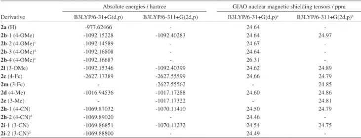
Table S1. Absolute energies (Hartree) and GIAO nuclear magnetic shielding tensors (ppm) for compounds 2a-2e and 2h-2i. All calculations were carried out on a optimized B3LYP/6-31G(d) geometry
Absolute energies / hartree GIAO nuclear magnetic shielding tensors / ppm
Derivative B3LYP/6-31+G(d,p) B3LYP/6-311+G(2d,p) B3LYP/6-31+G(d,p)a B3LYP/6-311+G(2d,p)b
2a (H) -977.62466 - 24.64
-2b-1 (4-OMe) -1092.15228 -1092.40283 24.64 24.97
2b-2 (4-OMe)c -1092.14589 - 24.67
-2b-3 (4-OMe)d -1092.16808 - 24.64
-2b-4 (4-OMe)e -1092.16687 - 26.31
-2l (3-OMe) -1092.15346 -1092.40399 24.62 24.89
2c (4-Fc) -2627.17389 -2627.55599 24.66 24.79
2m (3-Fc) - -2627.55562 - 24.85
2d (4-Me) -1016.94536 -1017.17288 24.60 24.86
2e (3-Me) - -1017.17322 - 24.81
2h-1 (4-CN) -1069.87032 -1070.11410 24.50 24.79
2h-2 (4-CN)d -1069.89020 - 24.46
-2i-1 (3-CN) -1069.86851 -1070.11232 24.54 24.75
2i-2 (3-CN)d -1069.88800 - 24.49
-aAt this level, the absolute GIAO nuclear magnetic shielding of TMS is 32.09 ppm. bAt this level, the absolute GIAO nuclear magnetic shielding of TMS
is 31.82 ppm. c Conformation with the methoxy group rotated by 90o. dCalculation with inclusion of solvent (CPCM, chloroform). The absolute GIAO nuclear magnetic shielding of TMS at this level is 31.63 ppm eAlternative conformation, with the 2-methyl-propenyl group rotated by 180o, including solvent effect (chloroform).
Figure S1. View of the ORTEP plot for 2b with labeled atoms and 50%
Francisco et al. S3 Vol. 21, No. 1, 2010
Figure S3. View of the self-assembly 1D by classical and non classical hydrogen bonds along the [100] crystallography direction. [Symmetry codes: (i) x−1, y, z; (ii) x+1, y, z].
Table S2. Crystal data and structure reinement for 2b.
Formula C21H19O3N
formula weight 333.37
crystal system, space group Triclinic, P-1
crystal size / mm 0.30 x 0.20 x 0.15
a /Å 7.8709(16)
b/Å 9.3748(19)
c/Å 12.117(2)
α / (o) 86.11(3)
β / (o) 81.60(3)
γ / (o) 78.26(3)
V / Å3 865.3(3)
Z 2
T / K 295
ρcalc. / (g cm-3) 1.279
μ / mm-1 0.086
2θ range 3.99 to 25.00
F(000) 352
Relections collected 10324
Relections unique 3023
Rint 0.0358
Max. and min. transmission 0.9873 and 0.9748
datab/restraints/parameters 3023 / 0 / 232
Sb,c 1.027
R1a,d 0.0430
wR2b,e 0.1031
largest difference peak and hole / (e Å-3) 0.153 and -0.180
aF
o2≥ 2σ(Fo2). bFo2≥ 3σ(Fo2). cS = [Σw(Fo2 – Fc2)2/(n – p)]1/2. dR1 = Σ||Fo| - |Fc||Σ|Fo|. ewR
2 = [Σw(Fo2 –Fc2)2/Σw(Fo4)]1/2
Table S3. Geometric parameters for molecule 2b (Å, °)
Bonds distances N(1)-C(11) 1.424(2) N(1)-C(2) 1.367(2) O(1)-C(4) 1.234(2) O(2)-C(1) 1.223(2) O(3)-C(14) 1.378(2) O(3)-C(17) 1.423(2) C(3)-C(18) 1.480(2) C(19)-C(18) 1.328(3) C(19)-C(20) 1.502(3) C(19)-C(21) 1.510(3) Bond angles C(2)-N(1)-C(11) 129.43(15) C(14)-O(3)-C(17) 116.93(16) O(1)-C(4)-C(3) 120.81(16) O(1)-C(4)-C(10) 119.19(16) O(2)-C(1)-C(9) 121.52(16) O(2)-C(1)-C(2) 119.54(16) C(19)-C(18)-C(3) 126.38(17) C(20)-C(19)-C(21) 115.62(18)
Table S4. Geometric parameters for non-classical H bonds for molecule (2b) (Å, °)
D-H···A D-H H···A D···A ∠D-H···A
N(1)—H(1) ···O(2) 0.87 (2) 2.18 (2) 2.620 (2) 111.2 (18)
N(1)—H(1) ···O(1)i 0.87 (2) 2.46 (2) 3.241 (2) 150.2 (18)
C(12)—H(12)···O(1)i 0.93 2.58 3.246 (2) 129
C(5)—H(5) ···O(2)ii 0.93 2.39 3.304 (2) 169
Novel 2-(R-phenyl)amino-3-(2-methylpropenyl)-[1,4]-naphthoquinones J. Braz. Chem. Soc.
S4
Table S5. Atomic coordinates (× 104) and equivalent isotropic displacement parameters (A2 × 103) for (2b). U(eq) is deined as one third of the trace of the orthogonalized Uij tensor
x y z U(eq)
C(19) −974(2) −1627(2) 5732(2) 43(1)
C(20) −2387(3) −494(2) 5299(2) 52(1)
C(21) −31(3) −2776(3) 4922(2) 70(1)
O(1) 1355(2) −120(2) 7804(1) 54(1)
O(2) −5563(2) 1227(2) 9030(1) 54(1)
C(2) −3178(2) −323(2) 8018(1) 33(1)
N(1) −4432(2) −1001(2) 7743(1) 38(1)
C(1) −3973(2) 916(2) 8784(1) 36(1)
C(9) −2800(2) 1709(2) 9233(1) 35(1)
O(3) −4254(2) −6772(1) 6747(1) 57(1)
C(10) −984(2) 1312(2) 8931(1) 35(1)
C(4) −239(2) 152(2) 8106(2) 36(1)
C(3) −1409(2) −604(2) 7634(1) 34(1)
x y z U(eq)
C(11) −4275(2) −2456(2) 7413(1) 34(1)
C(12) −5544(2) −2751(2) 6832(2) 40(1)
C(18) −559(2) −1641(2) 6757(2) 39(1)
C(16) −2999(2) −3610(2) 7728(2) 40(1)
C(8) −3498(2) 2840(2) 9955(2) 44(1)
C(14) −4310(2) −5325(2) 6920(2) 39(1)
C(15) −3008(2) −5025(2) 7473(2) 43(1)
C(5) 112(2) 2050(2) 9372(2) 44(1)
C(13) −5574(2) −4176(2) 6589(2) 43(1)
C(6) −601(3) 3166(2) 10106(2) 51(1)
C(7) −2398(3) 3565(2) 10390(2) 51(1)
C(17) −5788(3) −7133(2) 6440(2) 61(1)
3.
1H NMR and APT Spectra (CDCl
3
) of Compounds 2a-k
Francisco et al. S5 Vol. 21, No. 1, 2010
Figure S5. APT spectrumof 2-(phenyl)amino-3-(2-methylpropenyl)-1,4-naphthoquinone(2a).
Novel 2-(R-phenyl)amino-3-(2-methylpropenyl)-[1,4]-naphthoquinones J. Braz. Chem. Soc.
S6
Francisco et al. S7 Vol. 21, No. 1, 2010
Figure S10. 1H NMR spectrumof 2-(4-methyl-phenyl)amino-3-(2-methylpropenyl)-1,4-naphthoquinone (2d).
Novel 2-(R-phenyl)amino-3-(2-methylpropenyl)-[1,4]-naphthoquinones J. Braz. Chem. Soc.
S8
Figure S12. 1H NMR spectrumof 2-(3-methylphenylene)amino-3-(2-methylpropenyl)-1,4-naphthoquinone (2e).
Francisco et al. S9 Vol. 21, No. 1, 2010
Figure S14. 1H NMR spectrumof 2-(4-iodo-phenyl)amino-3-(2-methylpropenyl)-1,4-naphthoquinone (2f).
Novel 2-(R-phenyl)amino-3-(2-methylpropenyl)-[1,4]-naphthoquinones J. Braz. Chem. Soc.
S10
Figure S16. 1H NMR spectrumof 2-(3-iodo-phenyl)amino-3-(2-methylpropenyl)-1,4-naphthoquinone (2g).
Francisco et al. S11 Vol. 21, No. 1, 2010
Figure S18. 1H NMR spectrumof 2-(4-ciano-phenyl)amino-3-(2-methylpropenyl)-1,4-naphthoquinone (2h).
Novel 2-(R-phenyl)amino-3-(2-methylpropenyl)-[1,4]-naphthoquinones J. Braz. Chem. Soc.
S12
Figure S20. 1H NMR spectrumof 2-(3-ciano-phenyl)amino-3-(2-methylpropenyl)-1,4-naphthoquinone (2i).
Francisco et al. S13 Vol. 21, No. 1, 2010
Figure S22.1H NMR spectrumof 2-(4-nitro-phenylene)-3-(2-methylpropenyl)-1,4-naphthoquinone (2j).
Novel 2-(R-phenyl)amino-3-(2-methylpropenyl)-[1,4]-naphthoquinones J. Braz. Chem. Soc.
S14
Figure S24. 1H NMR spectrum of 2-(3-nitro-phenyl)amino-3-(2-methylpropenyl)-1,4-naphthoquinone (2k).
Francisco et al. S15 Vol. 21, No. 1, 2010
Figure S25. APT spectrum of 2-(3-nitro-phenyl)amino-3-(2-methylpropenyl)-1,4-naphthoquinone (2k).
Figure S26. VT 1H NMR spectra of compound 2a (range: 25oC to −90 oC) in CD 2Cl2.
Novel 2-(R-phenyl)amino-3-(2-methylpropenyl)-[1,4]-naphthoquinones J. Braz. Chem. Soc.
S16
4. Cyclic Voltammograms (100mV) of Compounds 2a-k
Figure S27. CV of 2-(phenyl)amino-3-(2-methylpropenyl)-1,4-naphthoquinone(2a).
Figure S28. CV of 2-(4-methoxy-phenyl)amino-3-(2-methylpropenyl)-1,4-naphthoquinone (2b).
Figure S29. CV of 2-(4-ferrocenyl-phenyl)amino-3-(2-methylpropenyl)-1,4-naphthoquinone (2c).
Figure S30. CV of 2-(4-methyl-phenyl)amino-3-(2-methylpropenyl)-1,4-naphthoquinone (2d).
Figure S31. CV of 2-(3-methyl-phenyl)amino-3-(2-methylpropenyl)-1,4-naphthoquinone (2e).
Francisco et al. S17 Vol. 21, No. 1, 2010
Figure S33. CV of 2-(3-iodo-phenyl)amino-3-(2-methylpropenyl)-1,4-naphthoquinone (2g).
Figure S34. CV of 2-(4-ciano-phenyl)amino-3-(2-methylpropenyl)-1,4-naphthoquinone (2h).
Figure S35. CV of 2-(3-ciano-phenyl)amino-3-(2-methylpropenyl)-1,4-naphthoquinone (2i).
Figure S36. CV of 2-(4-nitro-phenyl)amino-3-(2-methylpropenyl)-1,4-naphthoquinone (2j).
Novel 2-(R-phenyl)amino-3-(2-methylpropenyl)-[1,4]-naphthoquinones J. Braz. Chem. Soc.
S18
5. Antitumor Assays
The antitumor screening of compounds 2a-k was
carried out against three cancer cell lines: SF-295 (central
nervous system), HCT-8 (colon), MDAMB-435 (breast)
through an MTT assayref and the results are summarized
in Table S6.
Table S6. Screening of the cytotoxic activity (growth inhibition %) of compounds 2a-k and 1
Compounds SF295 GI% HCT-8 GI% MDA-MB435 GI%
Average SD Average SD Average SD
2a (H) 68.20 4.80 73.75 1.68 54.20 5.00
2b (4-OMe) -11.01 11.89 -2.95 13.01 24.54 9.15
2c (4-Fc) 2.57 7.43 5.45 6.24 102.31 1.09
2d (4-Me) 12.20 3.71 12.33 2.76 17.21 3.58
2e (3-Me) 58.59 0.40 71.52 1.01 43.86 3.54
2f (4-I) -4.93 23.88 10.29 2.13 -7.63 3.42
2g (3-I) 2.49 2.20 15.92 4.49 25.14 13.42
2h (4-CN) -5.79 0.56 2.65 0.84 10.72 0.93
2i (3-CN) -12.56 2.48 6.73 6.36 -20.51 4.35
2j (4-NO2) 8.78 0.90 17.00 1.44 -18.20 8.24
2k (3-NO2) -7.40 4.20 17.03 2.02 4.01 7.68
1 41.09 15.08 27.01 3.12 106.05 0.16
DOX 96.19 0.63 94.85 3.99 95.38 2.23

![Figure 5. View of the self-assembly 1D by classical and non-classical hydrogen bonds along the [100] crystallography direction](https://thumb-eu.123doks.com/thumbv2/123dok_br/18994953.461900/4.892.144.731.123.421/figure-view-assembly-classical-classical-hydrogen-crystallography-direction.webp)
![Figure S3. View of the self-assembly 1D by classical and non classical hydrogen bonds along the [100] crystallography direction](https://thumb-eu.123doks.com/thumbv2/123dok_br/18994953.461900/13.892.178.740.115.403/figure-view-assembly-classical-classical-hydrogen-crystallography-direction.webp)


