ABSTRACT
Bone quality describes aspects of bone composition and structure that contribute to bone strength independently of bone mineral density. These include bone turnover, microarchitecture, mineralisation, micro-damage and the composition of bone matrix and mineral. New tech-niques to assess these components of bone quality are being developed and should produce important insights into determinants of fracture risk in untreated and treated disease. (Arq Bras Endocrinol Metab 2006;50/4:579-585)
Keywords: Bone quality; Turnover; Mineralisation; Microarchitecture; Fracture; Bone strength
RESUMO
Qualidade Óssea: O Que Significa e Como Mensurá-la?
A qualidade do osso compreende os aspectos da composição e estru-tura óssea que contribuem para sua força, independentemente da densidade mineral óssea, as quais incluem: turnover ósseo, microar-quitetura, mineralização, microdanos e a composição da matriz óssea e mineral. Novas técnicas para avaliar estes componentes da qualidade óssea têm sido desenvolvidas e devem proporcionar importantes avanços para determinação do risco de fraturas nas doenças tratadas e não tratadas. (Arq Bras Endocrinol Metab 2006;50/4:579-585)
Descritores:Qualidade óssea; Turnover; Mineralização; Microarquitetura; Fratura; Força óssea
WHAT IS BONE QUALITY?
B
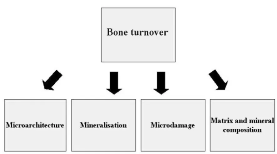
ONE STRENGTH IS DETERMINEDby bone mass, geometry and quality. The latter includes several aspects of bone structure and composition, including bone turnover, microarchitecture, the degree and distribution of mineralisation, the extent of microdamage and its repair and, finally, the composition of bone matrix and mineral (figure 1). These components are largely interdependent, so that a primary abnormality in one will often lead to changes in others. In particular, bone turnover is a major determinant of other components of bone quality and hence its measurement in clini-cal practice is of key importance.The recent interest in bone quality has arisen from observations that the traditional measure of bone strength in clinical practice, namely bone densitometry, does not always reliably predict fracture risk (1). This has stimulated the search for other aspects of bone composition and structure that contribute to bone fragility. This review describes some disease states in which abnormal bone quality is associated with increased fracture risk, sometimes despite increased bone mineral density. Techniques for the measurement of bone quality will be discussed together with how these
Juliet Compston
University of Cambridge School of Clinical Medicine, Cambridge CB2 2QQ, UK.
have advanced our understanding of the mechanisms by which bone strength may be improved by thera-peutic intervention.
ASSESSMENT OF BONE QUALITY
In vivo assessment of bone quality is limited to mea-surement of bone turnover and of some aspects of bone geometry and architecture. However, using bone biopsy or autopsy specimens, a number of approaches have been developed that have increased our under-standing of how bone quality contributes to bone strength in untreated and treated disease (table 1). Bone turnover
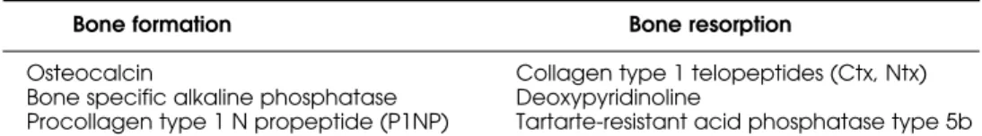
Bone turnover is most commonly assessed in clinical practice by measurement of biochemical markers of resorption and formation (table 2). The markers, which are mainly serum based, reflect whole body turnover and thus provide assessment predominantly of cortical bone, which constitutes 80–90% of the skeleton. They show considerable variability, both within and between individuals, and may be affected by diet, so blood or urine specimens should ideally be obtained in the fasting state and at a standard time of the day.
Bone turnover can also be assessed by histo-morphometric assessment of bone, using tetracycline labelling prior to the biopsy (2). The extent of tetra-cycline-labelled surfaces indicates bone turnover, pro-vided that bone remodelling is in a steady state and that bone resorption and formation are coupled.
However, bone turnover in iliac crest biopsies may not reflect turnover at other sites, since there are consider-able intra-individual variations in bone turnover throughout the skeleton (3). Thus it is not surprising that there may be differences between biochemical markers and histomorphometry in their assessment of bone turnover; in particular, the degree of suppression of bone turnover by anti-resorptive agents is generally greater when assessed by the latter technique. Tech-niques such as 18F-fluoride positron electron
phy and single photon emission computed tomogra-phy gamma camera imaging using technetium labelled bisphosphonate provide new approaches to the assess-ment of regional bone turnover at sites of clinical rel-evance, for example the spine.
Assessment of bone microarchitecture
Alterations in bone microarchitecture make an impor-tant contribution to bone strength that may not always be captured by bone mineral density measurements. Cortical and cancellous architecture are both important in this respect. In cancellous bone, the size and shape of trabeculae and their connectivity and orientation (anisotropy) contribute to bone strength whilst in cor-tical bone corcor-tical width, corcor-tical porosity and bone size are the main determinants. Although some of these architectural features can be assessed in histological sec-tions of bone biopsy specimens using 2-dimensional approaches (4), more sophisticated methods have now been developed that enable 3-dimensional visualisation and quantification. These include high-resolution mag-netic resonance imaging (HR-MRI), high resolution peripheral quantitative computed tomography
pQCT), micro-CT (µCT) and synchrotron radiation µCT (5). These are currently research tools and, in vivo, can only be applied to the peripheral skeleton although technological advances may eventually extend their use to the central skeleton.
Changes in bone microarchitecture in untreated and treated disease states result from the underlying alterations in bone remodelling. High turnover states and increased osteoclast activity predispose to trabecu-lar penetration, loss of connectivity, cortical thinning and increased cortical porosity, whereas low bone turnover states and reduced bone formation are asso-ciated with trabecular thinning and relative preserva-tion of bone microarchitecture.
Assessment of bone mineralisation
Mineralisation of bone matrix occurs in two phases. Pri-mary mineralisation occurs when the bone mineral is deposited during the bone remodelling cycle, whereas secondary mineralisation describes the process of further mineralisation after the remodelling cycle has been com-pleted. The degree of secondary mineralisation is criti-cally dependent on bone turnover; when this is low, there is more time for mineralisation to proceed where-as in high turnover states, recently formed bone is removed before there is time for prolonged secondary mineralisation (6). The degree of mineralisation and its distribution throughout bone can be measured ex vivo by several methods including microradiography, quan-titative back-scattered electron imaging and spectro-scopic techniques. The degree of mineralisation is cap-tured by bone mineral density measurements, but its contribution relative to other factors influencing bone mineral density cannot be directly deduced.
Assessment of bone matrix and mineral composition
Relatively little is known about how bone matrix and mineral composition contribute to bone strength. Changes in the cross-linking of type 1 col-lagen (7) and post-translational modifications such as lysyl-hydroxylation, glycosylation and beta-iso-merisation of aspartate residues in carboxyterminal telopeptides may have significant biomechanical implications (8,9), as may alterations in the size and structure of bone mineral. Since collagen structure and mineralisation are so closely associated it is like-ly that when changes occur in one, both are affect-ed (10).
New approaches to studying bone matrix and composition include Raman and Fourier transform infrared spectroscopy, transmission electron micros-copy, and small angle X-ray scattering (SAXS). These techniques can only be applied ex vivo to bone speci-mens; however, assays for the measurement of beta-isomerisation of CTX have recently been developed and this approach, together with the development of other biochemical measurements of changes in colla-gen composition in serum or urine, is an important area for research in the future.
Assessment of microdamage
Microdamage in bone consists of microcracks and microfractures. The relationship between these, if any, is unknown and although both forms of micro-damage increase with age their effects on bone strength are unclear (11). Assessment of microdam-age can currently be made only by histological tech-niques.
Table 1.Assessment of bone quality.
Variable Technique
Bone turnover Biochemical markers, histomorphometry Bone microarchitecture Histomorphometry, µCT, SR-µCT, HR-MRI, pQCT Bone mineralisation Microradiography, qBSEI, SAXS, spectroscopy Microdamage Histology, confocal microscopy
Matrix/mineral composite FTIR, TEM, SAXS, Raman spectroscopy, biochemistry
µCT – micro computed tomography; SR – synchrotron radiation; HR-MRI – magnetic resonance imaging; pQCT – peripheral computed computed tomography; qBSEI – quantitative backscattered electron imaging; FTIR – Fourier Transform Infrared; TEM – transmission electron microscopy; SAXS – small angle X-ray scattering
Table 2.Biochemical markers of bone turnover.
Bone formation Bone resorption
Osteocalcin Collagen type 1 telopeptides (Ctx, Ntx) Bone specific alkaline phosphatase Deoxypyridinoline
CHANGES IN BONE QUALITY IN BONE DISEASES
Reduced bone strength and increased fracture risk may be caused by a number of abnormalities in bone qual-ity and may occur despite increased bone mineral den-sity. Examination of these disease states emphasises the interdependence of the different components of bone quality and the importance of normal bone quality in the maintenance of bone health.
Changes in bone mineralisation
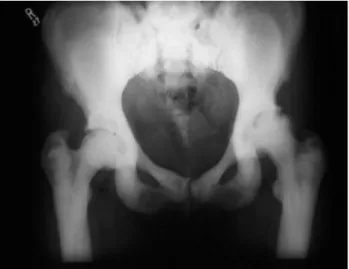
Both decreased and increased mineralisation may be associated with increased bone fragility. Osteomalacia is associated with reduced mineralisation of bone, leading to accumulation of osteoid. Osteomalacic bones are soft and bend easily, resulting in the charac-teristic skeletal deformities that are seen in rickets and in severe cases of adult osteomalacia. Pseudofractures and pathological fractures may also occur. In contrast, the condition of osteopetrosis is characterised by increased mineralisation as a result of absent or great-ly reduced osteoclastic activity (figure 2). Osteopetrot-ic bones are stiff and brittle and can absorb little ener-gy before breaking; thus despite greatly increased bone mineral density, fracture risk is increased.
Bone exposed to fluoride provides an example of a qualitative abnormality of bone mineral that is associ-ated with reduced bone strength. The size and compo-sition of hydroxyapatite crystals is changed as a result of substitution of the hydroxyl group of hydroxyapatite by fluoride; in addition, there may be accumulation of osteoid and the formation of woven bone (12).
Abnormalities of type 1 collagen
Osteogenesis imperfecta is a disease in which there is production of abnormal type 1 collagen. Depending on the genotype, there may be alterations in the bone matrix/mineral composite, decreased mineralisation of bone and abnormal bone modelling and architec-ture; these changes are associated with increased frac-ture risk (13).
Even subtle abnormalities in the structure of type 1 collagen may adversely affect bone strength and fracture risk. For example, a polymorphism affecting a binding site of the transcription factor Sp1 in the pro-moter region of the collagen type 1A1 gene is associ-ated with reduced spine bone mineral density and increased fracture risk (14). The increase in fracture risk cannot be explained solely on the basis of the decrease in bone mineral density, indicating that the abnormal collagen structure contributes independent-ly to reduced bone strength.
High bone turnover
Several high turnover states are associated with increased fracture risk including postmenopausal osteoporosis, Paget’s disease of bone, immobilisation-induced bone loss, post-transplantation bone disease and secondary hyperparathyroidism. High bone turnover reduces bone strength both through reduc-tion in bone mass and disrupreduc-tion of bone microarchi-tecture, an effect that is largely independent of the changes in bone mass. The degree of mineralisation of bone is also reduced in high turnover states. Cortical porosity and endosteal resorption increase, resulting in reduced cortical thickness and strength.
In Paget’s disease of bone, increased bone turnover and osteoclastic activity are associated with multiple alterations in bone quality. Bone matrix may have a mosaic structure due to the presence of both woven and lamellar bone, and mineralisation, architec-ture and geometry may also be abnormal. Post-trans-plantation bone loss affects both cortical and cancel-lous bone (15), whilst in secondary hyperparathy-roidism bone loss is predominantly cortical. Increased bone turnover is also likely to contribute to bone loss in the early stages of glucocorticoid therapy, although in the longer-term reduced bone turnover and forma-tion predominate. There is evidence that the increase in fracture risk associated with glucocorticoid therapy is to some extent independent of bone mineral densi-ty (16), consistent with a role for altered bone qualidensi-ty. Low bone turnover
The effects of low bone turnover on bone strength have not been established. In theory, low bone
turnover might be expected to increase bone fragility as a result of hypermineralisation, reduced osteocyte viability and accumulation of microdamage (17). However, whilst over-suppression of bone turnover in dogs causes significant accumulation of microdamage, adverse effects on bone strength have not been shown. In humans, adynamic renal bone disease (18,19) is associated with histological evidence of very low bone turnover, but robust evidence for increased fracture risk in this condition is lacking. Biochemical markers of bone turnover do not always reflect the suppression of bone turnover seen histologically and bone mineral density may be normal.
Concerns have been expressed about whether long-term treatment with potent anti-resorptive agents for osteoporosis might cause over-suppression of bone turnover and increased bone fragility. Clinical trials indi-cate that anti-fracture efficacy is maintained for up to five years of treatment; subsequently, the data are less robust but studies for up to 7 years of treatment with risedronate (20) and 10 years with alendronate (21) are consistent with continued efficacy and have not demon-strated an increase in fracture risk above that expected.
Odvina et al. (22) recently reported the presence of spontaneous fractures, often with evidence of impaired healing, in 9 patients treated with alendronate. Three were also taking hormone replacement therapy and two prednisolone. Bone biopsy in all cases showed complete absence of double tetracycline labelling, although biochemical markers of bone turnover were normal in many cases and only two patients had osteo-porosis as defined by densitometric criteria. Although firm conclusions cannot be drawn from these observa-tional data, these cases raise the possibility that very low bone turnover may be associated with increased bone fragility despite normal bone mineral density values.
A possible association between osteonecrosis of the jaw and bisphosphonate therapy has recently been reported (23). This condition often presents with a non-healing tooth extraction socket or painful exposed bone in the mandible or maxilla and is seen most commonly in individuals with malignant disease. Although not exclusively associated with bisphospho-nate therapy, the increased numbers of cases reported in such patients has raised the possibility that bisphos-phonates may contribute to osteonecrosis by several mechanisms including immunosuppression, inhibition of angiogenesis and suppression of bone turnover. It should be emphasised that the majority of cases have been described in association with malignant diseases for which high doses of intravenous bisphosphonates have been used.
EFFECTS OF PHARMACOLOGICAL INTERVENTIONS ON BONE QUALITY
Bone turnover
The degree of suppression of bone turnover induced by anti-resorptive drugs varies, whether measured by biochemical markers or bone histomorphometry. The most potent effects are seen with alendronate, zole-dronate and ibanzole-dronate, which reduce activation fre-quency in iliac crest bone biopsies by around 75–90% (24-26). Risedronate and hormone replacement ther-apy are of intermediate potency, with a reduction of around 50% (27,28) and the smallest effect is seen with raloxifene (approximately 20%) (29). These dif-ferences do not appear to be reflected by variations in anti-fracture efficacy, at least in the spine. However, different degrees of suppression of bone turnover may be relevant to anti-fracture efficacy at non-vertebral sites. Thus for weaker anti-resorptive agents such as raloxifene, whilst the modest reduction in bone turnover is sufficient to reduce fractures at cancellous bone sites where high bone turnover has a marked effect on bone strength, this may not be the case at cortical bone sites where the effects of bone turnover on bone microarchitecture are less prominent, and where larger increases in bone mineral density are required to provide protection against fracture (30).
The reduction in bone turnover induced by anti-resorptive agents has been shown to be a major and independent determinant of fracture reduction, at least at vertebral sites (31,32). This is attributable to the major role of high bone turnover in the pathogen-esis of vertebral fracture, which again is independent of bone mineral density (33) and the consequent preven-tion of microarchitectural changes by anti-resorptive drugs.
In the case of teriparatide (recombinant human parathyroid hormone peptide 1-34) activation fre-quency is increased in cancellous bone, but this is asso-ciated with a positive remodelling balance and thus bone mass increases.
Microarchitecture
unchanged cortical thickness (35) have also been reported in iliac crest bone obtained from women undergoing treatment with anti-resorptive therapy.
In contrast, teriparatide improves bone microarchitecture in both cancellous and cortical bone. Increased connectivity density of cancellous bone and increased cortical thickness have been demonstrated using µCT of iliac crest biopsy speci-mens (38,39). There is also some evidence for increased periosteal bone apposition, leading to an increase in bone size.
Mineralisation
Anti-resorptive therapy increases the degree of miner-alisation of bone as a result of the reduction in bone turnover. Three years alendronate therapy in post-menopausal women with osteoporosis increased the mean degree of mineralisation in iliac bone by around 11%, an effect that was seen both in cancellous and cortical bone (40). Similar, although smaller, changes have been reported in women treated with risedronate (35), hormone replacement therapy (41), and ralox-ifene (44). These changes are associated with increased homogeneity of mineralisation.
In postmenopausal women with osteoporosis treated with teriparatide, a small reduction in the degree of mineralisation of bone has been reported, reflecting the increased bone turnover that results from this treatment (42).
Bone matrix and mineral composition
Little is known about the effects of anti-resorptive and anabolic interventions on the bone matrix/mineral com-posite. There is some evidence that age-related changes in the ratio of non-reducible to reducible collagen cross-links and in bone mineral crystallinity are prevented by anti-resorptive therapy; however, the implications of these effects, if any, on bone strength are unclear.
The effects of strontium ranelate on bone miner-al structure are of particular interest since strontium is a bone-seeking element that is taken up mainly by adsorp-tion onto bone mineral, exchanging with a maximum of one in ten calcium ions in hydroxyapatite (43). These changes in mineral composition do not result in any change in the degree of mineralisation of bone.
SUMMARY AND CONCLUSIONS
Bone quality comprises a number of components that contribute to bone strength but are only partially cap-tured by measurements of bone mineral. Advances in
the assessment of bone quality in recent years have provided new insights into bone fragility in both untreated and treated bone disease. The translation of these into clinical practice is an important priority for future research and may eventually lead to better pre-diction of fracture risk and an improved understanding of the mechanisms by which pharmacological inter-ventions affect bone strength.
REFERENCES
1. Delmas PD, Seeman E. Changes in bone mineral density explain little of the reduction in vertebral or nonvertebral fracture risk with anti-resorptive therapy. Bone 2004 ;34:599-604.
2. Frost HM. Tetracycline-based histological analysis of bone remodeling. Calcif Tissue Res 1969;3:211-37.
3. Eventov I, Frisch B, Cohen Z, Hammel I. Osteopenia, hematopoiesis, and bone remodelling in iliac crest and femoral biopsies: a prospective study of 102 cases of femoral neck fractures. Bone 1991;12:1-6.
4. Croucher PI, Garrahan NJ, Compston JE. Assessment of cancellous bone structure: comparison of strut analysis, trabecular bone pattern factor and marrow space star volume. J Bone Miner Res 1996;11:955-61.
5. Jårvinen TLN, Sievånen H, Jokihaara J, Einhorn TA. Revival of bone strength: the bottom line. J Bone Miner Res 2005;20:717-20.
6. Meunier PJ, Boivin G. Bone mineral density reflects bone mass but also the degree of mineralization of bone: thera-peutic implications. Bone 1997;21:373-8.
7. Paschalis EP, Verdelis K, Doty SB, Boskey AL, Mendelsohn R, Yamauchi M. Spectroscopic characterization of collagen cross-links in bone. J Bone Miner Res 2001;16:1821-8. 8. Wang XF, Shen X, Li X, Agrawal CM. Age-related changes
in the collagen network and toughness of bone. Bone 2002;31:1-7.
9. Vashishth D, Gibson GJ, Khoury JI, Schaffler MB, Kimura J, Fhyrie DP. Influence of non-enzymatic glycation on bio-mechanical properties of cortical bone. Bone 2001;28:1-7. 10. Paschalis EP, Shane E, Lyritis G, Skarantavos G, Mendel-sohn R, Boskey AL. Bone fragility and collagen cross-links. J
Bone Miner Res 2004;19:2000-4.
11. Burr DB. Targeted and non-targeted remodelling. Bone
2002;30:2-4.
12. Boivin G, Duriez J, Chapuy M-C, Flautre B, Hardouin P, Meunier PJ. Relationship between bone fluoride content and histologic evidence of calcification defects in osteo-porotic women treated long term with sodium fluoride.
Osteoporos Int 1993;3:204-8.
13. Glorieux FH, Bishop NJ, Travers R. Bone histomorphometric analysis in osteogenesis imperfecta type IV: evidence for three discrete forms. Bone 1996;19:142S.
15. Compston J. Osteoporosis after liver transplantation. Liver Transpl 2003;9:321-30.
16. Kanis JA, Johansson H, Oden A, Johnell O, de Laet C, Melton III LJ, et al. A meta-analysis of prior corticosteroid use and fracture risk. J Bone Miner Res 2004;19:893-9. 17. Weinstein RS. Perspective. True Strength. J Bone Miner Res
2000;15:621-5.
18. Coco M, Rush H. Increased incidence of hip fractures in dialysis patients with low serum parathyroid hormone. Am
J Kidney Dis 2000;36:1115-21.
19. Atsumi K, Kushida K, Yamazaki K, Shimizu S, Ohmura A, Inoue T. Risk factors for vertebral fractures in renal osteody-strophy. Am J Kidney Dis 1999;33:287-93.
20. Mellström DD, Sorensen OH, Goemare S, Roux C, Johnson TD, Chines AA. Seven years of treatment with risedronate in women with postmenopausal osteoporosis. Calcif Tissue Int 2004;75:462-8.
21. Bone HG, Hosking D, Devogelaer J-P, Tucci JR, Emkey RD, Tonino RP, et al. Ten years’ experience with alendronate for osteoporosis in postmenopausal women. N Engl J Med
2004;350:1189-99.
22. Odvina CV, Zerwekh JE, Rao DS, Maalouf N, Gottschalk F, Pak CYC. Severely suppressed bone turnover: a potential complication of alendronate therapy. J Clin Endocrinol Metab 2005;90:1294-301.
23. Ruggiero SL, Mehrotra B, Rosenberg TJ, Engroff SL. Osteonecrosis of the jaws associated with the use of bis-phosphonates: a review of 63 cases. J Oral Maxillofac Surg
2004;62:527-34.
24. Chavassieux PM, Arlot ME, Roux JP, Portero N, Daifotis A, Yates AJ, et al. Effects of alendronate on bone quality and remodeling in glucocorticoid-induced osteoporosis: a his-tomorphometric analysis of transiliac biopsies. J Bone
Miner Res 2000;15:754-62.
25. Reid IR, Brown JP, Burckhardt P, Horowitz Z, Richardson P, Treschel U, et al. Intravenous zoledronic acid in post-menopausal women with low bone mineral density. N Engl J Med 2002;346:653-61.
26. Recker RR, Weinstein RS, Chesnut CH, Schimmer RC, Mahoney P, Hughes C, et al. Histomorphometric evalua-tion of daily and intermittent oral ibandronate in women with postmenopausal osteoporosis: results from the BONE study. Osteoporos Int 2004;15:231-7.
27. Eriksen EF, Melsen F, Sod E, Barton I, Chines A. Effects of risedronate on bone quality and bone turnover in women with postmenopausal osteoporosis. Bone 2002;31:620-5. 28. Vedi S, Skingle SJ, Compston JE. The effects of long-term
hormone replacement therapy on bone remodelling in postmenopausal women. Bone 1996;19:535-9.
29. Prestwood KM, Gunness M, Muchmore DB, Lu Y, Wong M, Raisz LG. A comparison of the effects of raloxifene and estrogen on bone in postmenopausal women. J Clin
Endocrinol Metab 2000;85:2197-202.
30. Riggs BL, Melton III LJ. Bone turnover matters: the raloxifene treatment paradox of dramatic decreases in vertebral fractures without commensurate increases in bone densi-ty. J Bone Miner Res 2002;17:11-4.
31. Eastell R, Barton I, Hannon RA, Chines A, Garnero P, Del-mas PD. Relationship of early changes in bone resorption to the reduction in fracture risk with risedronate. J Bone
Miner Res 2003;18:1051-6.
32. Bjarnason NH, Sarkar S, Duong T, Mitlak B, Delmas PD, Chris-tiansen C. Six and twelve-month changes in bone turnover are related to reduction in vertebral fracture risk during 3 years of raloxifene treatment in postmenopausal osteo-porosis. Osteoporos Int 2001;12:922-30.
33. Melton III LJ, Khosla S, Atkinson EJ, O’Fallon WM, Riggs BL. Relationship of bone turnover to bone density and frac-tures. J Bone Miner Res 1997;12:1083-91.
34. Vedi S, Croucher PI, Garrahan NJ, Compston JE. Effects of hormone replacement therapy on cancellous bone microstructure in postmenopausal women. Bone
1996;19:69-72.
35. Dufresne TE, Chmielewski PA, Manhart MD, Johnson TD, Borah B. Risedronate preserves bone architecture in early postmenopausal women in 1 year as measured by three-dimensional microcomputed tomography. Calcif Tissue Int 2003;73:423-32.
36. Borah B, Dufresne TE, Chmielewski PA, Johnson TD, Chines A, Manhart MD. Risedronate preserves bone architecture in postmenopausal women with osteoporosis as measured by three-dimensional microcomputed tomography. Bone 2004;34:736-46.
37. Roschger P, Rinnerthaler S, Yates J, Rodan GA, Fratzl P, Klaushofer K. Alendronate increases degree and uniformi-ty of mineralization in cancellous bone and decreases the porosity in cortical bone of osteoporotic women. Bone
2001;29:185-91.
38. Dempster DW, Cosman F, Kurland ES, Zhou H, Nieves J, Woelfert L, et al. Effects of daily treatment with parathyroid hormone on bone microarchitecture and turnover in patients with osteoporosis: a paired biopsy study. J Bone
Miner Res 2001;16:1846-53.
39. Jiang Y, Zhao JJ, Mitlak BH, Wang O, Genant HK, Eriksen EF. Recombinant human parathyroid hormone (1-34) [teri-paratide] improves both cortical and cancellous bone structure. J Bone Miner Res 2003;18:1932-41.
40. Boivin G, Chavassieux PM, Santora AC, Yates AJ, Meunier PJ. Alendronate increases bone strength by increasing the mean degree of mineralisation of bone tissue in osteo-porotic women. Bone 2000;27:687-94.
41. Boivin G, Vedi S, Purdie DW, Compston JE, Meunier PJ. Influence of estrogen therapy at conventional and high doses on the degree of mineralisation of iliac bone tissue: a quantitative microradiographic analysis in post-menopausal women. Bone 2005;36:562-7.
42. Roschger P, Grabner BM, Messmer P, Dempster DW, Cos-man F, Nieves J, et al. Influence of intermittent PTH treat-ment on mineral distribution in the human ilium: a paired biopsy study before and after treatment. J Bone Miner Res 2001;16:S179.
43. Boivin G, Deloffre P, Perrat B, Panczer G, Boudeulle M, Tsouderos Y, et al. Strontium distribution and interactions with bone mineral in monkey iliac bone after strontium salt (S12911) administration. J Bone Miner Res 1996;11:1302-11.
Address for correspondence:
JE Compston