ABSTRACT
The calcium-sensing receptor (CASR) adjusts the extracellular calcium set point regulating PTH secretion and renal calcium excretion. The receptor is expressed in several tissues and is also involved in other cellular functions such as proliferation, differentiation and other hormonal secretion. High extracellular calcium levels activate the receptor resulting in modulation of several signaling pathways depending on the target tissues. Mutations in the CASR gene can result in gain or loss of receptor function. Gain of func-tion mutafunc-tions are associated to Autossomal dominant hypocalcemia and Bartter syndrome type V, while loss of function mutations are associ-ated to Familial hypocalciuric hypercalcemia and Neonatal severe hyper-parathyroidism. More than one hundred mutations were described in this gene. In addition to calcium, the receptor also interacts with several ions and polyamines. The CASR is a potential therapeutic target to treatment of diseases including hyperparathyroidism and osteoporosis, since its inter-action with pharmacological compounds results in modulation of PTH secretion. (Arq Bras Endocrinol Metab 2006;50/4:628-639)
Keywords: Familial hypocalciuric hypercalcemia; Autosomal dominant hypocalcemia; Mutations; CASR; Bartter syndrome type V; Neonatal severe hyperparathyroidism
RESUMO
O Receptor Sensor de Cálcio e Doenças Associadas.
O receptor sensor de cálcio (CASR) ajusta o set pointdo cálcio extracelu-lar através da regulação da secreção de PTH e da excreção renal de cál-cio. O receptor é expresso em diversos tecidos e também está envolvido em outras funções celulares como proliferação, diferenciação e secreção de outros hormônios. Concentrações altas de cálcio extracelular ativam o receptor resultando em modulação de inúmeras vias de sinais intracelu-lares dependendo do tecido-alvo. Mutações no gene do CASR podem resultar em ganho ou perda de função do receptor. Mutações com ganho de função são associadas à Hipocalcemia autossômica domi-nante e à Síndrome de Bartter tipo V, enquanto que mutações com perda de função são associadas à Hipercalcemia hipocalciúrica familiar e ao Hiperparatireoidismo neonatal grave. Mais de cem mutações foram descritas neste gene. Além do cálcio, o receptor também interage com inúmeros íons e poliaminas. CASR é um alvo terapêutico potencial para tratamento de doenças incluindo hiperparatireoidismo e osteoporose, pois a sua interação com compostos farmacológicos resulta em modulação da secreção de PTH. (Arq Bras Endocrinol Metab 2006;50/4:628-639)
Descritores: Hipercalcemia hipocalciúrica familiar; Hipocalcemia autossômica dominante; Mutações; CASR; Síndrome de Bartter tipo V; Hiperparatireoidismo neonatal grave
Lília D’Souza-Li
Pediatric Endocrinology Laboratory, Center for Investigation in Pediatrics and Department of Pediatrics, Faculty of Medical Science, State University of Campinas, São Paulo, Brazil.
E
LECTROPHYSIOLOGICAL STUDIES SHOW thatpara-thyroid cells possess a cell surface [Ca2+
o] sensing
mechanism that results in changes in phosphoinositide turnover and cytosolic calcium to regulate PTH secre-tion (1). Extracellular calcium regulates itself by serv-ing as a first messenger and interactserv-ing with its recep-tor, the calcium-sensing receptor (CASR) on target tissues. The receptor was cloned in 1993 from bovine parathyroid (BoPCAR1) by expression cloning in
Xenopus laevisoocytes and is a member of the G pro-tein-coupled receptor super family (2). High calcium levels activate the CASR in the parathyroid cell surface to inhibit PTH secretion, and in the kidney to increase calcium excretion (3).
STRUCTURE OF THE CALCIUM-SENSING RECEPTOR
The human CASR gene is located on chromosome 3q13.3-21 (4,5) and spans over 50 kb of genomic DNA. It has a coding region of 3234 bp, which is contained within 6 exons (6). The human CASR is ~120 kDa pro-tein, consisting of 1078 amino acid, with 612 amino acids in the extracellular domain (ECD), 250 amino acids of which comprise seven transmembrane spanning domains (TM), intracellular (ICL) and extracellular loops (ECL), and 216 amino acids of a long C-terminus cytoplasmic tail (ICD) (6). The CASR belongs to the metabotropic glutamate receptor subfamily, which comprises the metabotropic glutamate receptors (mGluR) (7), the GABAB receptor (8), the Vomero-nasal (pheromone)
receptors (9), the taste receptors (10), the GPRC6A receptor (11) and five orphan receptors (12,13).
Studies, either with CASR cDNA transiently transfected in HEK293 cells (14) or expressed endogenously in rat inner medullary collecting duct endosomes (15), show that the CASR form homod-imers via intermolecular disulfide linkages within the ECD. Dimers are the most abundant species present on the cell surface and intermolecular interactions within the dimeric CASR are important for receptor function (16,17). The ECD of the CASR contains nine potential N-linked glycosylation sites (6). The native CASR, as well as recombinant receptors trans-fected in HEK293 cells, present as three forms: 1) a 120 kDa band, which represents the non glycosylated species; 2) a 140 kDa band, which represents the immature glycosylated receptor; and 3) a 160 kDa band, which is the mature fully glycosylated receptor (18). Although the immature glycosylated receptor can reach the plasma membrane to a low extent, only the fully glycosylated receptor is functional (19,20).
Calcium binding sites
Due to the lack of a high-affinity ligand binding assay for the CASR, all the positions where calcium binds are unknown. Also, it is not known how many calcium ions bind to each receptor since there is the possibility of different affinity of each ligand binding domain for the ligand, cooperativity between the ligand binding sites and dimerization of the receptor (21). The phar-macology of the CASR is unusual for a receptor, as it only responds to the ligand in the millimolar ion con-centration range, suggesting low affinity of the recep-tor for [Ca2+
o] (22). However, this affinity range of
the receptor is of physiological relevance as the free [Ca2+] range is 0.75 to 2.0 mM for extracellular fluids
(22). The ECD of the CASR has homologous regions that align with the mGluRs and the related bacterial periplasmic amino acid binding protein, suggesting that the CASR might have evolved from an ancient family of cell-surface proteins binding essential extra-cellular solutes, and suggesting the existence of addi-tional ion-sensing receptors (23). The Venus flytrap model is the proposed model of ligand receptor inter-action for the metabotropic glutamate receptor family (24). In this model two ligand-bound forms have been observed: an open conformation with the ligand ini-tially bound to one ligand pocket in the large ECD with low affinity, and a closed form in which the lig-and binds to a second domain stabilizing a high-affin-ity closed conformation, enclosed within the cleft. The liganded N-terminal segment interacts with the mem-brane-associated domain to generate a signal (24). Alignment of the extracellular domain of the CASR with the metabotropic glutamate receptor and the related bacterial amino-acid binding protein suggested that Ser 147 and Ser 170 correspond to residues in the binding pockets in the CASR (25). Further mutation analysis associated to molecular modeling studies indi-cated that calcium interact with polar residues in the binding pockets in the ECD of the receptor, with residues Ser 170, Asp 190, Gln193, Ser 296 and Glu 297 directly involved in Ca2+ coordination and
residues Tyr 218 and Phe 270 and Ser 147 contribut-ing to complete the coordination (26).
Calcium-sensing receptor agonists
Although calcium is the endogenous ligand for the CASR, it also shows affinity for a variety of di-, tri-, and polyvalent cations in vitrosuch as Mg2+, Ba2+, Sr2+, Gd3+,
La3+, neomycin, spermine, and protamine (6). The rank
order of potency of some agonists is: Gd3+> neomycin >
Ca2+> Mg2+, with a half-maximal response (EC
50) of 20
physiological relevance of the interaction of CASR with ions other than Ca2+is unknown. In addition to cations,
studies suggest that the CASR also senses sodium and ionic strength in parathyroid cells (27,28).
CALCIUM-SENSING RECEPTOR SIGNALING
When BoPCaR1 is expressed in Xenopus laevisoocytes, agonists elicit an increase in inositol 1,4,5-trisphosphate, which is completely blocked by treatment with pertussis toxin, indicating that the response is mediated through Gαi or Gαo(2). However, in bovine parathyroid cells, high levels of [Ca2+
o] activate phospholipase C (PLC) in
a pertussis toxin-insensitive manner, suggesting that the CASR is coupled to PLC through a member of the Gq family (29). Interaction with Gαq is followed by activa-tion of phospholipase Cβ, breakdown of phosphatidyli-nositol 4,5-bisphosphate with formation of 1,2-sn-dia-cylglycerol and of inositol 1,4,5-trisphosphate (IP3). The accumulation of IP3 leads to the release of intracel-lular pools of calcium contributing to intracelintracel-lular sig-naling and causing inhibition of PTH secretion through mechanisms that remain to be fully defined (30). The high [Ca2+
o] also induces a sustained rise in [Ca2+i] in
parathyroid and in CASR transfected HEK293 cells associated with activation of a Ca2+-permeable,
nonse-lective cation channel (31). Activation of the receptor mediates different signal transduction pathways, depend-ing on the cell line. In Chinese hamster ovary cells elicit phosphatidylinositol and arachidonic acid responses (32). In a mouse pituitary cell line (AtT-20) agonist-elicited increase in inositol phosphate is pertussis toxin-sensitive (33). In Madin-Darby canine kidney cell line, the CASR shows interactions of the receptor with Gα i-2, Gαi-3, and Gαq/11 (34). In the human astrocytoma cell line U87 (35), rat oligodendrocytes (36), rat microglia primary cultures (37), as well as in human lens-epithelial cells (38), the CASR activates an outwards K+
channel. In rat fibroblasts, CASR was shown to mediate cell proliferation through an increase in c-SRC and ERK1 tyrosine kinases activity (39). In the human colonic cell line Caco-2, low Ca2+(via interaction with
CASR) induces proliferation and c-myc proto-oncogene expression via PKC activation (40).
ROLE OF THE CASR IN DIFFERENT TISSUES
Regulation of parathyroid function
The highest cell surface expression levels of CASR are found in parathyroid cells. CASR plays a crucial role in regulating PTH secretion and the
parathy-roid cells recognize remarkably small perturbations in the [Ca2+
o], and respond by altering the secretion
of PTH (22). [Ca2+
o] has an inverse steep sigmoidal
relationship with PTH secretion, and most of the sensing of [Ca2+
o] in parathyroid cells occurs over
changes in free [Ca2+] of approximately 0.25 mM
(22). The set point of normal human parathyroid, defined as the calcium concentration at which PTH secretion is half-maximal, is ~1 mM, and it plays an important role in determining the level at which [Ca2+
o] is set by the homeostatic system.
Inactivat-ing mutations in the CASR result in a mild increase in the set point for [Ca2+
o]. In addition, [Ca2+o]
exerts several other actions on parathyroid function including modulation of the intracellular degrada-tion of PTH, cellular respiradegrada-tion and membrane voltage, but the role of the CASR in mediating these effects is not known (31). Bovine parathyroid cells maintained in culture for more than 24 hours reduce dramatically their responsiveness to [Ca2+
o] (2). This
is associated with a significant reduction in mRNA and protein levels of CASR (41).
Regulation of calcium excretion in kidney
Kidney is the major route for mineral ion excretion from the body and plays a key role in calcium home-ostasis. In addition to PTH, the CASR plays an impor-tant role in regulating renal divalent mineral transport processes by both direct (by regulating calcium and water handling) and indirect (by modulating PTH secretion) mechanisms (42). [Ca2+
o] modulates renal
tubular divalent mineral and water transport processes by interacting with the CASR (42).
The CASR has been localized within several segments of the rat tubule, but it is expressed at highest levels in the cortical thick ascending limb (CTAL) (43). It is found mostly on the basolateral surface of tubular cells, but also to a lesser extent on the apical surface (31). Elevated peritubular [Ca2+
o]
and [Mg2+
o] reduces the tubular reabsorption in
iso-lated microperfused segments of CTAL in vitro
(44). The reabsorption of Ca2+ and Mg2+ in CTAL
occurs mainly through a paracellular pathway driven by a lumen-positive, transepithelial potential gener-ated by the transport of Na+, K+, and Cl- by the
api-cal Na-K-2Cl co-transporter combined with recy-cling of K+ into the lumen via an apical K+ channel
(31). While PTH acts through its receptor in the kidney, stimulates cAMP accumulation, enhances the co-transport activity and results in an increase of Ca2+and Mg2+transport, [Ca2+
o] inhibits the
co-transporter activity and a reduction of Ca2+ and
Mg2+ transport (31). High [Ca2+
o] in the mouse
CTAL decreases hormone-dependent cAMP accu-mulation as a result of a direct inhibition of adenylyl cyclase (AC) activity (45). An increase in Arginine vasopressin (AVP)-elicited osmotic water permeabil-ity in collecting ducts stimulates water reabsorption selectively via aquaporin-2 (AQP-2) water channels (46). CASR and AQP-2 were also found to co-express in rat kidney inner medullary collecting ducts (IMCD) suggesting a direct effect of CASR in inhibition of AVP-elicited osmotic water permeabil-ity and the consequent increase in diuresis (46). CASR and Ca2+-inhibitable AC were found to
co-express and co-localize in the rat CTAL cells (47), and cAMP synthesis is inhibited by agents coupled to PLC or to Gαi protein-mediated process suggest-ing that the CASR contributes to the effect observed for high [Ca2+
o] (47). Additional evidence of the
role of CASR in regulating Ca2+and Mg2+transport
in CTAL is found in subjects with mutations in the
CASRgene. In subjects with FHH due to an inacti-vating mutation in the CASR there is a PTH-inde-pendent increase in tubular Ca2+ reabsorption (48),
while in ADH subjects there is increased urinary cal-cium excretion (31).
Role of the calcium-sensing receptor in other tissues
The CASR is widely distributed and is also found in tissues that are not directly involved in calcium homeostasis. In these tissues it appears that high [Ca2+
o], via interaction with CASR, regulates a series
of cellular functions such as increased cell prolifera-tion in fibroblasts (39), inducprolifera-tion of cell differentia-tion in keratinocytes (49) and human colon epithe-lial cells (50), prevention of apoptosis in AT-3 prostate carcinoma cells (51), and cataract formation in lens epithelial cell (38). CASR was detected in a murine bone marrow-derived stromal cell line (ST2) (52), in osteoblast-like cell lines (53) and in rabbit mature osteoclasts (54), however its role in bone is still debatable (55,56). In the mouse pituitary cell line, AtT-20 cells, it was shown that the CASR was implicated in adrenocorticotropic hormone (ACTH) (57) and α-MSH release (58). The CASR was also demonstrated in human insulinoma primary cul-tures, causing released insulin upon [Ca2+
o]
stimula-tion (59), in hepatocytes stimulating bile flow (60) and in antral gastric cells stimulating gastrin secre-tion (61).
DISEASES ASSOCIATED WITH MUTATIONS IN THE CASR
Disorders due to loss of the calcium-sensing receptor function
Two autosomal disorders, Familial Hypocalciuric calcemia (FHH) and Neonatal Severe Primary Hyper-parathyroidism (NSHPT), have been associated with loss of CASR function due to inactivating mutations.
Familial hypocalciuric hypercalcemia
FHH is characterized by moderate elevations of serum calcium concentration (hypercalcemia), lower urinary calcium excretion (hypocalciuria) and inappropriately normal parathyroid hormone (PTH) levels (62,63). This is not a life-threatening condition and most of the usual sequelae of hypercalcemia such as altered mental status, kidney stones, decreased urinary concentrating ability and hypertension are absent (64). Patients are usually asymptomatic or have nonspecific symptoms such as fatigue, weakness, painful joints and headache, with the diagnosis only suspected after a routine bio-chemical screening showing high blood calcium levels (63). Interestingly, some subjects with FHH present with an incomplete phenotype, lacking hypocalciuria. In some families a more severe phenotype suggestive of familiar isolated hyperparathyroidism is present (65,66). FHH is inherited as an autosomal dominant disorder, and all affected individuals with mutations in the CASR gene are heterozygous for the mutation (67). The dominant pattern of inheritance of this dis-ease has been attributed to haploinsufficiency of the
CASR gene, where protein receptor produced by a single normal allele cannot support normal function, although it may suffice for survival (68).
medical treatment for lowering the calcium level is not indicated (75). Surgical exploration of the parathyroid glands is also not indicated, as parathyroidectomy does not cure the disorder (63).
Neonatal Several Hyperparathyroidism
NSHPT (76,77) represents the most severe expres-sion of familial hypocalciuric hypercalcemia (68). In most patients in which mutations were found in the
CASR, the two gene copies are mutated, with both parents having passed on a mutated copy and pre-senting with FHH. There are three reports of muta-tions being found de novoin individuals with NSHPT with only one copy mutated and no mutation found in the parents (78,79). Neonatal severe hyper-parathyroidism causes a marked elevation in serum calcium and PTH levels. It appears very early, in the first days of life, and the baby presents with hypoto-nia, poor feeding, failure to thrive and respiratory distress associated with rib cage deformities (80). PTH concentrations are very high, associated with calcium levels that are life-threatening (80). In severe cases, surgical intervention is essential, with total parathyroidectomy still being the currently accepted method of treatment. However, there are reports of cases where symptoms are not life threatening and could be controlled using medical therapy to main-tain calcium at levels compatible with normal life (81,82).
Disorders due to gain of calcium-sensing receptor function
An autosomal dominant hypocalcemia (ADH) and Bartter syndrome type V have been associated with gain of CASR function due to activating mutations in the receptor.
Autosomal Dominant Hypocalcemia
ADH presents with a wide clinical spectrum, from severe hypocalcemia in the neonatal period to an inci-dental finding in adulthood (83). Associated problems include seizures, mental deficiency, orodental prob-lems, basal ganglia calcification, kidney stones and renal failure (84). Individuals present with hypocal-cemia, hyperphosphatemia, low serum PTH levels and hypercalciuria (84). Autosomal dominant hypocal-cemia was initially classified as familial isolated hypo-parathyroidism, a heterogeneous group of disorders characterized by PTH deficiency, hypocalcemia and hyperphosphatemia. Within this group, different modes of inheritance were identified with transmission patterns consistent with autosomal dominant,
autoso-mal recessive, and X-linked forms. Finegold et al. linked one form of autosomal dominant hypoparathy-roidism to chromosome 3q13 (85) and Pollak et al. (86) described the first activating mutation in the
CASRin a family with ADH, and the terminology was recommended to be changed to autosomal dominant hypocalcemia, as a direct contrast to the hypercalcemia in FHH (87). In most individuals where mutations have been found, familial inheritance is clear, with one parent being affected with the same mutation (67). However, de novo mutations found in individuals where no mutation was found in the parents have also been described (67). A careful treatment for this con-dition is required, as attempts to normalize blood cal-cium levels with regular doses of vitamin D tend to exacerbate urinary calcium levels and increase the risk of kidney stone and renal impairment (88). Treatment should be limited to symptomatic patients. Hydrochlorothiazide has been used to control hyper-calciuria in these patients (88). Recombinant human PTH to improve hypocalcemia symptoms has been described, however longer follow-up studies are required (89).
Bartter Syndrome type V
Bartter syndrome is a heterogeneous rare disease due to deficiency in sodium and chloride absorption. Bio-chemical profile is renal salt wasting, hypokalemic metabolic alkalosis, elevated renin and aldosterone lev-els with low blood pressure. In some individuals hypercalciuria is also present. Gain of function muta-tions in the CASR has been described in some patients with Batter syndrome associated to hypocalcemia and hypercalciuria (90,91). Functional studies showed that these mutations (L125P, C131W and A843E) result in a more severe receptor activation when compared to other activating mutations described (90,91). Of interest, the mutation A843E is the only constitutive mutation described in the CASR, presenting a high basal activity in the absence of [Ca2+
o] (92). Clinical
data in the literature may be biased towards the most severely affected individuals in both ADH and FHH/NSHPT and may not reflect the whole spec-trum of the disease.
Calcium-sensing abnormalities in other disorders
Autoimmune hypoparathyroidism
an integral part of type I autoimmune polyglandular syndrome, a rare disorder characterized by the pres-ence of AH, Addison’s disease, and mucocutaneous candidiasis and can be associated with female primary hypogonadism, keratopathy, alopecia, vitiligo, parietal cell atrophy, insulin-dependent diabetes mellitus, autoimmune hepatitis and hypothyroidism (93). In one study, an epitope within the ECD of the CASR was specifically recognized in 14 of 25 individuals (56%) with AH, suggesting that the CASR is a key antigen in directing the immune response against parathyroid tissue in this condition (94). The mecha-nism of the hypoparathyroidism is destruction of the parathyroid gland due to the inflammatory reaction and complement fixation.
Autoimmune hypocalciuric hypercalcemia The ECD of the CASR is also antigen for antibodies that instead of inducing parathyroid cell destruction, interferes with the normal activation of the receptor resulting in increase PTH levels (95). Patients may manifest clinically with hypercalcemia, not suppressed PTH levels and hypocalciuria similar of FHH patients. However, it is not associated to mutation in the CASR
gene (95). In addition to the hypercalcemia, patients may present other autoimmune disease such as thy-roiditis, celiac sprue, psoriasis, hypophysitis, uveitis and rheumatoid arthritis (95).
Mutations in the calcium-sensing receptor
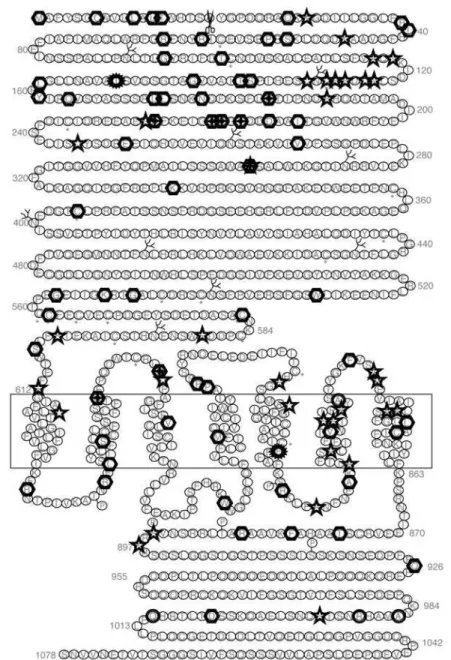
One hundred and twelve mutations (98 missense, 6 nonsense, 8 insertion and or deletion, and 1 splice mutation) have been described in the CASR mutation database (http://www.casrdb.mcgill.ca) related to FHH, NSHPT, ADH families or as de novo disease (figure 1) (96). In addition, 6 polymorphisms were found in samples from a normal population or in fam-ilies with FHH and ADH in which this base pair change was present in affected and unaffected mem-bers and did not segregate with the disease. Fifteen mutations were found more than once in the CASR
gene in apparently unrelated families. In several posi-tions two different mutaposi-tions were described in the same codon with the same receptor phenotype, with one exception. At position 297 an activation mutation (E297D) and an inactivating mutation (E297K) were described (26,70) confirming the crucial role of this position on receptor activation. Most of the mutations found in the ECD are located in the first third of the N-terminus suggesting the importance of this region in ligand binding. Activating mutations in the proxi-mal 1/3 of the ECD may facilitate the ligand-binding
interaction in the different binding sites, increasing the receptor affinity to the ligand, whereas inactivating mutations may have the opposite effect, disrupting the ligand binding pockets. This is supported by in vitro
functional analyses of mutations in this location that show ligand-dependent changes in the affinity of the receptor to extracellular calcium (67).
Mutation in the TM domain may abrogate con-straints, tilting the TM and locking the receptor in either an inactivating or activating conformation, as residues in the TM7 are critical for maintaining the receptor in an inactive conformation (97). From func-tional analyses of receptors with gain of function, only A843E showed the ability to activate the receptor in the absence of the ligand (92). The other activating mutations all showed a ligand-dependent shift of the dose-response curves to the left. This suggests that the mechanism of activation of the receptor in most of the TM domain mutations is to facilitate the TMD activa-tion, with the exception of A843E that most likely locks the TMD in an active conformation. Inactivating mutations resulting in total loss of function of the receptor may also be associated to total loss of ability of the ligand bind and activate the receptor, even though the receptor is well expressed in the plasma membrane or due to misfolding and retention within the cytoplasm resulting in lack of receptor at the mem-brane (98).
The ICD seems to be important for receptor traf-ficking to the cell membrane and for the interaction with intracellular proteins. Large deletion of the c-terminal tail was associated to gain of function in an ADH family (99). In vitrofunctional studies showed gain of function and increase mutant receptor cell surface expression level. Mutagenesis in the ICD confirms its involvement in degradation and processing of the receptor (99). Residues 962-981 in the c-terminal tail are critical for its interaction with filamin A and this interaction prevents the receptor degradation and facilitates MAPK signaling (100). In contrast, interaction of the ICD with dorfin targets the receptor for degradation (101).
Activating mutations
sim-ilar symptoms are found in different families.
Inactivating mutations
Of the 72 inactivating mutations in the CASR gene, 59 are missense, 6 are nonsense, 6 are insertions and/or deletion including an Alu element insertion (102) and one splice mutation (103). The gene dosage effect is clear in most FHH cases, with one mutated gene copy resulting in FHH with mild hyper-calcemia and two mutated copies resulting in NSHPT, a more severe phenotype that manifests very early in
life with severe hypercalcemia, bone demineralization and failure to thrive (68). However, the three cases of
de novo NSHTP reported in the literature were he-terozygous for missense mutations located in the extracellular domain, with only one mutated allele and no mutation found in the parents (79,104). One indi-vidual with de novo NSHPT was heterozygous for a previously described mutation in a FHH family (79).
Polymorphisms
Six polymorphisms were found in the CASRgene: one
Figure 1.Topography and positions of mutations in the CASR. Symbols indicate positions with one inactivating mutation, positions with two different inactivating mutations, positions with one activating mutation, positions with two different activating mutations and indicate positions with polymorphisms. Adapted from D’Souza-Li in CASR mutation database, 1999. Reproduced with permission.
"
# #
in intron 5 just before exon 6 (IVS 5 -88 t/c) and the remaining five in exon 7 in the coding region (one in the 6th TM [A/T826], one in the 7thTM [C/S851], and
three in the ICD [A/S986, R/G990 and Q/E1011]). The polymorphism in intron 5, IVS 5 -88 t/c, is very common (105) and, when analyzed in a large group of normal and affected individuals, no correlation was found between this mutation and the incidence of parathyroid adenoma or diabetes (106). The A/T 826 mutation was initially found in 4 parathyroid adenomas (107). Further analysis showed the same change in 16% of 50 normal subjects’ samples (108). The C/S 851 was found in an ADH family in both affected and unaffected members (109). They also found another mutation in this family (A116T), which segregates with the disease, and concluded that C/S 851 was a rare polymorphism. The frequency of the 3 common polymorphisms in the cytoplasmic tail varies in different populations. In a large series in a Caucasian population the incidence for A/S986 was 24%, for R/G990 was 4% and for Q/E1011 was 3% within 377 unrelated DNA samples (110). In addition, a study analyzing serum calcium lev-els in samples from a normal population found that the homozygous polymorphism 986S was associated to higher serum calcium levels when compared to the het-erozygous form, while the homozygous 986A had the lowest calcium levels (110,111).
INTERACTION OF THE CASR WITH PHARMACOLOGICAL COMPOUNDS
Calcimimetics
The CASR is also a target for pharmacological com-pounds that act synergistically with calcium as positive allosteric modulators of the receptor, such as NPS R-568 and cinalcalcet (112,113). However, these com-pounds require the presence of calcium and act by increasing the sensitivity of the receptor to [Ca2+
o]
(114). Their interaction sites are in the TMD of the receptor with Glu 837 critical for their action (115). Clinical studies using a calcimimetic in secondary hyperparathyroidism showed significant reduction on PTH, and in the calcium x phosphate product levels in patients treated for 26 weeks (116). Cinacalcet was approved by the US FDA for the treatment of sec-ondary hyperparathyroidism (113). Other potential uses for calcimimetics are co-adjuvant in the treatment of hyperparathyroidism and parathyroid carcinoma.
Calcilytic
Compounds that interact with the CASR as negative allosteric modulators have also been developed, such
as NPS-2143 (117) and compound 1 (118). These compounds are potent CASR antagonists resulting in transient increase PTH secretion and bone formation (117). Calcilytic compounds are potential therapeutic agents for the treatment of osteoporosis, since they can reproduce the anabolic effect in bone of transitory increase in PTH.
CONCLUSION
The CASR plays an important role in regulating [Ca+2
o]. The receptor is more versatile than ever
expected, being involved in a variety of cellular func-tion. It is also target to new pharmacological com-pounds that modifies its function with potential ther-apeutic applications.
ACKNOWLEDGEMENTS
Work supported by FAPESP (grant research #00/ 08587-0, fellowship 00/14775-4).
REFERENCES
1. Brown EM. Extracellular Ca2+ sensing, regulation of
parathyroid cell function, and role of Ca2+ and other
ions as extracellular (first) messengers. Physiol Rev 1991;71(2):371-411.
2. Brown EM, Gamba G, Riccardi D, Lombardi M, Butters R, Kifor O, et al. Cloning and characterization of an extra-cellular Ca(2+)-sensing receptor from bovine
parathy-roid. Nature 1993;366(6455):575-80.
3. Hauache OM. Extracellular calcium-sensing receptor: Structural and functional features and association with diseases. Braz J Med Biol Res 2001;34(5):577-84.
4. Janicic N, Soliman E, Pausova Z, Seldin MF, Riviere M, Szpirer J, et al. Mapping of the calcium-sensing receptor gene (CASR) to human chromosome 3q13.3-21 by fluo-rescence in situ hybridization, and localization to rat chromosome 11 and mouse chromosome 16. Mamm Genome 1995;6(11):798-801.
5. Aida K, Koishi S, Tawata M, Onaya T. Molecular cloning of a putative Ca(2+)-sensing receptor cDNA from human
kidney. Biochem Biophys Res Commun 1995;214(2):524-9.
6. Garrett JE, Capuano IV, Hammerland LG, Hung BC, Brown EM, Hebert SC, et al. Molecular cloning and func-tional expression of human parathyroid calcium recep-tor cDNAs. J Biol Chem 1995;270(21):12919-25.
7. Nakanishi S. Molecular diversity of glutamate receptors and implications for brain function. Science 1992;258 (5082):597-603.
9. Herrada G, Dulac C. A novel family of putative pheromone receptors in mammals with a topographi-cally organized and sexually dimorphic distribution. Cell 1997;90(4):763-73.
10. Hoon MA, Adler E, Lindemeier J, Battey JF, Ryba NJ, Zuker CS. Putative mammalian taste receptors: A class of taste-specific GPCRs with distinct topographic selec-tivity. Cell 1999;96(4):541-51.
11. Wellendorph P, Hansen KB, Balsgaard A, Greenwood JR, Egebjerg J, Brauner-Osborne H. Deorphanization of GPRC6A: A promiscuous L-alpha-amino acid receptor with preference for basic amino acids. Mol Pharmacol 2005;67(3):589-97.
12. Brauner-Osborne H, Krogsgaard-Larsen P. Sequence and expression pattern of a novel human orphan G-pro-tein-coupled receptor, GPRC5B, a family C receptor with a short amino-terminal domain. Genomics 2000;65(2):121-8.
13. Brauner-Osborne H, Jensen AA, Sheppard PO, Brodin B, Krogsgaard-Larsen P, O’Hara P. Cloning and character-ization of a human orphan family C G-protein coupled receptor GPRC5D. Biochim Biophys Acta 2001;1518(3): 237-48.
14. Bai M, Trivedi S, Brown EM. Dimerization of the extracel-lular calcium-sensing receptor (CaR) on the cell surface of CaR-transfected HEK293 cells. J Biol Chem 1998;273(36):23605-10.
15. Ward DT, Brown EM, Harris HW. Disulfide bonds in the extracellular calcium-polyvalent cation-sensing receptor correlate with dimer formation and its response to diva-lent cations in vitro. J Biol Chem 1998;273(23):14476-83.
16. Bai M, Trivedi S, Kifor O, Quinn SJ, Brown EM. Intermolec-ular interactions between dimeric calcium-sensing receptor monomers are important for its normal func-tion. Proc Natl Acad Sci USA 1999;96(6):2834-9.
17. Hauache OM, Hu J, Ray K, Spiegel AM. Functional inter-actions between the extracellular domain and the seven-transmembrane domain in Ca2+receptor
activa-tion. Endocrine 2000;13(1):63-70.
18. Bai M, Quinn S, Trivedi S, Kifor O, Pearce SHS, Pollak MR, et al. Expression and characterization of inactivating and activating mutations in the human Ca2+
o-sensing
receptor. J Biol Chem 1996;271(32):19537-45.
19. Oda Y, Tu CL, Pillai S, Bikle DD. The calcium sensing recep-tor and its alternatively spliced form in keratinocyte differ-entiation. J Biol Chem 1998;273 (36):2 3344-52.
20. Fan G, Goldsmith PK, Collins R, Dunn CK, Krapcho KJ, Rogers KV, et al. N-linked glycosylation of the human Ca2+receptor is essential for its expression at the cell
sur-face. Endocrinology 1997;138(5):1916-22.
21. Kubo Y, Miyashita T, Murata Y. Structural basis for a Ca2+
-sensing function of the metabotropic glutamate recep-tors. Science 1998;279(5357):1722-5.
22. Hebert SC, Brown EM, Harris HW. Role of the Ca(2+)
-sens-ing receptor in divalent mineral ion homeostasis. J Exp Biol 1997;200(Pt 2):295-302.
23. Conklin BR, Bourne HR. Homeostatic signals. Marriage of the flytrap and the serpent [news]. Nature 1994;367 (6458):22.
24. O’Hara PJ, Sheppard PO, Thogersen H, Venezia D, Haldeman BA, McGrane V, et al. The ligand-binding domain in metabotropic glutamate receptors is related to bacterial periplasmic binding proteins. Neuron 1993;11(1):41-52.
25. Brauner-Osborne H, Jensen AA, Sheppard PO, O’Hara P, Krogsgaard-Larsen P. The agonist-binding domain of the calcium-sensing receptor is located at the amino-termi-nal domain. J Biol Chem 1999;274(26):18382-6.
26. Silve C, Petrel C, Leroy C, Bruel H, Mallet E, Rognan D, et al. Delineating a Ca2+binding pocket within the Venus
flytrap module of the human calcium-sensing receptor. J Biol Chem 2005;280(45):37917-23.
27. Quinn SJ, Kifor O, Trivedi S, Diaz R, Vassilev P, Brown E. Sodium and ionic strength sensing by the calcium receptor. J Biol Chem 1998;273(31):19579-86.
28. Doroszewicz J, Waldegger P, Jeck N, Seyberth H, Waldegger S. pH dependence of extracellular calcium sensing receptor activity determined by a novel tech-nique. Kidney Int 2005;67(1):187-92.
29. Hawkins D, Enyedi P, Brown E. The effects of high extra-cellular Ca2+ and Mg2+concentrations on the levels of
inositol 1,3,4,5-tetrakisphosphate in bovine parathyroid cells. Endocrinology 1989;124(2):838-44.
30. Dare E, Kifor O, Brown EM, Weber G. Characterization of the phosphatidylinositol-specific phospholipase C isozymes present in the bovine parathyroid and in human kidney HEK293 cells stably transfected with the human parathyroid Ca2+-sensing receptor. J Mol
Endocrinol 1998;21(1):7-17.
31. Brown EM, Hebert SC. Calcium-receptor-regulated parathyroid and renal function. Bone 1997;20(4):303-9.
32. Ruat M, Snowman AM, Hester LD, Snyder SH. Cloned and expressed rat Ca2+-sensing receptor. J Biol Chem
1996;271(11):5972-5.
33. Emanuel RL, Adler GK, Kifor O, Quinn SJ, Fuller F, Krapcho K, et al. Calcium-sensing receptor expression and regu-lation by extracellular calcium in the AtT-20 pituitary cell line. Mol Endocrinol 1996;10(5):555-65.
34. Arthur JM, Collinsworth GP, Gettys TW, Quarles LD, Ray-mond JR. Specific coupling of a cation-sensing receptor to G protein alpha-subunits in MDCK cells. Am J Physiol 1997;273(1 Pt 2):129-35.
35. Chattopadhyay N, Ye CP, Yamaguchi T, Vassilev PM, Brown EM. Evidence for extracellular calcium-sensing receptor mediated opening of an outward K+ channel in a human astrocytoma cell line (U87). Glia 1999;26 (1):64-72.
36. Chattopadhyay N, Ye CP, Yamaguchi T, Kifor O, Vassilev PM, Nishimura R, et al. Extracellular calcium-sensing receptor in rat oligodendrocytes: Expression and poten-tial role in regulation of cellular proliferation and an out-ward K+ channel. Glia 1998;24(4):449-58.
37. Chattopadhyay N, Ye C, Yamaguchi T, Nakai M, Kifor O, Vassilev PM, et al. The extracellular calcium-sensing receptor is expressed in rat microglia and modulates an outward K+ channel. J Neurochem 1999;72(5):1915-22.
39. McNeil SE, Hobson SA, Nipper V, Rodland KD. Functional calcium-sensing receptors in rat fibroblasts are required for activation of SRC kinase and mitogen-activated pro-tein kinase in response to extracellular calcium. J Biol Chem 1998;273(2):1114-20.
40. Kallay E, Kifor O, Chattopadhyay N, Brown EM, Bischof MG, Peterlik M, et al. Calcium-dependent c-myc proto-oncogene expression and proliferation of Caco-2 cells: A role for a luminal extracellular calcium-sensing recep-tor. Biochem Biophys Res Commun 1997;232(1):80-3.
41. Mithal A, Kifor O, Kifor I, Vassilev P, Butters R, Krapcho K, et al. The reduced responsiveness of cultured bovine parathyroid cells to extracellular Ca2+is associated with
marked reduction in the expression of extracellular Ca(2+)-sensing receptor messenger ribonucleic acid and
protein. Endocrinology 1995;136(7):3087-92.
42. Hebert SC. Extracellular calcium-sensing receptor: Impli-cations for calcium and magnesium handling in the kid-ney. Kidney Int 1996;50(6):2129-39.
43. Chattopadhyay N, Baum M, Bai M, Riccardi D, Hebert SC, Harris HW, et al. Ontogeny of the extracellular calci-um-sensing receptor in rat kidney. Am J Physiol 1996;271(3 Pt 2):736-43.
44. Quamme GA. Control of magnesium transport in the thick ascending limb. Am J Physiol 1989;256(2 Pt 2):197-210.
45. Chabardes D, Firsov D, Aarab L, Clabecq A, Bellanger AC, Siaume-Perez S, et al. Localization of mRNAs encod-ing Ca2+-inhibitable adenylyl cyclases along the renal
tubule. Functional consequences for regulation of the cAMP content. J Biol Chem 1996;271(32):19264-71.
46. Sands JM, Naruse M, Baum M, Jo I, Hebert SC, Brown EM, et al. Apical extracellular calcium/polyvalent cation-sensing receptor regulates vasopressin-elicited water permeability in rat kidney inner medullary collecting duct. J Clin Invest 1997;99(6):1399-405.
47. de Jesus Ferreira MC, Helies-Toussaint C, Imbert-Teboul M, Bailly C, Verbavatz JM, Bellanger AC, et al. Co-expression of a Ca2+-inhibitable adenylyl cyclase and of
a Ca2+- sensing receptor in the cortical thick ascending
limb cell of the rat kidney. Inhibition of hormone-depen-dent cAMP accumulation by extracellular Ca2+. J Biol
Chem 1998;273(24):15192-202.
48. Attie MF, Gill JR Jr., Stock JL, Spiegel AM, Downs RW Jr., Levine MA, et al. Urinary calcium excretion in familial hypocalciuric hypercalcemia. Persistence of relative hypocalciuria after induction of hypoparathyroidism. J Clin Invest 1983;72(2):667-76.
49. Bikle DD, Ratnam A, Mauro T, Harris J, Pillai S. Changes in calcium responsiveness and handling during ker-atinocyte differentiation. Potential role of the calcium receptor. J Clin Invest 1996;97(4):1085-93.
50. Chakrabarty S, Wang H, Canaff L, Hendy GN, Appel-man H, Varani J. Calcium sensing receptor in huAppel-man colon carcinoma: interaction with Ca(2+)and
1,25-dihy-droxyvitamin D(3). Cancer Res 2005;65(2):493-8.
51. Lin KI, Chattopadhyay N, Bai M, Alvarez R, Dang CV, Baraban JM, et al. Elevated extracellular calcium can prevent apoptosis via the calcium-sensing receptor. Biochem Biophys Res Commun 1998;249(2):325-31.
52. Yamaguchi T, Chattopadhyay N, Kifor O, Brown EM. Extracellular calcium (Ca2+
(o))-sensing receptor in a
murine bone marrow-derived stromal cell line (ST2): Potential mediator of the actions of Ca2+(o)on the
func-tion of ST2 cells. Endocrinology 1998;139(8):3561-8.
53. Yamaguchi T, Kifor O, Chattopadhyay N, Brown EM. Expression of extracellular calcium (Ca2+
o)-sensing
receptor in the clonal osteoblast-like cell lines, UMR-106 and SAOS-2. Biochem Biophys Res Commun 1998;243(3):753-7.
54. Kameda T, Mano H, Yamada Y, Takai H, Amizuka N, Kobori M, et al. Calcium-sensing receptor in mature osteoclasts, which are bone-resorbing cells. Biochem Biophys Res Commun 1998;245(2):419-22.
55. Zaidi M, Shankar VS, Tunwell R, Adebanjo OA, Mackrill J, Pazianas M, et al. A ryanodine receptor-like molecule expressed in the osteoclast plasma membrane functions in extracellular Ca2+ sensing. J Clin Invest
1995;96(3):1582-90.
56. Quarles LD, Hartle JE2, Siddhanti SR, Guo R, Hinson TK. A distinct cation-sensing mechanism in MC3T3-E1 osteoblasts functionally related to the calcium receptor. J Bone Miner Res 1997;12(3):393-402.
57. Ferry S, Chatel B, Dodd RH, Lair C, Gully D, Maffrand JP, et al. Effects of divalent cations and of a calcimimetic on adrenocorticotropic hormone release in pituitary tumor cells. Biochem Biophys Res Commun 1997;238(3):866-73.
58. Van Den Hurk MJ, Jenks BG, Roubos EW, Scheenen WJ. The extracellular calcium-sensing receptor increases the number of calcium steps and action currents in pituitary melanotrope cells. Neurosci Lett 2005;377(2):125-9.
59. Kato M, Doi R, Imamura M, Furutani M, Hosotani R, Shi-mada Y. Calcium-evoked insulin release from insulinoma cells is mediated via calcium-sensing receptor. Surgery 1997;122(6):1203-11.
60. Canaff L, Petit JL, Kisiel M, Watson PH, Gascon-Barre M, Hendy GN. Extracellular calcium-sensing receptor is expressed in rat hepatocytes coupling to intracellular calcium mobilization and stimulation of bile flow. J Biol Chem 2001;276(6):4070-9.
61. Ray JM, Squires PE, Curtis SB, Meloche MR, Buchan AM. Expression of the calcium-sensing receptor on human antral gastrin cells in culture. J Clin Invest 1997;99(10):2328-33.
62. Foley TP Jr., Harrison HC, Arnaud CD, Harrison HE. Famil-ial benign hypercalcemia. J Pediatr 1972;81(6):1060-7.
63. Marx SJ, Attie MF, Levine MA, Spiegel AM, Downs RW Jr., Lasker RD. The hypocalciuric or benign variant of familial hypercalcemia: Clinical and biochemical features in fif-teen kindreds. Medicine (Baltimore) 1981;60(6):397-412.
64. Law WM Jr., Heath HD. Familial benign hypercalcemia (hypocalciuric hypercalcemia). Clinical and pathogenet-ic studies in 21 families. Ann Intern Med 1985;102(4):511-9.
65. Pidasheva S, Canaff L, Simonds WF, Marx SJ, Hendy GN. Impaired co-translational processing of the calcium-sensing receptor due to signal peptide missense muta-tions in familial hypocalciuric hypercalcemia. Hum Mol Genet 2005;14(12):1679-90.
66. Warner J, Epstein M, Sweet A, Singh D, Burgess J, Stranks S, et al. Genetic testing in familial isolated hyper-parathyroidism: Unexpected results and their implica-tions. J Med Genet 2004;41(3):155-60.
68. Pollak MR, Chou YH, Marx SJ, Steinmann B, Cole DE, Brandi ML, et al. Familial hypocalciuric hypercalcemia and neonatal severe hyperparathyroidism. Effects of mutant gene dosage on phenotype. J Clin Invest 1994;93(3):1108-12.
69. Chou YH, Brown EM, Levi T, Crowe G, Atkinson AB, Arn-qvist HJ, et al. The gene responsible for familial hypocal-ciuric hypercalcemia maps to chromosome 3q in four unrelated families. Nat Genet 1992;1(4):295-300.
70. Pollak MR, Brown EM, Chou YH, Hebert SC, Marx SJ, Steinmann B, et al. Mutations in the human Ca(2+)
-sens-ing receptor gene cause familial hypocalciuric hyper-calcemia and neonatal severe hyperparathyroidism. Cell 1993;75(7):1297-303.
71. Chou YH, Pollak MR, Brandi ML, Toss G, Arnqvist H, Atkin-son AB, et al. Mutations in the human Ca(2+)
-sensing-receptor gene that cause familial hypocalciuric hyper-calcemia. Am J Hum Genet 1995;56(5):1075-9.
72. Heath HD, Jackson CE, Otterud B, Leppert MF. Genetic linkage analysis in familial benign (hypocalciuric) hyper-calcemia: Evidence for locus heterogeneity. Am J Hum Genet 1993;53(1):193-200.
73. Lloyd SE, Pannett AA, Dixon PH, Whyte MP, Thakker RV. Localization of familial benign hypercalcemia, Okla-homa variant (FBHOk), to chromosome 19q13. Am J Hum Genet 1999;64(1):189-95.
74. Brown EM. Editorial: Mutant extracellular calcium-sens-ing receptors and severity of disease. J Clin Endocrinol Metab 2005;90(2):1246-8.
75. Spiegel AM. Mutations in G proteins and G protein-cou-pled receptors in endocrine disease. J Clin Endocrinol Metab 1996;81(7):2434-42.
76. Pratt EL, Geren BB, Neuhauser EBD. Hypercalcemia and idiopathic hyperplasia of parathyroid glands in an infant. J Pediat 1947;30:388-99.
77. Philips R. Primary diffuse parathyroid hyperplasia in an infant of four months. Pediatrics 1948;2:428-34.
78. Pearce SH, Bai M, Quinn SJ, Kifor O, Brown EM, Thakker RV. Functional characterization of calcium-sensing receptor mutations expressed in human embryonic kid-ney cells. J Clin Invest 1996;98(8):1860-6.
79. Bai M, Pearce SH, Kifor O, Trivedi S, Stauffer UG, Thakker RV, et al. In vivo and in vitro characterization of neona-tal hyperparathyroidism resulting from a de novo, het-erozygous mutation in the Ca2+-sensing receptor gene:
Normal maternal calcium homeostasis as a cause of secondary hyperparathyroidism in familial benign hypocalciuric hypercalcemia. J Clin Invest 1997;99 (1):88-96.
80. Marx SJ, Fraser D, Rapoport A. Familial hypocalciuric hypercalcemia. Mild expression of the gene in heterozy-gotes and severe expression in homozyheterozy-gotes. Am J Med 1985;78(1):15-22.
81. Harris SS, D’Ercole AJ. Neonatal hyperparathyroidism: The natural course in the absence of surgical interven-tion. Pediatrics 1989;83(1):53-6.
82. Aida K, Koishi S, Inoue M, Nakazato M, Tawata M, Onaya T. Familial hypocalciuric hypercalcemia associated with mutation in the human Ca(2+)-sensing receptor gene. J
Clin Endocrinol Metab 1995;80(9):2594-8.
83. Winter WE, Silverstein JH, Maclaren NK, Riley WJ, Chiaro JJ. Autosomal dominant hypoparathyroidism with vari-able, age-dependent severity. J Pediatr 1983;103 (3):387-90.
84. Pearce SH, Williamson C, Kifor O, Bai M, Coulthard MG, Davies M, et al. A familial syndrome of hypocalcemia with hypercalciuria due to mutations in the calcium-sensing receptor. N Engl J Med 1996;335(15):1115-22.
85. Finegold DN, Armitage MM, Galiani M, Matise TC, Pan-dian MR, Perry YM, et al. Preliminary localization of a gene for autosomal dominant hypoparathyroidism to chromosome 3q13. Pediatr Res 1994;36(3):414-7.
86. Pollak MR, Brown EM, Estep HL, McLaine PN, Kifor O, Park J, et al. Autosomal dominant hypocalcaemia caused by a Ca(2+)-sensing receptor gene mutation. Nat Genet
1994;8(3):303-7.
87. Heath D. Familial hypocalcemia — not hypoparathy-roidism. N Engl J Med 1996;335(15):1144-5.
88. Sato K, Hasegawa Y, Nakae J, Nanao K, Takahashi I, Taji-ma T, et al. Hydrochlorothiazide effectively reduces uri-nary calcium excretion in two Japanese patients with gain-of-function mutations of the calcium-sensing recep-tor gene. J Clin Endocrinol Metab 2002;87(7):3068-73.
89. Mittelman SD, Hendy GN, Fefferman RA, Canaff L, Mosesova I, Cole DE, et al. A hypocalcemic child with a novel activating mutation of the calcium-sensing recep-tor gene: Successful treatment with recombinant human parathyroid hormone. J Clin Endocrinol Metab 2006;91(7):2474-9.
90. Vargas-Poussou R, Huang C, Hulin P, Houillier P, Jeune-maitre X, Paillard M, et al. Functional characterization of a calcium-sensing receptor mutation in severe autoso-mal dominant hypocalcemia with a Bartter-like syn-drome. J Am Soc Nephrol 2002;13(9):2259-66.
91. Watanabe S, Fukumoto S, Chang H, Takeuchi Y, Hasegawa Y, Okazaki R, et al. Association between activating mutations of calcium-sensing receptor and Bartter’s syndrome. Lancet 2002;360(9334):692-4.
92. Zhao XM, Hauache O, Goldsmith PK, Collins R, Spiegel AM. A missense mutation in the seventh transmembrane domain constitutively activates the human Ca2+
recep-tor. FEBS Lett 1999;448(1):180-4.
93. Ahonen P, Myllarniemi S, Sipila I, Perheentupa J. Clinical variation of autoimmune polyendocrinopathy-candidi-asis-ectodermal dystrophy (APECED) in a series of 68 patients. N Engl J Med 1990;322(26):1829-36.
94. Li Y, Song YH, Rais N, Connor E, Schatz D, Muir A, et al. Autoantibodies to the extracellular domain of the calci-um sensing receptor in patients with acquired hypoparathyroidism. J Clin Invest 1996;97(4):910-4.
95. Pallais JC, Kifor O, Chen YB, Slovik D, Brown EM. Acquired hypocalciuric hypercalcemia due to autoan-tibodies against the calcium-sensing receptor. N Engl J Med 2004;351(4):362-9.
97. Hu J, McLarnon SJ, Mora S, Jiang J, Thomas C, Jacobson KA, et al. A region in the seven-transmembrane domain of the human Ca2+ receptor critical for response to
Ca2+. J Biol Chem 2005;280(6):5113-20.
98. D’Souza-Li L, Yang B, Canaff L, Bai M, Hanley DA, Baste-pe M, et al. Identification and functional characteriza-tion of novel calcium-sensing receptor mutacharacteriza-tions in familial hypocalciuric hypercalcemia and autosomal dominant hypocalcemia. J Clin Endocrinol Metab 2002;87(3):1309-18.
99. Lienhardt A, Garabedian M, Bai M, Sinding C, Zhang Z, Lagarde JP, et al. A large homozygous or heterozygous in-frame deletion within the calcium-sensing receptor’s carboxylterminal cytoplasmic tail that causes autoso-mal dominant hypocalcemia. J Clin Endocrinol Metab 2000;85(4):1695-702.
100.Zhang M, Breitwieser GE. High affinity interaction with fil-amin A protects against calcium-sensing receptor degradation. J Biol Chem 2005;280(12):11140-6.
101.Huang Y, Niwa JI, Sobue G, Breitwieser GE. Calcium sensing receptor ubiquitination and degradation medi-ated by the E3 ubiquitin ligase dorfin. J Biol Chem 2006.
102.Janicic N, Pausova Z, Cole DE, Hendy GN. Insertion of an Alu sequence in the Ca(2+)-sensing receptor gene in
familial hypocalciuric hypercalcemia and neonatal severe hyperparathyroidism. Am J Hum Genet 1995;56(4):880-6.
103.D’Souza-Li L, Canaff L, Janicic N, Cole DE, Hendy GN. An acceptor splice site mutation in the calcium-sensing receptor (CASR) gene in familial hypocalciuric hyper-calcemia and neonatal severe hyperparathyroidism. Hum Mutat 2001;18(5):411-21.
104.Pearce SH, Trump D, Wooding C, Besser GM, Chew SL, Grant DB, et al. Calcium-sensing receptor mutations in familial benign hypercalcemia and neonatal hyper-parathyroidism. J Clin Invest 1995;96(6):2683-92.
105.Lovlie R, Eiken HG, Sorheim JI, Boman H. The Ca(2+)
-sens-ing receptor gene (PCAR1) mutation T151M in isolated autosomal dominant hypoparathyroidism. Hum Genet 1996;98(2):129-33.
106.Koishi S, Aida K, Tawata M, Onaya T. Polymorphism of the human Ca(2+)-sensing receptor gene in Japanese
individuals: No relation to non-insulin dependent dia-betes mellitus. Horm Metab Res 1996;28(10):541-4.
107.Mutational analysis of the extracellular calcium-sensing receptor gene in human parathyroid tumors. 1997;97.
108.Cetani F, Pinchera A, Pardi E, Cianferotti L, Vignali E, Picone A, et al. No evidence for mutations in the calcium-sensing receptor gene in sporadic parathyroid adenomas [In Process Citation]. J Bone Miner Res 1999;14 (6):878-82.
109.Baron J, Winer KK, Yanovski JA, Cunningham AW, Laue L, Zimmerman D, et al. Mutations in the Ca(2+)-sensing
receptor gene cause autosomal dominant and sporadic
hypoparathyroidism. Hum Mol Genet 1996;5 (5):601-6.
110.Scillitani A, Guarnieri V, De Geronimo S, Muscarella LA, Battista C, D’Agruma L, et al. Blood ionized calcium is associated with clustered polymorphisms in the car-boxyl-terminal tail of the calcium-sensing receptor. J Clin Endocrinol Metab 2004;89(11):5634-8.
111.Cole DE, Peltekova VD, Rubin LA, Hawker GA, Vieth R, Liew CC, et al. A986S polymorphism of the calcium-sens-ing receptor and circulatcalcium-sens-ing calcium concentrations [see comments]. Lancet 1999;353(9147):112-5.
112.Nemeth EF, Steffey ME, Hammerland LG, Hung BC, Van Wagenen BC, DelMar EG, et al. Calcimimetics with potent and selective activity on the parathyroid calci-um receptor. Proc Natl Acad Sci USA 1998;95(7):4040-5.
113.Nagano N, Nemeth EF. Functional proteins involved in reg-ulation of intracellular Ca(2+) for drug development: The
extracellular calcium receptor and an innovative medical approach to control secondary hyperparathyroidism by calcimimetics. J Pharmacol Sci 2005;97(3):355-60.
114.Hammerland LG, Garrett JE, Hung BCP, Levinthal C, Nemeth EF. Allosteric activation of the Ca2+ receptor
expressed in Xenopus laevis oocytes by NPS 467 or NPS 568. Mol Pharmacol 1998;53(6):1083-8.
115.Petrel C, Kessler A, Dauban P, Dodd RH, Rognan D, Ruat M. Positive and negative allosteric modulators of the Ca2+-sensing receptor interact within overlapping but
not identical binding sites in the transmembrane domain. J Biol Chem 2004;279(18):18990-7.
116.Block GA, Martin KJ, de Francisco AL, Turner SA, Avram MM, Suranyi MG, et al. Cinacalcet for secondary hyper-parathyroidism in patients receiving hemodialysis. N Engl J Med 2004;350(15):1516-25.
117.Nemeth EF, DelMar EG, Heaton WL, Miller MA, Lambert LD, Conklin RL, et al. Calcilytic compounds: Potent and selective Ca2+ receptor antagonists that stimulate
secretion of parathyroid hormone. J Pharmacol Exp Ther 2001;299(1):323-31.
118.Arey BJ, Seethala R, Ma Z, Fura A, Morin J, Swartz J, et al. A novel calcium-sensing receptor antagonist transiently stimulates parathyroid hormone secretion in vivo. Endocrinology 2005;146(4):2015-22.
Address for correspondence:
Lília D’Souza-Li
Center for Investigation in Pediatrics Rua Tessália Vieira de Camargo 126 Cidade Universitária “Zeferino Vaz” Caixa Postal 6111