ABSTRACT
Technologies for the measurement of bone mineral density and other parameters of bone strength at peripheral skeletal sites have been stud-ied since the 1960s. Single-energy Photon Absorptiometry (SPA), Radi-ographic Absorptiometry (RA), Radiogrametry (RG), Single-energy X-ray Absorptiometry (SXA), Peripheral Dual-energy X-ray Absorptiometry (pDXA), and Quantitative Ultrasonometry (QUS) have been successively evaluated. These technologies and their clinical applications are dis-cussed in this article. The available scientific evidence supports the clini-cal use of these technologies at peripheral skeletal for assessment of fracture risk. Peripheral measurements other than the 33% (one-third) radius by DXA cannot be used to diagnose osteoporosis according to current standards. Peripheral skeletal sites are not clinically useful for monitoring changes in BMD with natural evolution of the disease and its treatment. Peripheral BMD measurement can theoretically be used to screen patients for selection to central DXA testing, although device-spe-cific cut-points should be developed before this is implemented. When central DXA testing is not available, peripheral BMD testing may be con-sidered to identify individuals who might benefit from pharmacological intervention. (Arq Bras Endocrinol Metab 2006;50/4:596-602)
Keywords:Bone mineral density; Clinical applications; Peripheral DXA
RESUMO
Densitometria Periférica: Aplicabilidade Clínica e Uso Atual.
Tecnologias para medidas de densidade (DMO) e outros parâmetros determinantes da resistência óssea no esqueleto periférico vêm sendo estu-dados desde os anos 60. Absorciometria Fotônica de Energia Única (SPA), Absorciometria Radiográfica (AR), Radiogrametria (RG), Absorciometria por Raios-X de Energia Única (SXA), Absorciometria por Raios-X de Dupla Energia (pDXA), e Ultrassonometria Quantitativa (QUS) têm sido sucessiva-mente avaliados. Tais tecnologias e suas aplicabilidades clínicas são discu-tidas neste artigo. As evidências científicas disponíveis apóiam o emprego clínico dessas tecnologias em sítios esqueléticos periféricos para avaliação prognóstica do risco de fraturas. Medidas periféricas além do radio 33% (um terço) medido por DXA não podem ser utilizadas para diagnóstico da osteoporose de acordo com os padrões atuais. Sítios esqueléticos não são úteis clinicamente para o monitoramento de mudanças na DMO com a evolução natural da doença e seu tratamento. Medidas de DMO periféri-ca podem, teoriperiféri-camente, ser utilizadas no rastreio de pacientes, para seleção daqueles que necessitem realizar exames DXA centrais, embora critérios de corte específicos para cada um desses equipamentos neces-sitem ainda ser desenvolvidos antes que isso seja incorporado à prática clínica. Em locais onde exames DXA centrais não sejam disponíveis, avali-ações de DMO do esqueleto periférico podem ser consideradas para iden-tificar indivíduos que poderão se beneficiar de intervenções farmacológi-cas. (Arq Bras Endocrinol Metab 2006;50/4:596-602)
Descritores:Densitometria periférica; Aplicabilidade clínica; DXA periférica
Sergio Ragi Eis
E. Michael Lewiecki
CEDOES – Centro de Diagnóstico e Pesquisa (SRE), Vitória, Brazil, and New Mexico Clinical
Research & Osteoporosis Center (EML), Albuquerque, USA.
I
N 1895, USING WILLIAM CROOKES’ X-ray tubedeveloped 20 years earlier, Sir Willelm Roentgen (1) presented to the world the first non-invasive periphe-ral bone evaluation: an X-ray of the hand of his wife, Bertha. Exactly 2 years later, Dennis described a method for measuring bone opacity and called it a “new method” (2). In 1901, the word “densitometry” was used for the first time to describe a technique in which an X-ray tube was placed inside a patients mouth with film (in fact, a piece of glass) placed out-side the patients cheek (3). In 1962, Garn published a bone densitometry bibliography (4). One year later, results of bone density testing with single-energy pho-ton densitometry (SPA) were released (5). SPA, the first peripheral technique to have potential clinical applications, was later succeeded by single-energy X-ray absorptiometry (SXA), and then peripheral dual-energy X-ray absorptiometry (pDXA).
The fingers, metacarpal bones, forearm, patella, and heel were the skeletal sites measured with the tech-nologies available in the 1960s and 1970s. It later became clear that measurement of bone density at the hip and spine might have greater clinical relevance, since fractures of those sites were associated with the greatest morbidity and mortality. By the end of the 1980s, with the development of dual-energy photon densitometry (DPA) and, just a few years later, dual-energy X-ray absorptiometry (DXA), clinical standardization of the bone density measurement technologies was emerging. Studies of bone mineral density (BMD) and fracture risk showed that low BMD at any skeletal site was associat-ed with increasassociat-ed risk of fracture at any other skeletal site, although hip BMD provided the best correlation with hip fracture risk (6). At the present time, central DXA measurement of BMD at the hip and spine is well established as the gold-standard method for diagnosing osteoporosis and monitoring changes in BMD over time. The role of peripheral BMD testing continues to evolve. This review covers the current understanding of peripheral BMD measurement and its potential applica-tions in clinical practice.
Potential Clinical Application #1: Prediction of Fracture Risk
It is widely recognized that BMD measurement pre-dicts fracture risk in the same way that serum choles-terol predicts cardiovascular disease. In fact, the strength of BMD measurement to predict fractures is approximately 3-fold higher than the strength of serum cholesterol to predict cardiovascular disease (6). Prospective observational trials have shown a relation-ship between peripheral BMD and the risk of fractures
at many skeletal sites (6-9). The portability and rela-tively low cost of peripheral BMD measurement devices makes them an attractive alternative to central DXA for stratifying patients according to fracture risk. It is also proven that the predictive value of peripheral BMD is enhanced when combined with clinical risk factors for fracture (8,9).
The best validated clinical application for peripheral bone densitometry technologies is fracture prediction. The site specificity concept (i.e., predicting fracture risk at a specific skeletal site by measuring BMD at that site) has limited the clinical applicability of the peripheral technologies. The more we recognize that the hip fracture is the most important event in osteoporosis and that the hip BMD measurement is the best predictor for hip fractures, the less likely we are to use peripheral technologies. This site specificity concept was very well documented by Marshall and collaborators (6) 10 years ago. Those data showed that hip and vertebral fracture risk is better predicted by measuring BMD at the hip and spine, respectively. Interestingly, when assessed by the ability to predict fractures occurring at any site, the risk ratios for differ-ent measuremdiffer-ent sites are comparable. From this observation it can be concluded that fracture predic-tion is similar for different skeletal sites. Within this sample (a set of Caucasian women above age 60), for each 1.0 standard deviation BMD decrease below the normal mean, measured at the heel or spine, predic-tion of vertebral fracture risk was similar. It follows that if a group of individuals above the age of 65 are to be tested for vertebral fracture risk, the heel evalua-tion could be considered as a predictor for vertebral fracture. In conclusion, it seems that the clinical utili-ty of peripheral technologies depend on the popula-tion as well as the skeletal site used. According to the exponential curves of fractures with age (10) it is to be recognized that to promote beneficial impact on human health, vertebral fractures need to be predicted before the age of 60.
Nevertheless, it could be advocated that certain population groups could, in some circumstances, be evaluated by peripheral technologies for fracture pre-diction purposes.
without a BMD test in the preceding 12 months; and not receiving medications for osteoporosis (except estrogen, vitamin D and calcium). These subjects were evaluated by heel SXA; forearm pDXA; finger pDXA or heel ultrasonometry. While recognizing that the WHO criteria are not applicable to the peripheral skeletal sites, they applied the -1.0 and -2.5 T-score cut off to stratify the studied population. T-scores were calculated using each the manufacturer’s young-normal Caucasian reference databases. Seven percent of the whole sample was classified as “osteoporosis”, a value substantially lower than the found in previous studies done with central DXA. Subjects with “periph-eral osteoporosis” had a 4-times higher rate of osteo-porotic fracture and almost 9 times higher rate of hip fracture compared with the group with “peripheral normal” T-scores. In this same sample, “peripheral osteopenic” subjects had a 1.8-times higher rate of osteoporotic fracture compared with patients with normal BMD. Patients with “peripheral osteopenia” had almost 3 times higher rate of hip fracture. Accord-ing to these results, we could start reviewAccord-ing our con-cepts about the clinical usefulness of peripheral tech-nologies for fracture prediction. Nevertheless, the amount of fractures per T-score strata is significantly different when sites and technologies are compared (11). One lesson that could be learned from the NORA data is that specific cut-offs should be deter-mined for each technology in order to positively iden-tify the true high-risk group.
Potential Application #2: Screening for Central DXA
The second potential clinical application of peripheral densitometry is screening for central DXA. The concept is to optimize the cost of identifying people at risk for fracture by initially performing a less expensive periph-eral BMD measurement and only considering DXA for those in whom fracture risk is still in doubt. Souza and his group evaluated 186 postmenopausal white women age 45 and older. They found that at least 30% of these women, evaluated by ultrasonometry, were classified in a different diagnostic strata when evaluated by central DXA, even using an ultrasonometric -1.0 SD criteria as the cut-off point. According to the author, this finding represents a problem for using this technology as a screening tool to refer patients for Central DXA (12). Blake and Fogelman published similar results several years ago, showing 38% misdiagnosis when both mea-surements were compared (13).
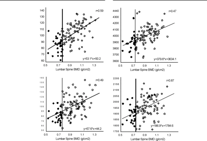
In an elegant study, a group from Switzerland studied 110 post menopausal healthy women to
eval-uate the ability of some peripheral technologies to pre-dict osteoporosis at the spine or the hip at a sensitivity level of 90% (14). Taking into account the results obtained by these different technologies using the same set of individuals, it is easily noticed that the T-score means were absolutely different when all six technologies were compared.
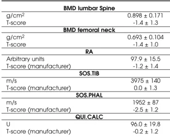
After adjusting each peripheral measurement for 90% sensitivity, according to Central DXA -2.5 SD criteria, four groups of results were then defined: True negatives, true positives, false negatives and false posi-tives. Table 1 shows the comparisons between Radi-ographic Absorptiometry at the Hand, Heel Ultra-sonometry (SOS and Broadband Ultrasound Attenua-tion), Tibial SOS and Phalangeal SOS and Lumbar Spine DXA. In figure 1, the shaded areas represent the false negative and the false positive results, where sub-jects can be misdiagnosed depending on the site and technology used.
Concentrating on the 90% Interval of Confi-dence data (table 1), where less than 10% of false neg-atives were present, it could be concluded that these technologies may be useful to pre-select from 25 to 54% of postmenopausal women in whom axial DXA is indicated to confirm or exclude osteoporosis at spine or hip. Unfortunately, it seems that a 29% difference, depending on the chosen technology and site, seems to be unacceptable.
To be cost-effective, the approach of identifying subjects to be referred to a central DXA scan, should demonstrate a lower cost than performing just central DXAs to all. These calculations are not the same for every country and must be done very carefully.
Table 1.Bone measurements using DXA, RA and QUS of Tibia, Phalanges and Heel (from reference 14).
BMD lumbar Spine
g/cm2 0.898 ± 0.171
T-score -1.4 ± 1.3
BMD femoral neck
g/cm2 0.693 ± 0.104
T-score -1.4 ± 1.0
RA
Arbitrary units 97.9 ± 15.5
T-score (manufacturer) -1.2 ± 1.4
SOS.TIB
m/s 3975 ± 140
T-score (manufacturer) 0.0 ± 1.3
SOS.PHAL
m/s 1952 ± 87
T-score (manufacturer) -2.5 ± 1.2
QUI.CALC
U 96.0 ± 19.8
Picard and collaborators (15) published, in 2004, results obtained with the forearm and phalanges evaluated by DXA and compared them to results obtained with Lumbar Spine and Femoral Neck.
The negative predictive value was nice (table 2), catching more than 95% of the true negatives at an optimal calculated BMD, but the positive predictive value was only ranged from 40 to 43% what is, again, distant from the ideal situation. Even recognizing that the amount of true osteopenic and normal individuals (according to central DXA classification) is high among all the “peripheral false positives individuals”, one question should be made at this point: Is it really important to have a peripheral technology or site that reproduces exactly the same results central DXA does? In general, it is recognized that the elder the subject is, the more likely to have agreement between a peripheral and a central measurement (16). In con-trast, the proposed strategy to use peripheral tech-nologies is (or should be) to improve primary predic-tion. So, it is an important limitation to propose this approach for clinical practice.
In conclusion, screening individuals using peripheral sites and technologies is not completely supported by current evidences. False positives and false negatives are unacceptably high in almost all pub-lished attempts so far.
Potential Application #3: Diagnosis of Osteoporosis
What about peripheral technologies and diagnosis? For almost two decades an uncountable number of experts have been trying to answer this question. We do not intend to answer this question either. It is our intention to show some relevant data and discuss its strengths and weaknesses.
As indicated by the WHO group in 1994 (17), all cut-off criteria for the diagnosis of osteoporosis are arbitrary and far from the ideal situation. This is not unique to bone densitometry. All reference ranges for almost all quantifiable biological variables are defined based on cut-offs that are somewhat arbitrarily estab-lished. So, to determine the reference (or the gold standard) is very important. The WHO did that in
1994, 1999 (18) and reviewed in 2003 (19). The hip (femoral neck or the total hip) and perhaps the lumbar spine (mainly for individuals between 50 and 65), measured by central DXA, are currently the gold stan-dard references for diagnosing purposes.
Each bone or even each region of interest has its own “behavioral” characteristics. The applied mechan-ical forces, amount of cortmechan-ical and trabecular tissue, shape, spatial organization and interactions with its neighbor structures determine the differences. So, each bone has its own personality and as an individual should be evaluated and perhaps diagnosed taking all these particularities into consideration. Our bones have different peak bone mass completion ages, rhythm of bone mass loss. Bone densitometry has dif-ferent accuracies as well as difdif-ferent artifacts that can interfere with each measured bone segment. If several sites of a subject are measured, regardless of the applied cut-off, different classifications will be fre-quently obtained.
Moreover, according to the Picard paper (15), the typical correlation between sites, no matter if they are peripheral or central, ranges between 0.6 and 0.7, leaving from 30 up to 42% of the individuals with dif-ferent classifications when difdif-ferent sites and technolo-gies are used.
As illustrated before, concordances between sites are more common as we become older. The dis-cordances are clearly more frequent in younger groups of individuals, even when central sites are measured. This is a reason that explains the better performance of peripheral DXA when used in elderly people.
The same situation could be demonstrated with Susan Greenspan’s data, published in 1996. The num-ber of subjects classified as having osteoporosis
accord-ing to the WHO definition depends directly on the skeletal site used and even when different regions of interests are used.
In a poster presented at ISCD meeting in New Orleans in 2005 (20), Paul Miller and his group looked at DXA results in a group of 685 women, aged 40 or older, in order to verify the intervention criteria differences (according to the NOF Guidelines) across three different sites. 329 women presented Hip T-Score equal or lower than -2.0 SD. 284 of the 685 women presented the same criteria at the Lumbar Spine. When the forearm was considered, 330 women had the same criteria. Only 170 of these women pre-sented the -2.0 criteria in all three sites and 141 women presented the same situation at only two of the three evaluated sites. About the same number — 151 women — had filled the densitometric criteria for intervention in only one of these sites.
So, according to these results, differences between sites are twice as common as or twice more common than concordances.
Looking again at the NORA results (7), all four technologies used were able to stratify the population but it is easy to see that the amount of individuals included in each diagnostic range differs according to the site/technology used.
Just as an example, in comparison to heel SXA, individuals measured with heel ultrasonometry had somewhat decreased odds of osteoporosis (OR 0.79), while those measured with Forearm or Finger pDXA had increased odds (OR 2.86 and 4.82 respectively).
By the epidemiological prospective, the total amount of fractures, seen in different populations, is not limited to the individuals classified as having osteoporosis. The results of the NORA study have
Table 2.Performance DXA as diagnostic test for presence of osteoporosis at either Lumbar Spine or Femoral Neck (T-score ≤
2.5)* (from reference 15).
Region Measured BMD Sensitivity Specificity False False Positive Negative
(g/cm2) (%) (%) (n) Positive (n) Negative (n) Predictive predictive
value (%) value (%)
Phalanx at 0.436 79 83 124 22 40 97
optimal BMD
Proximal forearm at 0.692 84 79 156 17 36 97
Optimal BMD
Distal forearm at 0.280 90 75 183 11 34 98
optimal BMD
Combined forearm 0.641
at optimal BMD and 83 84 119 18 43 97
0.252
confirmed this situation (although excluding people with previous diagnosis). It is exactly at the “peripher-al osteopenic” groups (between -1 and -2.5 SD at peripheral skeleton) where osteoporotic fractures were concentrated. These data could help on developing specific cut-offs for peripheral sites/technologies.
But what would happen if the NORA sample were reanalyzed using a more specific cut-off for peripheral densitometry, to say, -1.5 or 1.8 SD for osteoporosis?
Anyway, regarding the limitations of peripheral densitometry technologies to diagnose or even to identify individuals that need to be treated, we should point out some other limitations.
The site and age are important factors for bone loss measurement. Comparing the normal reference curves from several bone density measurement tech-nologies and sites, Faulkner and collaborators (21) noted that in some cases as it can be seen for Lateral Spine DXA and QCT Spine, people reach the 2.5 cut-off pretty early in life (around 60 to 65 years old) while when the Total Hip or Heel BMD are measured, this point can never be reached during a typical lifespan.
It can be concluded that, while peripheral den-sitometry has been showing good evidences for frac-ture risk prediction, the diagnosis criteria remains lim-ited to central measures as the standard reference.
Potential Application #4: Monitoring Changes Over Time
Age-related bone loss is a complex process that depends on several factors. It has been demonstrated that, for long bones, cortical bone becomes thinner and the cross sectional area increases, compensating to some extent the bone strength that accompanies aging (22). It is vital to keep in mind that BMD changes in peripheral bones such as phalanx and forearm must be very carefully interpreted, taking this phenomenon into consideration. Changes expected in BUA, SOS or even in the index calculated with these two parameters are far smaller and slower than BMD variations as mea-sured by central DXA (23). While with DXA of the hip, 30% BMD loss is expected from 50 to 70 years of age, only 5 to 15% are observed when ultrasound peripheral technologies are used. In other words, it is necessary to wait 2 to 6 times longer to repeat the test to detect the significant changes. Therefore, it is not practical to monitor the natural evolution of osteo-porosis with peripheral ultrasonometry technologies.
With a cohort of 1,042 postmenopausal women, with mean age of 56.6 years old, 860 white and 182 black (24) adjusted for age and weight, we
compared ultrasonometric results. All demographic parameters were very similar. When the mean stiffness for each age for the two ethnicities was compared, almost no significant difference between the two curves was found and, moreover, the total loss of stiff-ness over a 20-year period, between ages 50 and 70, was only about 12%. These data agree with previous publications showing that for monitoring purposes, a longer interval is necessary to detect bone changes when QUS is used.
Professor Larry Riggs and his group have recently compared the rate of change in BMD in some central and peripheral sites measured by DXA (22) and noted that, as expected, bigger BMD loss was noticed at central skeletal than at distal forearm and distal tibia during a 70-year period, from 20–90. As it can be seen while in the lumbar spine the total volumetric BMD shows a mean of 55% loss from 20 to 90 years of age, during that same interval of time the mean expected loss at the distal radius is 28% and 26% for the tibia.
Monitoring treatment is a very specific applica-bility but, even for treatment monitoring, distal sites are not completely accepted due, mainly, to the fact that the changes expected with the available therapies are also smaller than at central sites. As reviewed by the ORAG group (25), if the forearm was used for moni-toring treatment, only the treatment with Hormonal Therapy and Alendronate for 2 to 4 years could be ade-quately monitored. Peripheral densitometry seems to be inadequate for antiresorptive treatment monitoring. But what should we expect during treatment with bone forming agents? Is peripheral densitometry comparable to central DXA to monitor bone changes with this kind of treatment? If a bone loss at the fore-arm is detected after a 12-month treatment period, should it be considered a “good result” for the treat-ment? Having in mind that this agent increases the cross sectional and, perhaps, the areal size of the fore-arm as well as the bone remodeling rate, it would be very difficult to differentiate a response from an indi-vidual that continues to loose bone density.
CONCLUSIONS
• Peripheral devices are useful for assessing frac-ture risk.
• Peripheral BMD tests other than measure-ment of the 33% (one-third) radius by DXA cannot be used to diagnose osteoporosis.
•Before peripheral BMD testing can be used to screen patients for DXA, device-specific cut-points are needed to define a population in which there is at least 90% sensitivity for osteoporosis by central DXA. Indi-viduals with BMD below these cut-points should be referred to a central DXA testing.
•When central DXA testing is not available, peripheral testing may be considered to identify indi-viduals at high risk for fracture who might benefit from pharmacological therapy.
REFERENCES
1. Röntgen WC. [On a new kind of ray]. Würzburger Physik-Med Ges Jahrg 1895;132.
2. Dennis J. A new system of measurement of bone opac-ity. Dental Cosmos 1897;39:445.
3. Price WA. The Science of Dental Radiology. Dental Cos-mos 1901;43:483.
4. Garn SM. An annotated bibliography on bone densito-metry. Am J Clin Nutr 1962;10:59.
5. Cameron JR, Sorensen J. Measurement of bone mineral in vivo: an improved method. Science 1963;142:230-2. 6. Marshall D, Johnell O, Wedel H. Meta-analysis of how
well measures of bone mineral density predict occur-rence of osteoporotic fractures. BMJ 1996;312:1254-9. 7. Siris ES, Miller PD, Barrett-Connor E, Faulkner KG, Wehren
LE, Abbott TA, et al. Identification and fracture out-comes of undiagnosed low bone mineral density in postmenopausal women: results from the National Osteoporosis Risk Assessment. JAMA 2001;286:2815-22. 8. Cummings SR, Nevitt MC, Browner WS, Stone K, Fox KM,
Ensrud KE, et al. Risk factors for hip fractures in white women. N Eng J Med 1995;332(12):767-73.
9. Nevitt MC, Cummings SR, Stone KL, Palermo L, Black DM, Bauer DC, et al, Risk factors for a first-incident radi-ographic vertebral fracture in women > or = 65 years of age: the study of osteoporotic fractures. J Bone Miner Res 2005;20(1):131.
10. Melton LJ 3rd. Epidemiology of hip fractures: implica-tions of the exponential increase with age. Bone 1996;18(3 suppl.):121S-5.
11. Miller PD, Siris ES, Barrett-Connor E, Faulkner KG, Wehren LE, Abbott TA, et al Prediction of fracture risk in post-menopausal white women with peripheral bone densit-ometry: evidence from the National Osteoporosis Risk Assessment. J Bone Miner Res 2002;17:2222-30.
12. Souza AC. Abstracts presented at the III Brazilian Con-gress of SBDens, SBDens, 1999.
13. Blake GM, Fogelman I. Peripheral or central densitome-try: does it matter which technique we use? J Clin Den-sitom 2001;4(2):83-96.
14. Lippuner K, Fuchs G, Ruetsche AG, Perrelet R, Casez JP, Neto I. How well do radiographic absorptiometry and quantitative ultrasound predict osteoporosis at spine or hip? A cost-effectiveness analysis. J Clin Densitom 2000;3(3):241-9.
15. Picard D, Brown JP, Rosenthall L, Couturier M, Levesque J, Dumont M, et al. Ability of peripheral DXA measure-ment to diagnose osteoporosis as assessed by central DXA measurement. J Clin Densitom 2004;7(1):111-8. 16. Deng HW, Li JL, Li J, Davies KM, Recker RR.
Heterogene-ity of bone mineral densHeterogene-ity across skeletal sites and its clinical implications. J Clin Densitom 1998;1(4):339-53. 17. Kanis JA, Melton LJ 3rd, Christiansen C, Johnston CC,
Khaltaev N. The diagnosis of osteoporosis. J Bone Miner Res 1994;9(8):1137-41.
18. Genant HK, Cooper C, Poor G, Reid I, Ehrlich G, Kanis J, et al. Interim report and recommendations of the World Health Organization Task-Force for Osteoporosis. Osteo-poros Int 1999;10(4):259-64.
19. Kanis JA, et al. Report of a WHO Scientific Group. WHO Technical Report Series, no. 921, 2003.
20. Miller PD, et al. Abstracts from the 2005 ISCD Annual Meeting. J Clin Densitom 2005;8(2):228-49.
21. Faulkner KG, von Stetten E, Miller P. Discordance in patient classification using T-scores. J Clin Densitom 1999;2(3):343-50.
22. Riggs BL, Melton III LJ, Robb RA, Camp JJ, Atkinson EJ, Peterson JM, et al. Population-based study of age and sex differences in bone volumetric density, size, geome-try, and structure at different skeletal sites. J Bone Miner Res 2004;19(12):1945-54.
23. Sahota O, San P, Cawte SA, Pearson D, Hosking DJ. Comparison of the longitudinal changes in quantitative ultrasound with dual-energy X-ray absorptiometry: the four-year effects of hormone replacement therapy.
Osteoporos Int 2000;11(1):52-8.
24. Ragi S. Diferenças étnicas em ultrasonometria de cal-câneo entre mulheres brancas e negras. Diagnóstico en Osteologia, Oct/Nov/Dez, 2001, Buenos Aires.
25. Guyatt GH, Cranney A, Griffith L, Walter S, Krolicki N, Favus M, et al. Summary of meta-analyses of therapies for postmenopausal osteoporosis and the relationship between bone density and fractures. Endocrinol Metab Clin North Am 2002;31(3):659-79.
Address for correspondence:
Sergio Ragi Eis
CEDOES - Centro de Diagnóstico e Pesquisa Rua João da Silva Abreu 78