ABSTRACT
Primary hyperparathyroidism is a common disorder of mineral metabo-lism characterized by incompletely regulated, excessive secretion of parathyroid hormone from one or more of the parathyroid glands. In adults with the disease, a single, benign adenoma is seen approximate-ly 80 percent of the time, with multiple gland involvement comprising most of the remaining patients. Very rarely, a parathyroid cancer is responsible but it is seen in less than 0.5 percent of patients with primary hyperparathyroidism. In this article, we will review important clinical and diagnostic features of asymptomatic primary hyperparathyroidism as well as considerations for surgical or medical management of the dis-ease. (Arq Bras Endocrinol Metab 2006;50/4:647-656)
Keywords: Primary hyperparathyroidism; Osteoporosis; Vitamin D; Bone markers
RESUMO
Hiperparatiroidismo Primário Assintomático.
O hiperparatiroidismo primário é uma distúrbio comum do metabolismo mineral, caracterizado por um excesso de secreção do hormônio das paratiróides, de uma ou mais glândulas paratiroidianas. Nos pacientes adultos portadores da doença, um adenoma único é responsável por aproximadamente 80% dos casos, e o envolvimento de múltiplas glân-dulas pelo restante dos casos. Muito raramente um câncer de paratiróide é a causa, sendo visto em menos de 0,5% dos pacientes com hiperparatiroidismo. Neste artigo, revisaremos as características clínicas e diagnósticas do hiperparatiroidismo primário assintomático, bem como o tratamento clínico e cirúrgico. (Arq Bras Endocrinol Metab 2006;50/4:647-656)
Descritores:Hiperparatiroidismo primário; Osteoporose; Vitamina D; Mar-cadores ósseos
DIAGNOSIS
S
INCE IN PRIMARY HYPERPARATHYROIDISM, parathyroid hormone (PTH) is oversecreted, a reliable immunoassay for PTH is essential. The current-ly available immunoradiometric assay (IRMA) has the requisite sensitivity and specificity, revealing PTH levels that are either frankly elevated or in the upper range of normal. Even though the PTH is sometimes technically in the upper normal range, it is abnormal considering the presence of hyper-calcemia. Moreover, in malignancy, the other most common cause of hypercalcemia, the PTH level is classically suppressed even when the cause of the malignancy-related hypercalcemia is parathyroid hormone related protein (PTHrP). Despite the reliability of the IRMA for PTH in generalJohn P. Bilezikian
Mishaela Rubin
Shonni J. Silverberg
Division of Endocrinology, Department of Medicine, College of Physicians & Surgeons, Columbia University, New York, NY, USA.
use in the United States and throughout the world for over 20 years, it has recently been shown to detect not only the full-length (1-84) PTH molecule, but also forms of PTH foreshortened at the amino terminus (1). The most plentiful of these fragments, constituting up to 50% of the circulating species of PTH is PTH(7-84) (2). A newer IRMA for PTH that measures only the full length PTH molecule (1-84) has promise to be diagnostically more useful than the standard IRMA for PTH, but it remains to be seen whether this will be the case. (2-4). Vieira covers this topic completely in his article in this series. Although the differential diagnosis of hypercalcemia is long, the presumptive diagnosis of primary hyperparathyroidism in subjects whose calcium and PTH levels are elevated is generally accurate as long as the patient is not taking lithium or thiazide diuretics. In familial hypocalciuric hypercalcemia (FHH), the PTH can also be elevated, but it is distin-guished from primary hyperparathyroidism by family history and exceedingly low urinary calcium excretion. Silverberg and Bilezikian, as well as others, have recently observed patients whose PTH is elevated but the serum calcium is normal (5,6). These patients with
normocalcemic primary hyperparathyroidismare distinct from primary hyperparathyroidism that presents with overt hypercalcemia, the far more common clinical form, in which the serum calcium level can occasionally be normal. It is important to rule out all causes of sec-ondary hyperparathyroidism, such as vitamin D defi-ciency and hypercalciuria, in which the parathyroid glands oversecrete PTH through physiologically normal compensatory mechanisms. It is particularly important to be sure that the level of 25-hydroxyvitamin D, the major storage form of vitamin D, is greater than 30 ng/ml the revised new low limit of normal (7, see arti-cle by Bandeira elsewhere in this supplement).
Discussions in this article and others that focus upon asymptomatic primary hyperparathyroidism
gen-erally do not consider patients with normocalcemic primary hyperparathyroidism because we know so lit-tle about them. Some of these patients will develop more overt primary hyperparathyroidism with hyper-calcemia but it is also likely that some of these patients will continue to show elevated levels of PTH without hypercalcemia for years, having achieved a new steady state that does not progress. This article will not address specific issues with regard to normocalcemic primary hyperparathyroidism.
EVALUATION
Skeletal manifestations
In parts of the world where multichannel screening is readily available, primary hyperparathyroidism presents primarily as an asymptomatic disease. The classical skeletal manifestations are uncommon. It this setting, the demonstration of skeletal involvement in asympto-matic primary hyperparathyroidism depends upon dual energy X-ray absorptiometry (DXA). As described by Bandeira in this series, however, there are still regions of the world in which symptomatic disease exists and radiologically manifestations are present. By DXA, the classic pathophysiological effects of PTH are seen, namely to reduce bone density at the distal third of the radius, a site of cortical bone (8). The proclivity of PTH to be catabolic for cortical bone is in contrast to its protective effect on cancellous bone. At the lumbar spine, for example, a site of cancellous bone, bone mineral density tends to be reasonably normal. The hip, which contains a more even admixture of cortical and cancellous elements, shows bone mineral density that is intermediate between the distal 1/3 radius and the lumbar spine (figure 1). Although this classic den-sitometric profile is most commonly seen, a distinctive pattern characterized by vertebral osteopenia can also
Figure 1.The densitometric signature or primary hyperparathyroidism. Bone mineral density is shown at all
three skeletal sites in a cohort of patients with primary hyperparathyroidism. Note preferential involvement
be detected at the time of diagnosis (9). In more severe forms of primary hyperparathyroidism, general-ized reductions in bone density at all sites can be seen. Bone density is a very powerful predictor of fracture risk in osteoporosis. Whether it also predicts fracture risk in primary hyperparathyroidism remains to be seen because well-designed prospective studies are lacking. Surrogate data however raise interesting possibilities that argue for other considerations in primary hyperparathyroidism that may influence the predictive power of bone mineral density in this dis-ease. Histomorphometric analysis of the bone biopsy in primary hyperparathyroidism shows cortical thin-ning, maintenance of cancellous bone volume, and accelerated bone remodeling (10). Bone biopsy stud-ies have also shown that indices of trabecular con-nectivity are actually greater than expected. Even the expected age-dependent loss of cancellous bone is not seen in primary hyperparathyroidism (10,11). Based upon both the densitometric and histomor-phometric data, it might be expected that the corti-cal skeleton is at greater risk for fracture than the can-cellous skeleton. The data are conflicting, however, with some studies showing an increase and other studies no increase in incidence of vertebral fractures (12,13). The retrospective review of a 28-year Mayo Clinic experience concluded that overall fracture risk was increased (14).
If bone density were the only factor to consid-er, the distal forearm would be the site at greatest risk and the lumbar spine would be the site at smallest risk. Since PTH has important effects on other qualities of bone, such as bone size and microarchitecture, reduc-tions in bone mineral density may not directly relate to fracture risk. For example, cortical thinning through PTH-mediated endosteal resorption might be com-pensated in primary hyperparathyroidism by periosteal apposition, leading to bone that may be increased in cross sectional diameter as demonstrated by some (15) but not by others (16) using pQCT technology. In primary hyperparathyroidism, if there is an increase in cross sectional area of bone, it would be stronger, even though there is cortical thinning. Additionally, as described above, it is important to consider the gener-ally well-preserved cancellous microarchitecture in pri-mary hyperparathyroidism, another factor that might be protective. Thus, in primary hyperparathyroidism, certain skeletal features tend to compete with each other: cortical thinning favoring an increased fracture risk; increased bone size and preserved microarchitec-ture favoring a reduction in fracmicroarchitec-ture risk (17).
Renal involvement
Kidney stones remain the most common manifestation of primary hyperparathyroidism, but modern inci-dence figures of 15–20% are lower than they used to be. Other renal manifestations of primary hyperpara-thyroidism include hypercalciuria, which is seen in approximately 40 percent of patients, and nephrocalci-nosis, the frequency of which is unknown. An unex-plained reduction in the creatinine clearance has also been regarded to be a potential renal manifestation of primary hyperparathyroidism.
Other organ involvement
Neurologic and cognitive signs or symptoms
Still vexing are the nonspecific complaints of patients with primary hyperparathyroidism such as weakness and easy fatigability (18). These complaints must be distinguished from the rare classic neuromuscular syn-drome in which type II muscle cell dysfunctional is apparent (19). Nevertheless, nonspecific complaints of patients are noteworthy. They are often accompanied by some degree of constitutional, behavioral and/or psychiatric symptomatology. Prospective studies have reported apparent improvement after successful parathyroid surgery (20,21), while others have not been able to document changes postoperatively (21,22). This issue remains unsettled.
Cardiovascular system
Interest in the effect of primary hyperparathyroidism on cardiovascular function is rooted in pathophysio-logic observations of hypercalcemia in which hyper-tension, left ventricular hypertrophy and arrhythmias are common (23). When primary hyperparathyroidism is more severe, cardiovascular involvement has been noted, particularly in some European centers (24). This discussion excludes the multiple endocrine neo-plasia (MEN) syndromes in which the presence of pheochromocytoma or hyperaldosteronism could, of course, lead to cardiovascular manifestations. Whether any cardiovascular abnormalities are demonstrable in mild asymptomatic primary hyperparathyroidism will require highly sensitive measurements of cardiovascu-lar function that are currently ongoing (25,26).
classic presentations of primary hyperparathyroidism overtly involving the skeleton and the kidneys are described as common occurrences (27). Lack of rou-tine screening tests helps, in part, to explain these find-ings but it is not an adequate explanation by itself. Vit-amin D deficiency is also common in countries where primary hyperparathyroidism is a symptomatic disor-der, an observation that fits with the proposal made years ago by Lumb and Stanbury that primary hyper-parathyroidism is worse in the presence of vitamin D deficiency (28). Even in mild, asymptomatic primary hyperparathyroidism, it has been shown that low 25(OH)D levels are associated with increased indices of disease activity (29). In these patients with primary hyperparathyroidism, it seems reasonable to consider restoring vitamin D to sufficient levels. By restoring vitamin D levels to normal, that component of the increased PTH secretory drive would be ameliorated. Despite the attractiveness of this approach, providing vitamin D to these individuals could raise their serum calcium further. To address this question, Grey et al. (30) recently reported on 21 patients with mild pri-mary hyperparathyroidism whose 25(OH)D levels were < 20 ng/ml. After vitamin D repletion in which the mean 25-hydroxyvitamin D level rose from 11 ± 5 ng/ml to 31 ± 6 ng/ml after 12 months, the serum PTH level declined by 25 percent without any change in the serum calcium concentration. This report pro-vides some support for the hypothesis that vitamin D deficiency worsens the hyperparathyroid state and vit-amin D repletion improves some biochemical features of the disease. If subjects are to be given vitamin D, it should be done with care and at low doses.
TREATMENT OPTIONS IN ASYMPTOMATIC PRIMARY HYPERPARATHYROIDISM
In the opinion of most experts, it is quite reasonable to question the advisability of surgery in all patients with asymptomatic disease. Since many patients with primary hyperparathyroidism are known to have the disease only because an incidental serum calcium determination was obtained, it is possible the disease would have remained unrecognized without the serum calcium measurement was made. Silverberg and Bilezikian have shown, in fact, that some patients with asymptomatic primary hyperparathyroidism have a very uneventful course for many years (31). Given this observation, non-surgical approaches would appear to be a reasonable alternative in some patients. On the other hand, asymptomatic patients may have levels of
serum and/or urinary calcium that are sufficiently above normal to give concern as to the wisdom of fol-lowing them long term. In addition, if bone mineral density measurements are low, they could be at increased fracture risk.
The management of asymptomatic primary hyperparathyroidism has been addressed at two NIH conferences, one held in 1990 (32) and another more recently in 2002 (33). At the more recent conference (33), the 1990 guidelines were revised modestly from those resulting from the earlier conference (32). The latest guidelines for surgery are noted here for asymp-tomatic patients: (1) serum calcium concentration greater than 1 mg/dl above the upper limits of nor-mal; (2) marked hypercalciuria (> 400 mg/day) or reduction in creatinine clearance by more than 30 per-cent below age- and sex-matched reference values; (3) markedly reduced bone density (T-score < -2.5 at any site); and (4) age less than 50 years. Table 1 summa-rizes these guidelines. The intent of the guidelines is to define a surgical candidate if 1 of these guidelines is met. The patient does not have to meet all or even more than 1. Although these guidelines have been very useful for clinicians who are faced with the dilem-ma: to operate or not, it should be noted that surgery for asymptomatic patients who do not meet any of these guidelines is not necessarily ill advised. Surgery for asymptomatic primary hyperparathyroidism is always a valid option as long as there are no medical contraindications.
Surgery
An expert parathyroid surgeon will find all abnormal parathyroid tissue 95 percent of the time (34). Advances in surgery have popularized the minimally invasive procedure under local anesthesia. It requires successful preoperative localization of the abnormal parathyroid gland and capability to measure PTH rapidly in the operating room (35). A preoperative blood sample is obtained for comparison of the PTH concentration with an intraoperative sample obtained minutes after removal of the “abnormal” parathyroid gland. If the PTH level falls by more than 50 percent immediately following resection, the gland that has
Table 1. Indications for surgery in asymptomatic primary
hyperparathyroidism [from reference #33].
Serum calcium > 1.0 mg/dL (above normal)
24-h Urinary Ca > 400 mg/24 hours Creatinine clearance >30% below expected Bone density T-score < -2.5 at any site
been removed is considered to be the sole source of overactive parathyroid tissue and the operation is ter-minated. If the PTH level does not fall by more than 50 percent, the operation is extended to a more tradi-tional one in a search for other overactive parathyroid tissue. With advances in imaging technology and growing experience with minimally invasive parathy-roid surgery, it is likely that these newer approaches will become more widely used (36).
Results of surgery
Successful surgery cures primary hyperparathyroidism. Long-term follow up of these patients indicates a 10 to 12 percent improvement in bone density at the lumbar spine and femoral neck over 10 years (31,37). In patients who have nephrolithiasis, surgery is of clear benefit in reducing the incidence of recurrent stones (38). Vague or constitutional symptoms may or may not improve after surgery, while hypertension and pep-tic ulcer disease, if present, are unlikely to remit. Recent-ly, several cohorts have been observed with respect to changes in fracture incidence following successful parathyroid surgery. Vestergaard and Mosekilde report that fracture incidence might be reduced after parathy-roid surgery (39). It is not clear how comparable such cohorts of operated patients are with subjects who were followed conservatively, without surgery (40).
CONSERVATIVE MANAGEMENT OF ASYMPTOMATIC HYPERPARATHYROIDISM
If the guidelines offered by the expert panel convened after the Workshop on Aymptomatic Primary hyparathyroidism are followed (33), about 40–50 per-cent of patients with primary hyperparathyroidism in the United States will fit into the non-surgical, con-servative management category. The natural history of this cohort indicates that over a 10-year period,
patients in general are stable (40). The group data show that serum and urine biochemical parameters do not change. Similarly, in the majority of patients, bone mineral density is unchanged at lumbar spine, hip and distal radius after 10 years. When these data for the group as a whole are analyzed more closely, however, it is clear that some patients who are followed without surgery will show changes and thus meet criteria for surgery. In about 25 percent of these patients followed without surgery over 10 years, guidelines for surgery were met eventually by virtue of an increasing serum or urinary calcium or declining bone mineral density (40). The fact that an appreciable number of asympto-matic patients will progress to meet criteria for surgery emphasizes the need for all these patients to be moni-tored.
Monitoring asymptomatic patients who are not to undergo surgery
A plan for monitoring was offered by the Panel of the 2002 Workshop on Asymptomatic Primary Hyper-parathyroidism and is shown in table 2. A serum calci-um measurement should be obtained every six months. Once the urinary calcium has been ascer-tained to be acceptable, the urinary calcium excretion is not measured on a regular basis. The panel empha-sized the utility of measuring annually bone density at the lumbar spine, hip and distal 1/3 radius site.
Medical management
Hydration
It is important that patients maintain adequate hydra-tion, particularly in summer and warm climates when fluid losses can be substantial. Thiazide diuretics are to be avoided because they can worsen hypercalcemia.
Diet
Many patients are advised by their physicians to limit their dietary calcium intake. A counter argument,
how-Table 2. Monitoring asymptomatic primary hyperparathyroidism [from
refer-ence #33].
Measurement Guidelines
Serum calcium Semiannually
Urinary Ca Not recommended*
Creatinine Clearance Not recommended*
Serum Creatinine Annually
Bone density Annually (3-sites)
Abdominal X-ray ± ultrasound Not recommended*
ever, suggests that low calcium diets could lead to fur-ther increases in PTH levels (41). Given the fact that patients with primary hyperparathyroidism maintain some sensitivity to ambient calcium concentrations, even though the sensitivity is impaired, diets restricted in calcium could fuel processes associated with increased production of PTH. The logic of this argu-ment could lead to diets that are enriched in calcium with the idea that such a challenge could suppress PTH levels in primary hyperparathyroidism, as shown by Insogna et al. (42) Levels of 1,25(OH)D have to be taken into account in this discussion, since patients with primary hyperparathyroidism typically have con-centrations of this active metabolite that are at or above the upper limit of normal. The data of Locker et al. (43) are of interest in this regard. In subjects with ele-vated levels of 1,25(OH)2D3, high calcium diets were
associated with worsening hypercalciuria.
The prudent advice is that dietary calcium intake should be in the lower end of the range that is generally recommended for the non-hyperparathyroid population, namely approximately 1000 mg/day. If the 1,25(OH)2D level is increased, calcium intake
should be more limited. Similarly, calcium-enriched diets to levels greater than 1500 mg should also be avoided.
Pharmacological approaches
In certain patients, pharmacological approaches to the management of hyperparathyroidism are particularly desirable, such as those who meet guidelines for parathyroid surgery but refuse surgery or have medical contraindications. For those who do not meet surgical guidelines, approaches that safely lower serum calcium or increase bone density are also attractive. These pharmacological approaches to control hypercalcemia are additionally useful in patients with parathyroid car-cinoma when repeated attempts to resect malignant tissue have ultimately failed.
Selective Estrogen Receptor Modulators
(SERMS)
Rubin et al. (44) studied 18 postmenopausal women with primary hyperparathyroidism. They were ran-domly allocated to an 8-week course of raloxifene (60 mg/day) or placebo. There was a 4-week follow up period off therapy. In the raloxifene group, the aver-age serum calcium fell significantly from 10.8 ± 0.2 mg/dl to 10.4 ± 0.2 mg/dl (P< 0.05). Along with the reduction in serum calcium, markers of bone forma-tion and bone resorpforma-tion fell significantly. Raloxifene administration was not associated with any changes in
serum PTH, 1,25(OH)2D or urinary calcium
excre-tion (44). The placebo group did not show any change in serum calcium or in bone turnover markers. In a open pilot study of only three patients, but carried out for a longer period of time, one year, Zanchetta and Bogado (45) showed a similar reduction in serum cal-cium with Raloxifene and increases in bone mineral density of the lumbar spine and femoral neck.
Bisphosphonates
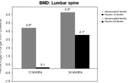
The conceptual basis for expecting that bisphospho-nates have potential as a medical approach to primary hyperparathyroidism is evident. Primary hyperparathy-roidism, even in patients who are asymptomatic, is fre-quently associated with increases in bone turnover (46). By reducing bone resorption, without affecting PTH secretion directly, bisphosphonates could reduce serum and urinary calcium levels. An additional poten-tial benefit of bisphosphonates in primary hyper-parathyroidism would be to increase bone mineral density. Two well-controlled double-blinded random-ized clinical trials have been conducted (47,48). The study of Khan et al. (48) was a randomized, placebo-controlled, study of 44 patients who were adminis-tered either alendronate 10 mg daily or placebo. In comparison to baseline, treatment with alendronate was associated with a significant 7% increase (P< 0.001) in bone mineral density of the lumbar spine after two years. Total hip bone mineral density also increased significantly in comparison to baseline by 4% (P< 0.001). As expected, bone turnover markers fell significantly in the group that received alendronate. There were no changes in serum or urine calcium or serum PTH in either group (figure 2). The only major difference between this experience and that of Chow et al. (47) was a significant alendronate-associated reduction in the serum calcium, by 0.34 mg/dl (P< 0.02). These encouraging results suggest there may be a role for bisphosphonate therapy, especially in patients who are poor operative risks.
Calcimimetics
gained with the calcimimetic, cinacalcet. This agent has been studied in the secondary roidism of renal failure, in primary hyperparathy-roidism and in parathyroid cancer (49). Shoback et al. (50) studied 22 patients with primary hyper-parathyroidism in a dose-ranging study. In all dose groups, ranging from 30–50 mg twice daily, cinacal-cet was associated with a normalization of the serum calcium after the second dose and remained within normal limits for the entire 2-week period. Reduc-tions in PTH, over 50 percent, occurred 2 to 4 hours after dosing in all cinacalcet-treated groups. There were no significant changes in urinary calcium excretion. Both serum calcium and PTH returned toward baseline by seven days after cinacalcet was stopped. Peacock et al. (51) have recently reported their experience in a longer study of cinacalcet. This multicenter, randomized, double-blind, placebo-controlled trial was designed to evaluated the longer term actions of cinacalcet in 78 patients with prima-ry hyperparathyroidism. Cinacalcet was titrated from 30–50 mg twice daily during a 12-week period lowed by a 12-week maintenance and 28-week fol-low-up period. Most patients treated with cinacalcet achieved normocalcemia, the primary endpoint (fig-ure 3). Normal calcium concentrations were main-tained for the entire duration of the study. Modest, but significant, reductions in the PTH concentration were observed in the cinacalcet group. Similar to the
study of Shoback, PTH levels fell quickly within hours after the administration of drug.
CONCLUSIONS
As experience with asymptomatic primary hyperparathy-roidism has grown over the past three decades, new approaches in diagnosis, evaluation, and treatment have emerged. Greater knowledge about the natural history of this disorder suggests that certain patients can be safely followed without surgery. On the other hand, in all patients with the disease, if medical status permits, surgery is always an acceptable option, especially with improvements in minimally invasive parathyroidectomy and the knowledge that bone mineral density improves significantly as a result of surgical correction of the dis-ease. Revised guidelines should help to direct physicians and patients in their choice of treatment options while balancing risks and benefits of the decision made. Increasing experience with specific pharmacological treatments shows promise as yet another alternative to surgery or simple medical monitoring.
ACKNOWLEDGMENTS
This work was supported in part by a grant from the National Institutes of Health (NIDDK 32333).
Figure 2.Alendronate in primary hyperparathyroidism. Bone mineral density (BMD) of the lumbar spine in patients treated with
alendronate. The group that was treated initially showed a significant increase after 1 year, which was maintained in year 2. In the group that received placebo, there were significant gains only when they were crossed over to alendronate in year 2. Significance from baseline is noted by the asterisks. [Adapted from reference #48]
Percentage
change
from
REFERENCES
1. Lepage R, Roy L, Brossard JH, Rousseau L, Dorais C, Lazure C, et al. A non (1-84) circulating parathyroid hor-mone (PTH) fragment interferes significantly with intact PTH commercial assay measurements in uremic sam-ples. Clin Chem 1998;44:805-9.
2. D’Amour P, Brossard J-H, Rousseau L, Nguyen-Yamamo-to L, Nassif E, Lazure C, et al. Structure of non-(1-84) PTH fragments secreted by parathyroid glands in primary and secondary hyperparathyroidism. Kidney Int. 2005;68:998-1007.
3. Gao, P, Scheibel S, D’Amour P, John MR, Rao SD, Schmidt-Gayk H, et al. Development of a novel immunoradiometric assay exclusively for biologically active whole parathyroid hormone 1-84. Implications for improvement of accurate assessment of parathyroid function. J Bone Miner Res 2001;16:605-14.
4. Silverberg SJ, Brown I, LoGerfo P, Gao P, Cantor T, Bilezikian JP. Clinical utility of an immunoradiometric assay for whole PTH (1-84) in primary hyperparathy-roidism. J Clin Endocrinol Metab 2003;88:4725-30.
5. Maruani G, Hertig A, Paillard M, Houillier P. Normocal-cemic primary hyperparathyroidism: evidence for a generalized target-tissue resistance to parathyroid hor-mone. J Clin Endocrinol Metab 2003;88:4641-8.
6. Silverberg SJ, Bilezikian JP. “Incipient” primary hyper-parathyroidism: a “forme fruste” of an old disease. J Clin
Endocrinol Metab 2003;88:5348-52.
7. Heaney RP. The vitamin D requirement in health and dis-ease. J Steroid Biochem Molec Biol 2005;97:13-9.
8. Silverberg SJ, Shane E, de la Cruz L, Dempster DW, Feld-man F, Seldin D, et al. Skeletal disease in primary hyper-parathyroidism. J Bone Miner Res 1989;4(2):283-91.
9. Silverberg SJ, Locker FG, Bilezikian JP. Vertebral osteope-nia: A new indication for surgery in primary hyperparathy-roidism. J Clin Endocrinol Metab 1996;81:4007-12.
10. Parisien M, Silverberg SJ, Shane E, de la Cruz L, Lindsay R, Bilezikian JP, et al. The histormorphometry of bone in pri-mary hyperparathyroidism: Preservation of cancellous bone structure. J Clin Endocrinol Metab 1990;70:930-8.
11. Dempster DW, Parisien M, Silverberg SJ, Liang XG, Schnitzer M, Shen V, et al. On the mechanism of can-cellous bone preservation in postmenopausal women with mild primary hyperparathyroidism. J Clin Endocrinol
Metab 1999;84:1562-6.
12. Wilson RJ, Rao S, Ellis B, Kleerekoper M, Parfitt AM. Mild asymptomatic primary hyperparathyroidism is not a risk factor for vertebral fractures. Ann Intern Med 1988;109:959-62.
13. Larsson K, Ljunghall S, Krusemo UB, Naessen T, Lindh E, Persson I. The risk of hip fractures in patients with primary hyperparathyroidism: A population-based cohort study with a follow-up of 19 years. J Intern Med 1993 ;234:585-93.
14. Khosla S, Melton LJ 3rd, Wermers RA, Crowson CS, O’Fal-lon W, Riggs B. Primary hyperparathyroidism and the risk of fracture: A population-based study. J Bone Miner Res 1999;14:1700-7.
15. Chen Q, Kaji H, Iu M-F, Nomur R, Sowa H, Yamauchi M, et al. Effects of an excess and a deficiency of endoge-nous parathyroid hormone on volumetric bone mineral density and bone geometry determined by peripheral quantitative computed tomography in female subjects.
J Clin Endocrinol Metab 2003;88:4655-8.
16. Charopoulos I, Tournis S, Trovas G, Raptou P, Kaldrymides P, Skarandavos G, et al. Effect of primary hyperparathyroidism on volumetric bone mineral densi-ty assessed by peripheral quantitative computed tomography in postmenopausal women. J Clin
Endocrinol Metab 2006;91(5):1748-53.
17. Bilezikian JP. Bone strength in primary hyperparathy-roidism. Osteoporos Int 2003;14(suppl 5):5113-7.
18. Silverberg SJ. Non-classical target organs in primary hyperparathyroidism. J Bone Min Res 2003;17(suppl 2):N117-25.
Figure 3.Cinacalcet in primary hyperparathyroidism. Serum calcium of a group of subjects is shown
19. Patten BM, Bilezikian JP, Mallette LE, Prince A, Engel WK, Aurbach GD. The neuromuscular disease of hyper-parathyroidism. Ann Intern Med 1974;80:182-94.
20. Solomon BL, Schaaf M, Smallridge RC. Psychologic symptoms before and after parathyroid surgery. Am J
Med 1994;96:101-6.
21. Talpos GB, Bone HG, Kleerekoper M, Phillips ER, Alam M, Honasoge M, et al. Randomized trial of parathyroidec-tomy in mild asymptomatic primary hyperparathy-roidism: Patient description and effects on the SF-36 health survey. Surgery 2000;128:1013-20.
22. Brown GG, Preisman RC, Kleerekoper MD. Neurobehav-ioral symptoms in mild hyperparathyroidism: related to hypercalcemia but not improved by
parathyroidecto-my. Henry Ford Med J 1987;35:211-5.
23. Stefenelli T, Abela C, Frank H, Koller-Strametz J, Globits S, Berger-Klein J, et al. Cardiac abnormalities in patients with PHPT: Implications for follow-up. J Clin Endocrinol
Metab 1997;82:106-12.
24. Stefenelli T, Mayr H, Berger-Klein J, Globits S, Wolosczuk W, Niederle B. Primary hyperparathyroidism: incidence of cardiac abnormalities and partial reversibility after suc-cessful parathyroidectomy. Am J Med 1993;95:197-202.
25. Silverberg SJ. Non-classical target organs in primary hyperparathyroidism. J Bone Miner Res 2002;17(suppl 2):N117-25.
26. Smith JC, Page MD, Wheeler MH, Cockroft JR, Scanlon MF, Davies JS. Augmentation of central arterial pressure in mild primary hyperparathyroidism. J Clin Endocrinol
Metab 2000;85:3515-9.
27. Rubin MR, Bilezikian JP, Silverberg SJ. Vascular stiffness is increased in patients with primary hyperparathyroidism.
J Bone Mineral Res 2002;17:S381.
28. Harinarayan DV, Gupta N, Kochupillai N. Vitamin D sta-tus in primary hyperparathyroidism in India. Clin
Endocrinol 1995;43:351-8.
29. Lumb GA, Stanbury SW. Parathyroid function in vitamin D deficiency in primary hyperparathyroidism. Am J Med 1974;54:833-9.
30. Silverberg SJ, Shane E, Dempster DW, Bilezikian JP. Vita-min D deficiency in primary hyperparathyroidism. Am J
Med 1999;107:561-7.
31. Grey A, Lucas J, Horne A, Gamble G, Davidson JS, Reid I. Vitamin D repletion in patients with primary hyper-parathyroidism and coexistent vitamin D insufficiency. J
Clin Endocrinol Metab 2005;90:2122-6.
32. Bilezikian JP, Silverberg SJ. Management of asympto-matic primary hyperparathyroidism. N Eng J Med 2004;350:1746-51.
33. National Institutes of Health. Consensus development conference statement on primary hyperparathyroidism.
J Bone Miner Res 1991;6:S9-13.
34. Bilezikian JP, Potts JT Jr, El-Hajj Fuleihan G, Kleerekoper M, Neer R, Peacock M, et al. Summary statement from a workshop on asymptomatic primary hyperparathy-roidism: a perspective for the 21st century. J Bone Min
Res 2002;17(suppl 2):N2-11.
35. Clark OH. How should patients with primary hyper-parathyroidism be treated? J Clin Endocrinol Metab. 2003;88:3011-4.
36. Udelsman R, Donovan POI, Sokoll LT. One hundred con-secutive minimally invasive parathyroid explorations.
Ann Surg 2000;232:331-9.
37. Gallagher SF, Denham DW, Murr MM, Norman JG. The impact of minimally invasive parathyroidectomy on the way endocrinologists treat primary hyperparathy-roidism. Surgery 2003;134:910-7.
38. Nomura R, Sugimoto T, Tsukamoto R, Yamauchi M, Sowa H, Chen Q, et al. Marked and sustained increase in bone density after parathyroidectomy in patients with primary hyperparathyroidism: a six-year longitudinal study with or without parathyroidectomy in a Japanese population. Clin Endocrinol (Oxf) 2004;60:335-42.
39. Deaconson TF, Wilson SD, Lemann J. The effect of parathyroidectomy on the recurrence of nephrolithiasis.
Surgery 1987;215:241-51.
40. Vestergaard P, Mosekilde L. Parathyroid surgery is asso-ciated with a decreased risk of hip and upper arm frac-tures in primary hyperparathyroidism: a controlled cohort study. J Internal Med 2004;255:108-18.
41. Silverberg SJ, Shane E, Jacobs TP, Siris E, Bilezikian JP. Pri-mary hyperparathyroidism: 10-year course with or with-out parathyroid surgery. N Engl J Med 1999;341:1249-55.
42. Barger-Lux MJ, Heaney RP. Effects of calcium restriction on metabolic characteristics of premenopausal women. J Clin Endocrinol Metab 1993;76:103-7.
43. Insogna KL, Mitnick ME, Stewart AF, Burtis WJ, Mallette LE, Broadus AE. Sensitivity of the parathyroid hormone-1, 25-dihydroxyvitamin D axis to variations in calcium intake in patients with primary hyperparathyroidism. N Engl J Med 1985;313:1126-30.
44. Locker FG, Silverberg SJ, Bilezikian JP. Optimal dietary calcium intake in primary hyperparathyroidism. Am J
Med 1997;102:543-50.
45. Rubin MA, Lee KH, McMahon DJ, Silverberg SJ. Ralox-ifene lowers serum calcium and markers of bone turnover in postmenopausal women with primary hyper-parathyroidism. J Clin Endocrinol Metab 2003;88:1174-8.
46. Zanchetta JR, Bogado CE. Raloxifene reverses bone loss in postmenopausal women with mild asymptomatic pri-mary hyperparathyroidism. J Bone Min Res 2001 ;16:189-90.
47. Silverberg SJ, Bilezikian JP. Primary hyperparathyroidism. In: Seibel MJ, Robins SP, Bilezikian JP (eds). Dynamics of
Bone and Cartilage Metabolism. San Diego: Elsevier
Press, 2006.
48. Chow CC, Chan WB, Li JKY, Chan NN, Chan MHM, Ko GTC, et al. Oral alendronate increases bone mineral density in postmenopausal women with primary hyper-parathyroidism. J Clin Endocrinol Metab 2003;88 (2):581-7.
49. Khan AA, Bilezikiam JP, Kung AW, Ahmed MM, Dubois SJ, Ho AY, et al. Alendronate in primary hyper-parathyroidism: a double-blind, randomized, place-bo-controlled trial. J Clin Endocrinol Metab 2004; 89:3319-25.
51. Shoback DM, Bilezikian JP, Turner SA, McCary LC, Guo MD, Peacock M. The calcimimetic AMG 073 normalizes serum calcium in patients with primary hyperparathy-roidism. J Clin Endocrinol Metab 2003;88:5644-9.
52. Peacock M, Bilezikian JP, Klassen PS, Guo MD, Turner SA, Shoback DM. Cinacalcet hydrochloride maintains long-term normocalcemia in patients with primary hyper-parathyroidism. J Clin Endocrinol Metab 2005;90:135-41.
Address for correspondence:
John P. Bilezikian Division of Endocrinology Department of Medicine
Columbia University College of Physicians & Surgeons 630 W. 168th Street
New York, NY 10032 Fax: 212-305-6486

![Table 1. Indications for surgery in asymptomatic primary hyperparathyroidism [from reference #33].](https://thumb-eu.123doks.com/thumbv2/123dok_br/19016780.469810/4.892.460.806.1004.1095/table-indications-surgery-asymptomatic-primary-hyperparathyroidism-reference.webp)
![Table 2. Monitoring asymptomatic primary hyperparathyroidism [from refer- refer-ence #33].](https://thumb-eu.123doks.com/thumbv2/123dok_br/19016780.469810/5.892.215.678.931.1052/table-monitoring-asymptomatic-primary-hyperparathyroidism-refer-refer-ence.webp)