ABSTRACT
With the ageing population in most countries, disorders of bone and mineral metabolism are becoming increasingly relevant to every day clinical practice. Consequently, the interest in, and the need for effec-tive measures to be used in the screening, diagnosis and follow-up of such pathologies have markedly grown. Together with clinical and imag-ing techniques, biochemical tests play an important role in the assess-ment and differential diagnosis of metabolic bone disease. In recent years, the isolation and characterisation of cellular and extracellular components of the skeletal matrix have resulted in the development of molecular markers that are considered to reflect either bone formation or bone resorption. These biochemical indices are non-invasive, com-paratively inexpensive and, when applied and interpreted correctly, helpful tools in the diagnostic and therapeutic assessment of metabolic bone disease. This review provides an overview of the current evidence regarding the clinical use of biochemical markers of bone remodelling in bone disease, with an emphasis on osteoporosis. (Arq Bras Endocrinol
Metab 2006;50/4:603-620)
Keywords:Remodelling; Markers; Osteoporosis; Fracture; Treatment
RESUMO
Marcadores Bioquímicos da Remodelação Óssea.
Tendo em vista o crescimento da população idosa na maioria dos paí-ses, os distúrbios do metabolismo ósseo e mineral estão tornando-se cada vez mais relevantes na prática clínica diária. Conseqüentemente, o interesse e a necessidade de medidas efetivas para serem usadas no rastreamento, diagnóstico e seguimento de tais patologias vêm crescendo acentuadamente. Além da avaliação clínica e de técnicas de imagens, os marcadores bioquímicos desempenham um importante papel na avaliação e diagnóstico das doenças ósseas metabólicas. Recentemente, a melhor caracterização dos componentes intracelu-lares e extraceluintracelu-lares da matriz óssea resultou no desenvolvimento dos marcadores moleculares, os quais refletem tanto a formação como a reabsorção óssea. Estes marcadores bioquímicos não são invasivos e comparativamente são de mais baixo custo, e quando aplicados e interpretados corretamente são instrumentos úteis no diagnóstico e tratamento das doenças ósseas metabólicas. Esta revisão abordará evidências atuais, levando em consideração o uso clínico dos mar-cadores bioquímicos da remodelação óssea nas doenças metabólicas
ósseas, com ênfase na osteoporose. (Arq Bras Endocrinol Metab
2006;50/4:603-620)
Descritores:Remodelação; Marcadores; Osteoporose; Fratura; Tratamento.
Markus J. Seibel
ANZAC Research Institute &
Dept. of Endocrinology,
University of Sydney at
Concord Campus,
Australia.
BACKGROUND
B
ONE IS A METABOLICALLY ACTIVEtissue and
under-goes continuous remodelling, a process that largely
relies on the activity of osteoclasts to remove bone and
of osteoblasts to form bone. Under normal conditions,
bone resorption and formation are coupled to each
other, and the long-term maintenance of skeletal
bal-ance is achieved through the action of systemic
hor-mones and local mediators. In contrast, metabolic bone
diseases, states of increased or decreased mobility, and
therapeutic interventions are characterised by more or
less pronounced imbalances in bone turnover (1,2).
With the increasing awareness of disorders of
bone and mineral metabolism in clinical practice, the
interest in, and the need for effective measures to be
used in the screening, diagnosis and follow-up of such
pathologies have markedly grown. Along with clinical
and imaging techniques, laboratory tests play an
inte-gral role in the assessment and differential diagnosis of
metabolic bone disease.
In recent years, the isolation and
characterisa-tion of cellular and extracellular components of the
skeletal matrix have resulted in the development of
biochemical markers that specifically reflect either
bone formation or bone resorption [for review: (3)].
These biochemical indices have greatly enriched the
spectrum of analytes used in the assessment of
skele-tal pathologies. They are non-invasive, comparatively
inexpensive and, when applied and interpreted
cor-rectly, helpful tools in the diagnostic and therapeutic
assessment of metabolic bone disease. Although the
various serum and urinary markers of bone turnover
include both cellular derived enzymes and
non-enzy-matic peptides, they are usually classified according
to the metabolic process they are considered to
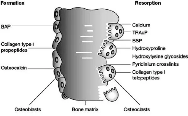
reflect. For clinical purposes, therefore, markers of
bone formation are distinguished from indices of
bone resorption (figure 1, table 1). It should be born
in mind, however, that some of these compounds
may reflect, at least in part, both bone formation and
resorption (e.g., urinary hydroxyproline). Also, most
marker components are present in other tissues than
bone and may therefore be influenced by
non-skele-tal processes. Thirdly, changes in bone markers are
usually not disease specific, but reflect alterations in
skeletal metabolism independent of the underlying
cause.
This review provides an overview of the current
evidence regarding the clinical use of biochemical
markers of bone remodelling in bone disease, with an
emphasis on osteoporosis.
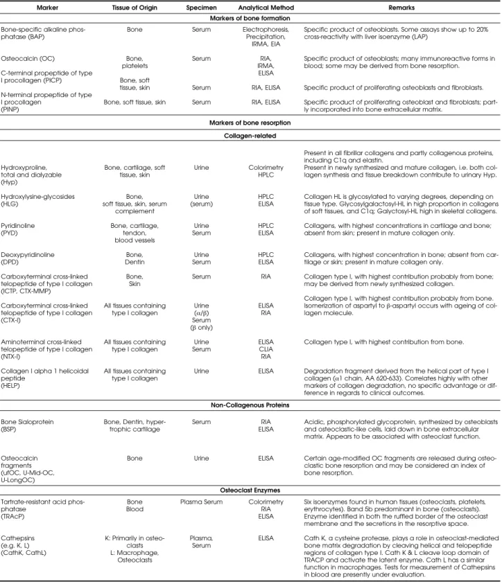
Marker
Bone-specific alkaline phos-phatase (BAP)
Osteocalcin (OC)
C-terminal propeptide of type I procollagen (PICP)
N-terminal propeptide of type I procollagen
(PINP)
Hydroxyproline, total and dialyzable (Hyp) Hydroxylysine-glycosides (HLG) Pyridinoline (PYD) Deoxypyridinoline (DPD) Carboxyterminal cross-linked telopeptide of type I collagen (ICTP, CTX-MMP)
Carboxyterminal cross-linked telopeptide of type I collagen (CTX-I)
Aminoterminal cross-linked telopeptide of type I collagen (NTX-I)
Collagen I alpha 1 helicoidal peptide (HELP) Bone Sialoprotein (BSP) Osteocalcin fragments (ufOC, U-Mid-OC, U-LongOC)
Tartrate-resistant acid phos-phatase
(TRAcP)
Cathepsins (e.g. K, L) (CathK, CathL)
Tissue of Origin
Bone
Bone, platelets
Bone, soft tissue, skin
Bone, soft tissue, skin
Bone, cartilage, soft tissue, skin
Bone, soft tissue, skin, serum
complement Bone, cartilage, tendon, blood vessels Bone, Dentin Bone, Skin
All tissues containing type I collagen
All tissues containing type I collagen
All tissues containing type I collagen
Bone, Dentin, hyper-trophic cartilage
Bone
Bone Blood
K: Primarily in osteo-clasts L: Macrophage, Osteoclasts Specimen Serum Serum Serum Serum Urine Urine (serum) Urine Serum Urine Serum Serum Urine
(α/β)
Serum
(βonly)
Urine Serum Urine Serum Urine Plasma Serum Plasma, Serum Analytical Method Electrophoresis, Precipitation, IRMA, EIA RIA, IRMA, ELISA RIA, ELISA RIA, ELISA Colorimetry HPLC HPLC ELISA HPLC ELISA HPLC ELISA RIA ELISA RIA ELISA CLIA RIA ELISA RIA ELISA ELISA Colorimetry RIA ELISA ELISA Remarks
Specific product of osteoblasts. Some assays show up to 20% cross-reactivity with liver isoenzyme (LAP)
Specific product of osteoblasts; many immunoreactive forms in blood; some may be derived from bone resorption.
Specific product of proliferating osteoblasts and fibroblasts.
Specific product of proliferating osteoblast and fibroblasts; part-ly incorporated into bone extracellular matrix.
Present in all fibrillar collagens and partly collagenous proteins, including C1q and elastin.
Present in newly synthesized and mature collagen, i.e. both col-lagen synthesis and tissue breakdown contribute to urinary Hyp.
Collagen HL is glycosylated to varying degrees, depending on tissue type. Glycosylgalactosyl-HL in high proportion in collagens of soft tissues, and C1q; Galyctosyl-HL high in skeletal collagens.
Collagens, with highest concentrations in cartilage and bone; absent from skin; present in mature collagen only.
Collagens, with highest concentration in bone; absent from car-tilage or skin; present in mature collagen only.
Collagen type I, with highest contribution probably from bone; may be derived from newly synthesized collagen.
Collagen type I, with highest contribution probably from bone.
Isomerization of aspartyl to β-aspartyl occurs with ageing of
col-lagen molecule.
Collagen type I, with highest contribution from bone.
Degradation fragment derived from the helical part of type I
collagen (α1 chain, AA 620-633). Correlates highly with other
markers of collagen degradation, no specific advantage or dif-ference in regards to clinical outcomes.
Acidic, phosphorylated glycoprotein, synthesized by osteoblasts and osteoclastic-like cells, laid down in bone extracellular matrix. Appears to be associated with osteoclast function.
Certain age-modified OC fragments are released during osteo-clastic bone resorption and may be considered an index of bone resorption.
Six isoenzymes found in human tissues (osteoclasts, platelets, erythrocytes). Band 5b predominant in bone (osteoclasts). Enzyme identified in both the ruffled border of the osteoclast membrane and the secretions in the resorptive space.
Cath K, a cysteine protease, plays a role in osteoclast-mediated bone matrix degradation by cleaving helical and telopeptide regions of collagen type I. Cath K & L cleave loop domain of TRACP and activate the latent enzyme. Cath L has a similar function in macrophages. Tests for measurement of Cathepsins in blood are presently under evaluation.
Markers of bone formation
Markers of bone resorption
Collagen-related
Non-Collagenous Proteins
Osteoclast Enzymes
MENOPAUSE AND AGEING
Once somatic growth subsides, the serum and urinary
concentrations of most bone markers return to a level
much below those seen during normal puberty and
growth. This stabilisation usually occurs during the 3
rddecade and in healthy men, levels of practically all
markers remain more or less unchanged until 70 years
of age. After that, a slight increase is usually seen in
both formation and resorption markers (4-7). In
con-trast, menopause is associated with a substantial
accel-eration in bone turnover, mirrored by a 50–100%
increase in both markers of bone formation and
resorption (4,5,8-14). In early postmenopausal
women, this increase in bone turnover can be
attenu-ated by oral calcium supplementation (15-19).
Long-term treatment of women with oestrogen was shown
to reduce resorption markers such as DPD and NTx to
premenopausal levels (9,12,14,16,20-24). A
prospec-tive study covering the peri-menopausal transition in
healthy women suggests that changes in bone
turnover occur during the late pre-menopause with a
decrease in bone formation, which only later is
fol-lowed by a rise in bone resorption (25). It is now
widely accepted that the accelerated rate of bone loss
seen after the menopause is mainly due to an
uncou-pling in bone turnover and an increase in bone
resorp-tion (26,27). Studies employing specific bone markers
indicate that bone turnover continues to be increased
(and to be associated with bone loss) during late
menopause (28-33). In some postmenopausal women
(34), but particularly in the very elderly (35-37), this
increase in bone turnover is often, but not always,
found to be due to vitamin D and/or calcium
defi-ciency and secondary hyperparathyroidism.
OSTEOPOROSIS
Bone turnover in osteoporosis
Osteoporosis is a heterogeneous disease. It is therefore
not surprising that in untreated patients with this
dis-order, rates of bone turnover tend to vary over wide
range. Although most cross-sectional studies show
accelerated bone turnover in a certain proportion of
postmenopausal osteoporotic women, there is usually
broad overlap between diseased and healthy
popula-tions (11-13,38-41). In this context, it is important to
bear in mind that research studies usually include
highly selective patient populations, which may not
always represent the population seen in the typical
clinical setting. Using a population-based data set, and
therefore avoiding this selection bias, we have
previ-ously shown that none of the major biochemical
mark-ers of bone turnover provide sufficient diagnostic
information to be useful in the screening for vertebral
osteopenia or osteoporosis (13). However, another
population-based study showed that urinary levels of
NTX could discriminate between older individuals
with normal hip bone density, osteopenia and
osteo-porosis (42). Again, this association did not hold true
for men at the level of the spine.
In retrospective population-based studies,
Akesson and co-workers (28,29,43,44) have
demon-strated that previous fractures were associated with
abnormal bone turnover. After adjustment for age and
BMD, women with fractures occurring within six years
prior to the study were characterised by lower serum
levels of OC and PICP, but normal rates of bone
resorption. In another investigation, the same authors
found decreased serum levels of OC, and elevated
uri-nary concentrations of collagen crosslinks in elderly
women at the time of admission for a newly sustained
hip fracture (2).
Taken together, these data suggest that a
long-term imbalance of bone metabolism may lead to
increased fragility. Together with the fact that high
bone turnover may be sustained for long periods and
bone loss may increase with age (44), these findings
may provide a rationale for designing more effective
intervention strategies. However, other factors such as
age (see above), medication (6,18,46-51),
immobilisa-tion (32,35), thyroid funcimmobilisa-tion (52), co-morbidity (35)
and the fracture itself (40,53,54) do influence bone
metabolism and therefore need to be considered in the
interpretation of biochemical data and their use in
individual patients. Clearly, none of the biochemical
markers of bone turnover has proven useful as a single
diagnostic index of osteoporosis.
Bone turnover and bone loss
endogenous sex steroid induces both high bone
turnover and rapid bone loss. Conversely, markers of
bone metabolism return to premenopausal levels
dur-ing hormone replacement therapy (HRT) (9,12,
13,16,20-24). Other biochemical studies suggest that
high rates of bone turnover may be sustained well into
advanced ages (10,13,31,58,59). However, it is
unclear whether this applies to all women.
Most longitudinal studies support the notion
that individuals with high rates of bone turnover lose
bone at a faster rate than subjects with normal or low
bone turnover (16,20,30,60-65). Following a small
group of early postmenopausal women, Christiansen
and colleagues demonstrated that the combined
mea-surement of serum total alkaline phosphatase,
osteo-calcin, fasting urinary calcium, hydroxyproline or
deoxypyridinoline can predict 60–70% of the
variabili-ty in bone loss (60,61). These studies also showed that
the correlation between baseline markers of bone
turnover and the subsequent rate of postmenopausal
bone loss is possibly consistent over a period of at least
twelve years (55,61). Less optimistic estimates were
reported by other groups using different combinations
of markers (30,62). For example, a study in elderly
women demonstrated that urinary NTX, serum
osteo-calcin and serum parathyroid hormone together
explained only 43% of the variability of bone loss at the
hip (30). Markers of bone resorption seemed to be
stronger predictors of future bone loss than markers of
bone formation, and correlations were stronger in
elderly than in younger women (62-65). In a
retro-spective study of 354 women (mean observation
peri-od: 13 years), Ross and Knowlton (65) showed a
con-tinuous relationship between the measured levels of
various bone turnover markers and the risk of rapid
bone loss at the calcaneus: the odds of rapid bone loss
(> 2.2%/year) doubled for each standard deviation
increase in serum bone specific alkaline phosphatase,
serum osteocalcin, urinary free pyridinoline or
deoxypyridinoline (65). In a study of 227 early
post-menopausal women treated with calcium alone or
HRT plus calcium, Chesnut et al. (20) and Rosen et
al. (16) reported that women with high baseline rates
of bone resorption were at higher risk of losing bone
than women with normal turnover rates (20) (figure
2). Different results were reported by Keen et al. (66),
who in a four-year prospective study were unable to
detect any correlation between rates of bone turnover
and changes in lumbar or hip BMD. Other groups
argue that due to the high degree of variability in
uri-nary markers of bone turnover, predicting either bone
density or changes therein for an individual patient
from a single marker measurement may not be
possi-ble (62,67). Vestergaard and colleagues showed that
serum OC, BSAP and hydroxyproline are poor
predic-tors of lumbar and hip bone loss in individual
peri-menopausal women (68).
Taken together, there is evidence that rates of
bone remodelling are associated with bone loss.
How-ever, the strength of this association seems to depend
on a number of factors, such as menopausal age,
skele-tal site and gender. Bone remodelling markers are no
substitute for individual bone mass measurements, or
for a careful assessment of the patient’s personal and
family history.
Bone turnover and fracture risk
Bone turnover is an independent predictor of fracture
risk. Earlier post-hoc analysis of data from clinical
tri-als suggested that in untreated osteoporotic women,
vertebral fracture rates increase as a direct function of
either increased bone turnover or of decreased
verte-Figure 2.Association between rates of bone resorption and
bone loss in early postmenopausal women. High baseline rates of bone resorption (stratified by quartiles, Y-Axis) are associated with a higher risk of bone loss (% change in lum-bar BMD after 1 year, X-Axis). When compared to women on hormone replacement therapy, the relative risk (RR) of bone loss in untreated women in the highest quartile (Q4) of baseline urinary NTX (> 67 BCE) was 17.3 (CI: 2.5–119). The RR (CI) in the three lower quartiles were Q1: 1.4 (0.8–2.5), Q2: 2.5 (1.0–6.1) and Q3: 3.8 (1.6–9.1), demonstrating a relative-ly flat gradient of risk. The association between bone loss and turnover seems to apply only to a small proportion of the entire population studied, and was not as clear in other markers of bone turnover such as free DPD, serum BAP or serum OC. [From: Rosen CJ et al. (16), with permission.] BMD (% Change)
bral BMD (69). Thus, at a given level of vertebral
BMD, the rate of vertebral fractures increases with the
rate of bone turnover. When bone turnover is normal,
however, the main determinant of vertebral fractures is
vertebral BMD (69).
Using the large population-based sample of the
Rotterdam study (7,983 individuals, 60% women aged
55 years and over), van Daele et al. (70) showed that
women with increased urinary DPD levels had an
increased risk of hip fracture. The relative risk per
stan-dard deviation increase in urinary DPD was 3.0 (95%
CI 1.3–8.6). Interestingly, part of this association
appeared to be related to disability at baseline.
How-ever, when the data were corrected for disability, a
rel-ative risk of 1.9 (95% CI 0.6–5.6) remained. This
number is very similar to the increase in fracture risk
calculated for 1 SD decrease in BMD at the lumber
spine. Later analyses of the same study revealed that
low
serum osteocalcin concentrations were also
associ-ated with an increased risk of hip fracture (odds ratio:
3.1; 95% CI 1.0–9.2).
In a 5-year follow-up of the same population,
Wheel et al. later showed that an increase in baseline
uri-nary DPD above the pre-menopausal mean value was
associated with an increased future risk of osteoporotic
fractures (71). All types of non-vertebral fractures, but
especially fractures of the hip (OR 5–6) and the upper
humerus (OR 3–5) were associated with urinary levels
of DPD above the premenopausal mean, independent
of bone mineral density and disability. Fracture risk
increased dramatically when elevated rates of bone
resorption were combined with low BMD.
Similar results have been published for the
French EPIDOS study (72). The relative fracture risks
as defined by either BMD or marker measurements
were similar (RR ~2) to those reported earlier by van
Daele. Again, combined measurements of hip bone
density and of bone resorption markers increased
pre-dictive power for hip fractures (RR 5–6). Thus, in
elderly women, the relative risk of hip fracture seems
to be highest in individuals with both low hip BMD
and high rates of bone resorption.
A nested case control study from the same group
later suggested that levels of serum under-carboxylated
osteocalcin (ucOC), but not of total osteocalcin, were
predictive of future hip fractures (odds-ratio 2.0; 95%
confidence interval: 1.2–3.2) (73). These data confirm
and extend previous reports, which suggest that
increased serum levels of ucOC are predictive of hip
fractures in elderly, institutionalised women (74-76).
These earlier results, however, may merely indicate an
association between poor nutritional status and hip
fracture risk among institutionalised subjects, and not a
general biological mechanism possibly relevant to a
more representative sampling of the population. The
significance of vitamin K deficiency to the
under-car-boxylation of osteocalcin had been demonstrated
earli-er by Price et al. (77), and subsequent clinical studies
showed that overt vitamin K deficiency may lead to a
disproportionate increase in ucOC in the circulation
(78,79). In addition, vitamin K
2levels have been
shown to be lower in women with osteoporotic
frac-tures than in healthy individuals (78). Although
mea-surement of ucOC may be useful in providing an
inte-grated assessment of the factors that are responsible for
the gamma-carboxylation of osteocalcin, such as
vita-mins K and D, the underlying biochemical mechanisms
by which ucOC could be associated with impaired
bone metabolism are, as yet, unknown.
In another prospective study from Sweden, low
serum levels of both the carboxyterminal propeptide
and telopeptide of type I collagen were associated with
an increased risk of hip fracture, independent of age
and BMD (43). Thus, increased rates of bone
resorp-tion or decreased rates of bone formaresorp-tion seem to be
associated with future osteoporotic fractures.
Recently, Meier and colleagues demonstrated in
a case-cohort control study of 151 elderly men followed
prospectively over 6.3 years that accelerated bone
resorption was associated with increased risk of
osteo-porotic fracture, independent of BMD. Combining
measurements of BMD and bone turnover improved
fracture prediction in elderly men (80) (figure 3).
Prospective data from the Australian FREE
study of 1,112 frail elderly men and women indicate
that high bone turnover is also an independent
predic-tor of all cause mortality. This association appeared to
be mainly manifested in deaths from cardiovascular
causes (33) (figure 4).
In summary, data from several independent and
large prospective studies indicate that in both
post-menopausal women and healthy men, increased rates
of bone resorption are associated with an increased risk
of vertebral and non-vertebral fractures, independent
of BMD, age and disability. In the future, markers of
bone turnover, in combination with other risk factors
for osteoporotic fracture, may be used to define
frac-ture risk and intervention thresholds.
Pre-treatment bone turnover and therapeutic
effect
abnormally low bone turnover at the time of
diagno-sis; hence, a patient presenting with high rates of bone
resorption may benefit from anti-resorptive therapy,
whereas in an individual with low bone turnover, a
stimulator of bone formation may yield better
long-term results. So, are pre-treatment bone marker
mea-surements helpful in guiding the selection of therapy
for individual patients? Some studies (81-83) have
shown that in osteoporotic patients treated with
sub-cutaneous calcitonin, increases in lumbar (but not
nec-essarily in hip) BMD were significantly greater in
indi-viduals with high than with normal or low baseline
rates of bone turnover. Similar results were later
reported for short-term Alendronate treatment (84),
although one report (with an equally small number of
subjects) suggests that changes in BMD during
treat-ment with Alendronate are independent of
pre-thera-peutic bone turnover rates (31).
Figure 3.Fracture risk and bone turnover in men. Incidence of any osteoporotic
frac-ture according to serum ICTP (S-ICTP) levels and femoral neck BMD. Case-cohort control study of 151 older men from the Dubbo Epidemiological Study (DOES) fol-lowed prospectively over 6.3 years. [From: Meier et al. 2004 (80), with permission.]
Figure 4.High bone turnover is associated with all-cause mortality in the frail elderly. In a prospective analysis of 1,112 frail
per-sons (79% female, mean age 86 years) recruited into the Australian FREE-Study, high bone turnover was an independent pre-dictor of all cause mortality. This association appeared to be mainly manifested in deaths from cardiovascular causes. Upper panel: Age- and sex-adjusted mortality curves by serum CTX-I, a bone resorption marker. Lower panel: Age- and sex-adjust-ed mortality curves by serum PINP, a bone formation marker. [From: Sambrook et al. 2006 (33), with permission.]
Chesnut et al. (20) and Rosen et al. (16)
de-monstrated in 227 women treated with either calcium
alone or a combination of HRT plus calcium, that
indi-viduals within the highest quartile for baseline measures
of bone turnover also experienced the greatest gain in
BMD after six and twelve months of treatment with
HRT and calcium. In this study, baseline urinary NTX
and serum OC showed the highest predictive values for
a change in spinal BMD after one year of either HRT or
calcium. In reverse, those women showing a gain in
BMD after one year of HRT had significantly higher
baseline rates of bone resorption (as determined by
uri-nary NTX) than non-responders or subjects losing bone
during HRT (16) (figure 2). This observation is in
agreement with the hypothesis that the rate of bone
turnover influences the likelihood of vertebral fractures
only if accelerated (69). In contrast, Stevenson et al. (85)
in a three-year prospective study on the effect of HRT on
spine and hip BMD were unable to distinguish between
responders and non-responders by means of either
base-line or follow-up measures of bone turnover. Both
groups showed the same pre-treatment values of bone
formation and resorption, and the change in bone
mark-ers in response to HRT was identical in the affected and
unaffected groups (85).
Post-hoc analyses of the Risedronate clinical
pha-se III programs show that the reduction in fracture risk
during one and three years of Risedronate treatment is
similar in patients with baseline urinary DPD below or
above the premenopausal median (i.e. with normal or
accelerated bone resorption) (86) (figure 5). However,
the number of patients needed to treat (NNT) to avoid
one fracture during one and three years of treatment
with Risedronate is significantly lower in patients with
elevated baseline bone turnover as compared to patients
with low baseline bone turnover. Thus, although the
re-duction in overall fracture risk seems to occur
indepen-dent of baseline bone turnover, patient stratification by
pre-treatment bone resorption rates seems to make some
sense from a pharmaco-economic point of view (86).
A similar post-hoc analysis of the Fracture
Intervention Trial (FIT), examining the influence of
pre-treatment bone turnover on the anti-fracture
effi-cacy of daily alendronate in postmenopausal women
found that the non-spine fracture efficacy of
alen-dronate was significantly greater among both and
osteoporotic and non-osteoporotic women with
high-er baseline levels of the bone formation markhigh-er PINP.
However, no such association was observed for
verte-bral fractures, and changes in both serum bone
alka-line phosphatase and CTX-I were not associated with
fracture outcomes at any site (87) (figure 6).
Taken together, it remains unclear whether
there is a clinically relevant relationship between bone
turnover at baseline and the response to
anti-resorp-tive treatment. Drugs even of the same class may
dif-fer in this respect.
Bone turnover markers and therapeutic
monitoring
Bisphosphonates, raloxifene, denosumab, strontium
ranelate, oestrogens, calcium, calcitonin and
teripara-tide all improve bone mineral density (BMD) to
vary-ing degrees. In contrast, the effects of these
anti-osteo-porotic drugs on bone turnover differ greatly:
bispho-sphonates (figure 7), oestrogens, denosumab (figure
8), calcitonin, raloxifene tend to reduce bone
resorp-tion and bone formaresorp-tion in a dose dependent manner
(88-96). Strontium ranelate, in contrast, has only
sub-tle effects on bone turnover, showing a slight
reduc-Figure 5.Pre-treatment bone turnover and change of non-vertebral fracture risk in response to risedronate treatment. Post-hoc
analysis of a subset of the risedronate phase III clinical programs using urinary deoxypyridinoline as an index of pre-treatment bone resorption (PBR). A total of 1593 women with postmenopausal osteoporosis were pooled, in similar proportions, from the rise-dronate multinational and North American VERT, and from the riserise-dronate HIP trials. Patients from treatment and placebo groups were stratified by the deoxypyridinoline premenopausal normative median. The figures show the cumulative incidence of new vertebral fractures by treatment group. The dotted lines represent patients with a PBR rate below the normative mean (< NM), while the solid lines represent those with a PBR above the normative mean (> NM). [From: Seibel et al. (86), with permission.]
≤NM > NM
However, the observed reduction in fracture risk is
only partly explained by the documented changes in
BMD (103-106), with the reduction in fracture risk
being much greater than predicted from
improve-ments in BMD only. Hence, it has been estimated that
changes in BMD explain only 4% to 28% of the
reduc-tion in vertebral fracture risk attributed to
antiresorp-tive treatments (107-109). It is therefore likely that
changes in other determinants of bone strength,
including the rate of bone turnover and its changes
during antiresorptive therapy, may be better predictors
anti-fracture efficacy. In fact, several studies confirmed
that short-term reductions in bone turnover were
asso-ciated with a reduction in vertebral and/or
non-verte-bral fracture risk in women treated with HRT (69),
raloxifene (94,95,111), risedronate (112), alendronate
(92) and ibandronate (110).
The relationship between 6 and 12 months
changes in bone turnover markers and vertebral
frac-ture risk after 3 years of raloxifene treatment in
post-menopausal women clearly favours bone turnover
markers as the better predictor of outcome (111). A
decrease of 9.3 pg/mL in serum OC after one year of
raloxifene treatment was associated with an odds ratio
for new vertebral fractures after 3 years of 0.69 (CI
0.54–0.88; p= 0.003). Similarly, for a decrease of 5.91
µg/L in serum bone alkaline phosphatase the odds
ratio was 0.75 (CI 0.62–0.92; p= 0.005).
Important-ly, these relationships remained after adjustment for
baseline vertebral fracture status and BMD. Two
sub-sequent analyses including postmenopausal women
with osteoporosis from the same cohort (MORE trial)
extended and confirmed these results showing that
both, 1-year percentage changes in serum PINP (95)
and OC (94) are able to predict the reduction in
ver-tebral fracture risk after 3 years of treatment.
Two studies have investigated the change in
bone turnover markers and fracture risk in
bisphos-phonate treated postmenopausal women (92,112).
Post-hoc analyses of data from the VERT studies
including postmenopausal women with at least one
vertebral fracture demonstrated that reductions in
uri-nary CTX-I (by 60%) and NTX-I (by 51%) at 3-6
months of risedronate treatment were significantly
associated with the reduction in vertebral and
non-ver-tebral fracture risk after 3 years (112). The change in
bone resorption markers explained 50–60% of the
rise-dronate-related fracture risk reduction for both,
verte-bral and non-verteverte-bral fractures. Bauer et al. (92)
reported that in alendronate-treated women, greater
reductions in bone turnover were associated with
fewer osteoporotic fractures. In their study, each SD
Figure 6.Pre-treatment bone turnover and change of
non-vertebral fracture risk in response to alendronate treatment (Fracture Intervention Trial). Osteoporotic women treated with alendronate are represented by dotted lines, whereas the placebo group is denoted by solid lines. The analysis was performed by tertile of pre-treatment serum PINP levels. P values refer to comparisons between alendronate versus placebo groups within each tertile. No relationships were found for CTX-I as a bone resorption marker, or for vertebral fractures with any of the markers used in this study. [From: Bauer et al. (87), with permission.]
tion in bone resorption markers and a mild increase in
bone formation markers (97) (figure 9). Finally,
teri-paratide strongly increases both bone formation and,
after a certain gap period, bone resorption markers
(98-102) (figure 10).
Most bisphosphonates, raloxifene, strontium
ranelate, oestrogens and teriparatide have also been
shown to reduce the risk of osteoporotic fractures.
Years to first non-vertebral fracture
Proportion
without
non-spine
Figure 7.Change in markers of bone turnover following treatment with intravenous zoledronate. Patients were injected with varying doses of zoledronate as shown in the legend. Of note, a single dose of 4 mg zoledronate resulted in a suppression of bone turnover markers for up to 12 months, without any signs of recovery at that time point. Newer data suggest that this sup-pression may last up to 18 months. [From: Reid et al. (90), with permission.]
S-TAP: serum total alkaline phosphatase, U-DPD: urinary deoxypyridinoline, S-Ca: serum calcium. p versus baseline: *< 0.05, *** < 0.001.
Figure 8.Change in markers of bone turnover following a 3-monthly dosing of sc. Denosumab. Patients received 3-monthly
sc. injection of varying doses of denosumab (AMG 162), as shown in the legend. The large panels show the percentage change from baseline in serum levels of C-telopeptide (upper panel) and in bone-specific alkaline phosphatase (lower panel). The small insert shows the percentage change from baseline in serum levels of C-telopeptide (CTX-I) in patients treat-ed with 6-monthly injections of denosumab. In the group of patients treattreat-ed with 14 mg of denosumab, a pronounctreat-ed rise in CTX-I levels can be seen before the next dose is administered. A similar, but much smaller rise in CTX-I levels can be seen with the 3-monthly 6 mg dose (large panel). The formation marker bone-specific alkaline phosphatase decreases with a delay, but exhibits no major dose-time dependent changes. [From: McClung et al. 2006 (88), with permission.]
Figure 9.Change in markers of bone turnover during treatment with oral strontium ranelate. The graph shows the differences in biochemical markers between the two treatment groups over time. For each marker (upper panel: bone alkaline phos-phatase; lower paner: CTX-I), and time point, mean (± SE) values of the placebo group were subtracted from the mean (± SE) values of the strontium ranelate group. Absolute changes in marker levels were small: max. change in bone alkaline phos-phatase: +2.5 ng/mL in the treated group at 24 months; max. change in CTX-I: -400 pmol/L in the treated group at 6 months, and +600 pmol/L in the placebo group at 24 months. [From: Meunier et al. 2004 (97), with permission.]
Figure 10.Change in markers of bone turnover during therapy with daily s.c. teriparatide and/or alendronate. Patients
reduction in the change in serum BALP at 1 year was
associated with fewer spine (odds ratio 0.74; CI: 0.63,
0.87), non-spine (relative hazard [RH] 0.89; CI: 0.78,
1.00) and hip fractures (RH 0.61; CI: 0.46, 0.78).
Furthermore, alendronate-treated women with at least
a 30% reduction in serum BALP had a lower risk of
non-spine (RH 0.72; CI: 0.55, 0.92) and hip fractures
(RH 0.26; CI: 0.08, 0.83) relative to those with
reductions < 30%. Again, this effect was at least as
strong as anti-fracture effect observed with 1-year
change in BMD (92).
In summary, changes in bone turnover during
raloxifene and bisphosphonate therapy seem to be
related to subsequent fracture risk with a far greater
effect on fracture reduction as what has been
attrib-uted to treatment-induced changes in BMD. These
data suggest that biochemical markers of bone
turnover are useful tools to evaluate therapeutic effects
after a relatively short period of time, and that serial
measurements of bone markers may help to decide
whether or not a patient responds to a specific
antire-sorptive treatment. Whether changes in bone turnover
during treatment with agents such as strontium
ranelate, denosumab or teriparatide predict fracture
outcomes is presently not clear.
Monitoring patient compliance using bone
markers
Long-term compliance with treatment for
osteoporo-sis is usually poor (113). Several studies reported that
up to 50% of postmenopausal women were not
adher-ent to their treatmadher-ent after one to 5 years of HRT
(114-118). A major cause of non-compliance were
unwanted side-effects or fear of side-effects,
inconve-nience caused by medication, and high drug costs
(119,120). Hence, monitoring patients on
antiresorp-tive medication is an eminent part of patient
manage-ment in order to improve adherence and persistence to
therapy, and ultimately treatment effectiveness.
Biochemical markers of bone turnover have
been advocated to facilitate follow-up of patients
receiving antiresorptive treatments for osteoporosis.
As bone turnover markers, in particular indices of
bone resorption, decrease rapidly after initiation of
treatment within 3–6 months, they might represent
useful surrogate markers for monitoring patient
com-pliance. Only few data, however, are available to
sup-port this theoretical approach. Using a decision
analy-sis model, Chapurlat et al. (121) compared two
strate-gies of follow-up: a) treatment of a woman without
specific monitoring, and b) treatment of this woman
with measurement of a serum marker of bone
resorp-tion after 3 months of treatment, with change of
treat-ment if response to treattreat-ment as assessed by this
mark-er was not satisfactory. It has been suggested that the
approach of monitoring osteoporotic women with
measurements of bone markers early during treatment
course may increase effectiveness of treatment with
greater quality adjusted life years than no follow-up. In
another study of 75 postmenopausal women treated
with raloxifene, Clowes et al. (122) examined whether
monitoring (nurse-monitoring or marker-monitoring)
enhances adherence and persistence with
antiresorp-tive therapy, and whether presenting information on
the biochemical response to therapy provided
addi-tional benefit. Survival analyses showed that in the
group being monitored, cumulative adherence to
ther-apy increased by 57% compared with no monitoring;
also, there was a trend for the monitored group to
per-sist with therapy for longer periods of time. However,
presentation of results of effects on NTX-I levels did
not improve compliance to therapy compared with
nurse-monitoring alone. Nevertheless, results from the
IMPACT study in postmenopausal women on
rise-dronate have shown that a reinforcement message
based on bone marker response influences persistence
with long-term treatment. In patients in whom a
ver-bal feedback on the change of urinary NTX-I was
pro-vided, one-year persistence was higher than in
non-reinforced subjects. Interestingly, the message given to
patients with a bone turnover marker response
consid-ered “good” was associated with significant
improve-ment in persistence, whereas the information given to
those with a poor resorption marker response led to a
lower persistence (123). Another large study
investi-gating patient compliance using measurements of
uri-nary CTX-I is under way and should give further
evi-dence whether monitoring osteoporosis treatment
using bone turnover markers should be encouraged in
clinical practice (124).
OTHER CONDITIONS
bowel disease (134,135), chronic starvation (136),
thyroid disorders (52,137) as well as the
pharmaco-logical effects of glucocorticosteroids (48,139,140),
androgens (6,7,141), gonadotropin-releasing
mone agonists (142), warfarin (143), growth
hor-mone or insulin-like growth factors (144). Bone
turnover markers may be useful in the diagnosis and
management of certain of the above conditions, but in
most cases has not been rigorously examined.
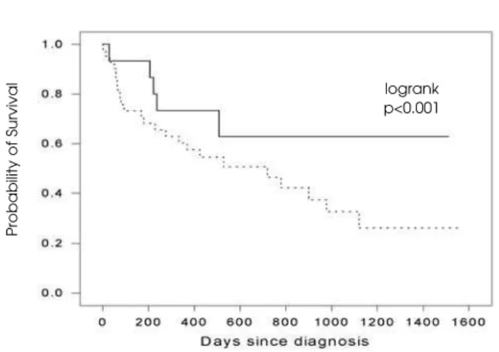
The situation is somewhat different in cancer: as
bone metastases profoundly perturb normal bone
remodelling, biochemical markers of bone turnover
have been shown to reflect these tumour-induced
changes in bone remodelling and may therefore be
useful in the diagnosis, follow-up and prognosis of
patients with malignant (bone) disease (145,146)
(fig-ure 11). Most markers of bone turnover, particularly
those of bone resorption, are elevated in patients with
established bone metastases [recently reviewed in
(147)]. While this may indicate a role of bone markers
as diagnostic tools in cancer patients, available
evi-dence does not provide any final conclusions as to the
accuracy and validity of the presently used markers in
the early diagnosis of bone metastases.
Markers of bone resorption respond promptly
and profoundly to bisphosphonate and anti-neoplastic
therapy, and this response appears to be associated
with a favourable clinical outcome in patients with
bone metastases (148-149). Recent evidence indicates
that the aim of bisphosphonate therapy should be to
normalize increased rates of bone remodelling
(150,151). However, it remains unknown whether the
use of bone markers in the routine clinical setting has
any defined beneficial effects on overall outcome in
cancer patients. In particular, no study has addressed
the question whether patients with bone metastases
should be treated according to their rate of bone
turnover, and what the treatment goals are in this
respect. While it is unlikely that bone turnover
mark-ers have sufficient diagnostic or prognostic value to be
used in isolation, the combination of these markers
with other diagnostic techniques may be the way
for-ward to improve the clinical assessment of patients
with bone seeking cancers.
Although the above-mentioned studies
repre-sent only a small selection of the available literature,
they all demonstrate that markers of bone turnover are
extremely helpful tools in evaluating the physiology
and pathophysiology of bone metabolism, and in
elu-cidating the pathogenesis of bone disease.
REFERENCES
1. Seibel MJ. Bone metabolism. In: Offermanns S, Rosenthal W (eds.). Encyclopaedic Reference of Molecular
Phar-macology. Nova York: Springer Heidelberg, 2003. pp.
457-65.
2. Dunstan CR, Seibel MJ. Bone and mineral metabolism. In: Encyclopedia of Medicinal Chemistry. In press. 3. Seibel MJ. Biochemical markers of bone turnover. Part I:
Biochemistry and Variability. Clin Biochem Rev 2005;26:97-122.
4. Beardsworth LJ, Eyre DR, Dickson IR. Changes with age in the urinary excretion of lysyl- and hydroxylysylpyridino-line, two new markers of bone collagen turnover. J Bone Miner Res 1990;5:671.
5. Midtby M, Magnus J, Joakimsen R. The Tromso Study: a population-based study on the variation in bone forma-tion markers with age, gender, anthropometry and sea-son in both men and women. Osteoporos Int 2001;12:835-43.
6. Meier C, Liu PY, Handelsman DJ, Seibel MJ. Endocrine regulation of bone turnover in men. Clin Endocrinol 2005;63:603-16.
7. Meier C, Liu PY, Seibel MJ, Handelsman DJ. Sex steroids and skeletal health in men. In: Lane NE, Sambrook PN (eds.). Osteoporosis and the Osteoporosis of Rheumatic Diseases. Nova York: Mosby, 2006.
8. Epstein S, McClintock R, Bryce G, Poser J, Johnston C, Hui S. Differences in serum bone gla protein with age and sex.Lancet 2006;11:307-10.
9. Hassager C, Risteli J, Risteli L, Christiansen C. Effect of the menopause and hormone replacement therapy on the carboxy-terminal pyridinoline cross-linked telopeptide of type I collagen. Osteoporos Int 1994;4:349-52.
Figure 11.Probability of survival according to serum BSP
val-ues in patients with multiple myeloma. Patients were strati-fied by serum BSP concentrations at baseline (solid line: serum BSP < 21 ng/mL, dashed line: serum BSP > 21 ng/mL). In a multivariate multivariate Cox proportional analysis, serum BSP (p= 0.012) and monoclonal protein (p= 0.028) were the only predictors of survival in this patient group. From: Woitge et al. 2001 (145), with permission.]
Probability
o
f
Survival
10. Koshla S, Atkinson E, Melton LJ, Riggs BL. Effects of age and estrogen status on serum parathyroid hormone lev-els and biochemical markers of bone turnover in women: a population based study. J Clin Endocrinol Metab 1997;82:1522-7.
11. Kushida K, Takahashi M, Kawana K, Inoue T. Comparison of markers for bone formation and resorption in pre-menopausal and postpre-menopausal subjects, and osteo-porosis patients. J Clin Endocrinol Metab 1995;80:2447-50.
12. Seibel MJ, Cosman F, Shen V, Ratcliffe A, Lindsay R. Uri-nary hydroxy-pyridinium crosslinks of collagen as markers of bone resorption and estrogen efficacy in post-menopausal osteoporosis. J Bone Min Res 1993;8:881-9.
13. Seibel MJ, Woitge H, Scheidt-Nave C, Leidig-Bruckner G, Duncan A, Nicol P, et al. Urinary hydroxypyridinium crosslinks of collagen in population-based screening for overt vertebral osteoporosis: results of a pilot study.J Bone Miner Res 1994;9:1433-40.
14. Uebelhart D, Schlemmer A, Johansen JS, Gineyts E, Christiansen C, Delmas PD. Effect of menopause and hormone replacement therapy on the urinary excretion of pyridinium crosslinks. J Clin Endocrinol Metab 1991;72:367-73.
15. Cleghorn DB, O’Loughlin PD, Schroeder BJ, Nordin BE. An open, crossover trial of calcium-fortified milk in pre-vention of early postmenopausal bone loss. Med J Aust 2001;175(5):242-5.
16. Rosen C, Chesnut CH III, Mallinak NJS. The predictive value of biochemical markers of bone turnover for bone mineral density in early postmenopausal women treat-ed with hormone replacement or calcium supplemen-tation. J Clin Endocrinol Metab 1997;82:1904-10.
17. Rosen HN, Parker RA, Greenspan SL, Iloputaife ID, Book-man L, Chapin D, et al. Evaluation of ability of biochem-ical markers of bone turnover to predict a response to increased doses of HRT. Calcif Tissue Int 2004;74:415-23.
18. Meier C, Woitge HW, Witte K, Lemmer B, Seibel MJ. Sup-plementation with oral vitamin D3 and calcium during winter prevents seasonal bone loss: a randomized con-trolled open-label prospective trial. J Bone Miner Res 2004;19:1221-30.
19. Meunier PJ, Jenvrin C, Munoz F, de la Gueronniere V, Garnero P, Menz M. Consumption of a high calcium mineral water lowers biochemical indices of bone remodeling in postmenopausal women with low calci-um intake. Osteoporosis Int 2005;16:1203-9.
20. Chesnut CH, Bell N, Clark G, Drinkwater BL, English SC, Johnson CC Jr, et al. Hormone replacement therapy in postmenopausal women: Urinary N-telopeptide of type I collagen monitors therapeutic effect and predicts response of bone mineral density. Am J Med 1997;102:29-37.
21. Harris ST, Eriksen EF, Davidson M, Ettinger MP, Moffett Jr AH, Baylink DJ, et al. Effect of combined risedronate and hormone replacement therapies on bone mineral density in postmenopausal women. J Clin Endocrinol Metab 2001;86(5):1890-7.
22. Heikkinen AM, Parviainen M, Niskanen L, Komulainen M, Tuppurainen MT, Kroger H, et al. Biochemical bone markers and bone mineral density during
post-menopausal hormone replacement therapy with and without vitamin D3: A prospective, controlled, random-ized study. J Clin Endocrinol Metabolism 1997;82:2476-82.
23. Prestwood K, Pilbeam C, Burleson J, Woodiel FN, Delmas PD, Deftos LJ, et al. The short-term effects of conjugated estrogen on bone turnover in older women. J Clin Endocrinol Metab 1994;79:366-71.
24. Greenspan SL, Resnick NM, Parker RA. Early changes in biochemical markers of bone turnover are associated with long-term changes in bone mineral density in elder-ly women on alendronate, hormone replacement ther-apy, or combination therapy: a three-year, double-blind, placebo-controlled, randomized clinical trial. J Clin Endocrinol Metab 2005;90:2762-7.
25. Seifert-Klauss V, Mueller JE, Luppa P, Probst R, Wilker J, Hoss C, et al. Bone metabolism during the peri-menopausal transition: a prospective study. Maturitas 2002;41:23-33.
26. McKane W, Khosla S, Risteli J, Robins S, Muhs J, Riggs B. Role of estrogen deficiency in pathogenesis of sec-ondary hyperparathyroidism and increased bone resorption in elderly women. Proc Assoc Am Physicians 1997;109:174-80.
27. Wasnich RD, Bagger Y, Hosking D, McClung M, Wu M, Mantz AM, et al; Early Postmenopausal Intervention Cohort Study Group. Changes in bone density and turnover after alendronate or estrogen withdrawal. Menopause 2004;11:622-30.
28. Akesson K, Ljunghall S, Gardsell P, Sernbo I, Obrant KJ. Serum osteocalcin and fracture susceptibility in elderly women. Calcif Tissue Int 1993;53:86-90.
29. Akesson K, Vergnaud P, Gineyts E, Delmas PD, Obrant KJ. Impairment of bone turnover in elderly women with hip fracture. Calcif Tissue Int 1993;53:162-9.
30. Dresner-Pollak R, Parker RA, Poku M, Thompson J, Seibel MJ, Greenspan SL. Biochemical markers of bone turnover reflect femoral bone loss in elderly women.
Calcif Tissue Int 199659:328-33.
31. Garnero P, Sornay-Rendu E, Chapuy M-C, Delmas PD. Increased bone turnover in late postmenopausal women is a major determinant of osteoporosis. J Bone Min Res 1996;11:337-49.
32. Chen JS, Cameron ID, Cumming RG, March LM, Sam-brook PN, Simpson JM, et al. Effect of age-related chronic immobility on markers of bone turnover. J Bone Miner Res 2006;21:324-31.
33. Sambrook PN, Chen JS, March L, Cameron ID, Cumming RG, Lord S, et al. High bone turnover is an independent predictor of mortality in the frail elderly. J Bone Miner Res 2006;21:549-55.
34. Mezquita-Raya P, Munoz-Torres M, Luna JD, Luna V, Lopez-Rodriguez F, Torres-Vela E, et al. Relation between vitamin D insufficiency, bone density, and bone metab-olism in healthy postmenopausal women.J Bone Miner Res 2001;16(8):1408-15.
36. Sambrook PN, Chen JS, March LM, Cameron ID, Cum-ming RG, Lord SR, et al. Serum parathyroid hormone is associated with increased mortality independent of 25-hydroxy vitamin D status, bone mass, and renal function in the frail and very old: A cohort study. J Clin Endocrinol Metab 2004;89:5477-81.
37. Zochling J, Chen JS, Seibel M, Schwarz J, Cameron ID, Cumming RG, et al. Calcium metabolism in the frail elderly. Clin Rheumatol 2005;24:576-82.
38. Charles P, Hasling C, Risteli L, Risteli J, Mosekilde L, Eriksen E. Assessment of bone formation by biochemical mark-ers in metabolic bone disease: separation between osteoblastic activity at the cell and tissue level. Calc Tis-sue Int 1992;51:406-11.
39. Garnero P, Shih WJ, Gineyts E, Karpf DB, Delmas PD. Comparison of new biochemical markers of bone turnover in late postmenopausal osteoporotic women in response to alendronate treatment. J Clin Endocrinol Metab 1994;79(6):1693-700.
40. McLarren AM, Hordon LD, Bird HA, Robins SP. Urinary excretion of pyridinium crosslinks of collagen in patients with osteoporosis and the effects of bone fracture. Ann Rheum Dis 1992;51:648-51.
41. Meier C, Meinhardt U, Greenfield JR, Nguyen TV, Dun-stan CR, Seibel MJ. Serum cathepsin K levels reflect osteoclastic activity in women with postmenopausal osteoporosis and patients with Paget’s disease. Clin Lab 2006;52:1-10.
42. Schneider DL, Barrett-Connor EL. Urinary N-telopeptide levels discriminate normal, osteopenic, and osteoporotic bone mineral density. Arch Intern Med 1997;157:1241-5.
43. Akesson K, Ljunghall S, Jonsson B, Sernbo I, Johnell O, Gardsell P, et al. Assessment of biochemical markers of bone metabolism in relation to the occurrence of frac-ture: A retrospective and prospective population-based study in women. J Bone Miner Res 1995;10:1823-9.
44. Akesson K, Vergnaud P, Delmas PD, Obrant KJ. Serum osteocalcin increases during fracture healing in elderly women with hip fracture.Bone 1995;6:427-30.
45. Ensrud KE, Palermo L, Black DM, Cauley J, Jergas M, Orwoll ES, et al. Hip and calcaneal bone loss increase with advancing age: Longitudinal results from the study of osteoporotic fractures. J Bone Min Res 1995;10:1778-87.
46. Meier C, Liu PY, Ly LP, de Winter-Modzelewski J, Jimenez M, Handelsman DJ, et al. Recombinant human chorion-ic gonadotropin, but not dihydrotestosterone alone stimulates osteoblastic collagen synthesis in older men with partial age related androgen deficiency. J Clin Endocrinol Metab 2004;89:3033-41.
47. Reid IR, Lucas J, Wattie D, Horne A, Bolland M, Gamble GD, et al. Effects of a beta-blocker on bone turnover in normal postmenopausal women: a randomized con-trolled trial. J Clin Endocrinol Metab 2005;90:5212-6.
48. Ton FN, Gunawardene SC, Lee H, Neer RM. Effects of low-dose prednisone on bone metabolism. J Bone Miner Res 2005;20:464-70.
49. Jamal SA, Cummings SR, Hawker GA. Isosorbide monon-itrate increases bone formation and decreases bone resorption in postmenopausal women: a randomized trial. J Bone Miner Res 2004;19:1512-7.
50. Rejnmark L, Buus NH, Vestergaard P, Heickendorff L, Andreasen F, Larsen L, et al. Effects of simvastatin on bone turnover and BMD: a 1-year randomized con-trolled trial in postmenopausal osteopenic women. J Bone Miner Res 2004;19:737-44.
51. Parkinson C, Kassem M, Heickendorff L, Flyvbjerg A, Trainer PJ. Pegvisomant-induced serum insulin-like growth factor-I normalization in patients with acromegaly returns elevated markers of bone turnover to normal. J Clin Endocrinol Metab 2003;88:5650-5.
52. Meier C, Beat M, Guglielmetti M, Christ-Crain M, Staub JJ, Kraenzlin M. Restoration of euthyroidism accelerates bone turnover in patients with subclinical hypothy-roidism: a randomized controlled trial. Osteoporos Int 2004;15:209-16.
53. Mallmin H, Ljunghall S, Larsson K. Biochemical markers of bone metabolism in patients with fracture of the distal forearm. Clin Orthop 1993;295:259-63.
54. Obrant KJ, Merle B, Bejui J, Delmas PD. Serum bone-gla protein after fracture. Clin Orthop 1990;258:300-3.
55. Hansen M. Assessment of age and risk factors on bone density and bone turnover in healthy premenopausal women. Osteoporos Int 1994;4:123-8.
56. Stepan JJ, Pospichal J, Presl J, Pacovsky V. Bone loss and biochemical indices of bone remodeling in surgi-cally induced postmenopausal women. Bone 1987; 8:279-84.
57. Stepan JJ, Presl J, Broulik P, Pacovsky V. Serum osteocal-cin levels and bone alkaline phosphatase isoenzyme after oophorectomy and in primary hyperparathy-roidism. J Clin Endocrinol Metab 1987;64:1079-82.
58. Ledger GA, Burritt MF, Kao PC, O’Fallon WM, Riggs BL, Khosla S. Role of parathyroid hormone in mediating noc-turnal and age-related increases in bone resorption. J Clin Endocrinol Metab 1995;80:3304-10.
59. Melton L, Khosla S, Atkinson EJ, O’Fallon WM, Riggs BL. Relationship of bone fractures to bone density and frac-tures. J Bone Mineral Res 1997;12:1083-91.
60. Christiansen C, Riis BJ, Rodboro P. Prediction of rapid bone loss in postmenopausal women. Lancet 1987; 1:1105-8.
61. Christiansen C, Riis BJ, Rodbro P. Screening procedure for women at risk of developing postmenopausal osteo-porosis. Osteoporos Int 1990;1:35-40.
62. Cosman F, Nieves J, Wilkinson C, Schnering D, Shen V, Lindsay R. Bone density change and biochemical indices of skeletal turnover. Calcif Tissue Int 1996;58:236-43.
63. Mole PA, Walkinshaw MH, Robins SP, Paterson CR. Can urinary pyridinium crosslinks and urinary oestrogens pre-dict bone mass and rate of bone loss after the menopause? Eur J Clin Invest 1992;22:767-71.
64. Reeve J, Pearson J, Mitchell A, Green J, Nicholls A, Jus-tice J, et al. Evolution of spinal bone loss and bio-chemical markers of bone remodeling after menopause in normal women. Calcf Tissue Int 1995;57:105-10.
66. Keen RW, Nguyen T, Sobnack R, Perry LA, Thompson PW, Spector TD. Can biochemical markers predict bone loss at the hip and spine? A 4-year prospective study of 141 early postmenopausal women. Osteoporos Int 1996;6:399-406.
67. Blumsohn A, Eastell R. Prediction of bone loss in post-menopausal women.Eur J Clin Invest 1992;22:764-6. 68. Vestergaard P, Hermann AP, Gram J, Jensen LB, Eiken P,
Abrahamsen B, et al. Evaluation of methods for predic-tion of bone mineral density by clinical and biochemical variables in perimenopausal women. Maturitas 2001;40(3):211-20.
69. Riggs BL, Melton LJ III, O’Fallon WM. Drug therapy for ver-tebral fractures in osteoporosis: Evidence that decreas-es in bone turnover and increasdecreas-es in bone mass both determine antifracture efficacy.Bone 1996;18:197-201. 70. Van Daele PL, Seibel MJ, Burger H, Hofman A, Grobbee
DE, van Leeuwen JP, et al. Case control analysis of bone resorption markers, disability and hip fracture risk: the Rotterdam study.BMJ 1996;312:482-3.
71. Weel A, Seibel MJ, Pols HAP, et al. Bone resorption and risk of non-vertebral fractures – a populations-based study: The Rotterdam study. J Bone Miner Res 2002;17:S356.
72. Garnero P, Hausherr E, Chapuy MC, Marcelli C, Grand-jean H, Muller C, et al. Markers of bone resorption pre-dict hip fractures in elderly women. The EPIDOS study. J Bone Miner Res 1996;11:1531-8.
73. Vergnaud P, Garnero P, Meunier PJ, Breart G, Kamihagi K, Delmas PD. Undercarboxylated osteocalcin mea-sured with a specific immunoassay predicts hip fracture in elderly women. The EPIDOS study. J Clin Endocrinol Metab 1997;82:719-24.
74. Szulc P, Arlot M, Chapuy MC, Duboeuf F, Meunier PJ, Delmas PD. Serum undercarboxylated osteocalcin cor-relates with hip bone mineral density in elderly women. J Bone Miner Res 1994;9:1591-5.
75. Szulc P, Chapuy MC, Meunier P, Delmas P. Serum under-carboxylated osteocalcin is a marker of the risk of hip fracture in elderly women. J Clin Invest 1993;91:1769-74.
76. Szulc P, Chapuy MC, Meunier P, Delmas P. Serum under-carboxylated osteocalcin is a marker of the risk of hip frac-ture: a three year follow-up study.Bone 1996;18:487-8. 77. Price PA, Kaneda Y. Vitamin D counteracts the effect of
warfarin in liver but not in bone. Thromb Res 1987;46:121-31.
78. Hodges SJ, Pilkington MJ, Stamp TC, Catterall A, Shearer MJ, Bitensky L, et al. Depressed levels of circulating menaquinones in patients with osteoporotic fractures of the spine and femoral neck. Bone 1991;12:387-9.
79. Knapen MH, Manulyak K, Vermeer C. The effect of vita-min K supplementation on circulating osteocalcin and urinary calcium excretion. Ann Int Med 1989;111:1001-5.
80. Meier C, Nguyen TV, Center JR, Seibel MJ, Eisman JA. Bone resorption and osteoporotic fractures in elderly men: The Dubbo Osteoporosis Epidemiology Study. J Bone Miner Res 2005;20:579-87.
81. Civitelli R, Gonnelli S, Zacchei F, Bigazzi S, Vattimo A, Avi-oli LV, et al. Bone turnover in postmenopausal osteo-porosis. J Clin Invest 1988;82:1268-74.
82. Nielsen NM, Von der Recke P, Hansen MA, Overgaard K, Christensen C. Estimation of the effect of salmon calci-tonin in established osteoporosis by biochemical bone markers. Calcif Tissue Int 1994;55:8-11.
83. Overgaard K, Hansen MA, Nielsen VH, Riis BJ, Chris-tiansen C. Discontinuous calcitonin treatment of estab-lished osteoporosis. Effects of withdrawal and treatment. Am J Med 1990;89:1-6.
84. Gonnelli S, Cepollaro C, Pondrelli C, Martini S, Montag-nani A, Monaco R, et al. Bone turnover and the response to alendronate treatment in postmenopausal osteoporosis. Calcif Tissue Int 1999;65(5):359-64.
85. Stevenson JC, Hillard TC, Lees B, Whitcroft SI, Ellering-ton MC, Whitehead MI. Postmenopausal bone loss: does HRT always work? Int J Fertil Stud 1993;38 (suppl.2):88-91.
86. Seibel MJ, Naganathan V, Barton I, Grauer A. Vertebral fracture incidence in postmenopausal osteoporotic women treated with risedronate: the role of pre-treat-ment bone turnover. J Bone Mineral Res 2004;19:323-9.
87. Bauer DC, Garnero P, Hochberg MC, Santora A, Delmas P, Ewing SK, et al.; for the Fracture Intervention Research Group. Pretreatment levels of bone turnover and the antifracture efficacy of alendronate: the fracture inter-vention trial.J Bone Miner Res 2006;21(2):292-9.
88. McClung MR, Lewiecki EM, Cohen SB, Bolognese MA, Woodson GC, Moffett AH, et al.; for the AMG 162 Bone Loss Study Group. Denosumab in postmenopausal women with low bone mineral density. N Engl J Med 2006;354(8):821-31.
89. Nenonen A, Cheng S, Ivaska KK, Alatalo SL, Lehtimaki T, Schmidt-Gayk H, et al. Serum TRACP 5b is a useful mark-er for monitoring alendronate treatment: comparison with other markers of bone turnover. J Bone Miner Res 2005;20(10):1804-12.
90. Reid IR, Brown JP, Burckhardt P, Horowitz Z, Richardson P, Trechsel U, et al. Intravenous zoledronic acid in post-menopausal women with low bone mineral density. N Engl J Med 2002;346:653-61.
91. Tahtela R, Seppanen J, Laitinen K, Katajamaki A, Risteli J, Valimaki MJ. Serum tartrate-resistant acid phosphatase 5b in monitoring bisphosphonate treatment with clo-dronate: a comparison with urinary N-terminal telopep-tide of type I collagen and serum type I procollagen amino-terminal propeptide. Osteoporosis Int 2005;16:1109-16.
92. Bauer DC, Black DM, Garnero P, Hochberg M, Ott S, Orloff J, et al.; for the Fracture Intervention Trial Study Group. Change in bone turnover and hip, non-spine, and vertebral fracture in alendronate-treated women: the fracture intervention trial. J Bone Miner Res 2004;19:1250-8.
93. Recker R, Stakkestad JA, Chesnut CH 3rd, Christiansen C, Skag A, Hoiseth A, et al. Insufficiently dosed intra-venous ibandronate injections are associated with sub-optimal antifracture efficacy in postmenopausal osteo-porosis. Bone 2004;34:890-9.
95. Reginster JY, Sarkar S, Zegels B, Henrotin Y, Bruyere O, Agnusdei D, et al. Reduction in PINP, a marker of bone metabolism, with raloxifene treatment and its relation-ship with vertebral fracture risk. Bone 2004;34:344-51.
96. Tanko LB, Mouritzen U, Lehmann H, Warming L, Moel-gaard A, Christgau S, et al. Oral ibandronate: changes in markers of bone turnover during adequately dosed continuous and weekly therapy and during different suboptimally dosed treatment regimens. Bone 2003;32:687-93.
97. Meunier PJ, Roux C, Seeman E, Ortolani S, Badurski JE, Spector TD, et al. The effects of strontium ranelate on the risk of vertebral fracture in women with postmenopausal osteoporosis. N Engl J Med 2004;350:459-68.
98. Black DM, Greenspan SL, Ensrud KE, Palermo L, McGowan JA, Lang TF, et al.; for the PaTH Study Investi-gators. The effects of parathyroid hormone and alen-dronate alone or in combination in postmenopausal osteoporosis. N Engl J Med 2003;349:1207-15.
99. Cosman F, Nieves J, Zion M, Woelfert L, Luckey M, Lind-say R. Daily and cyclic parathyroid hormone in women receiving alendronate. N Engl J Med 2005;353:566-75.
100.Black DM, Bilezikian JP, Ensrud KE, Greenspan SL, Paler-mo L, Hue T, et al.; PaTH Study Investigators. One year of alendronate after one year of parathyroid hormone (1-84) for osteoporosis. N Engl J Med 2005;353:555-65.
101.Arlot M, Meunier PJ, Boivin G, Haddock L, Tamayo J, Correa-Rotter R, et al. Differential effects of teriparatide and alendronate on bone remodeling in post-menopausal women assessed by histomorphometric parameters. J Bone Miner Res 2005;20:1244-53.
102.Chen P, Satterwhite JH, Licata AA, Lewiecki EM, Sipos AA, Misurski DM, et al. Early changes in biochemical markers of bone formation predict BMD response to teri-paratide in postmenopausal women with osteoporosis.J Bone Miner Res 2005;20:962-70.
103.Ettinger B, Black DM, Mitlak BH, Knickerbocker RK, Nick-elsen T, Genant HK, et al. Reduction of vertebral fracture risk in postmenopausal women with osteoporosis treat-ed with raloxifene: results from a 3-year randomiztreat-ed clin-ical trial. Multiple Outcomes of Raloxifene Evaluation (MORE) Investigators. JAMA 1999;282:637-45.
104.Harris ST, Watts NB, Genant HK, McKeever CD, Hangart-ner T, Keller M, et al. Effects of risedronate treatment on vertebral and nonvertebral fractures in women with postmenopausal osteoporosis: a randomized controlled trial. Vertebral Efficacy With Risedronate Therapy (VERT) Study Group.JAMA 1999;282:1344-52.
105.Storm T, Thamsborg G, Steiniche T, Genant HK, Sorensen OH. Effect of intermittent cyclical etidronate therapy on bone mass and fracture rate in women with post-menopausal osteoporosis. N Engl J Med 1990;322:1265-71.
106.Cummings SR, Black DM, Thompson DE, Applegate WB, Barrett-Connor E, Musliner TA, et al. Effect of alen-dronate on risk of fracture in women with low bone den-sity but without vertebral fractures: results from the Frac-ture Intervention Trial. JAMA 1998;280:2077-82.
107.Cummings SR, Karpf DB, Harris F, Genant HK, Ensrud K, LaCroix AZ, et al. Improvement in spine bone density and reduction in risk of vertebral fractures during treatment with antiresorptive drugs. Am J Med 2002;112:281-9.
108.Sarkar S, Mitlak BH, Wong M, Stock JL, Black DM, Harper KD. Relationships between bone mineral density and incident vertebral fracture risk with raloxifene therapy. J Bone Miner Res 2002;17:1-10.
109.Li Z, Meredith MP. Exploring the relationship between surrogates and clinical outcomes: analysis of individual patient data vs. meta-regression on group-level summa-ry statistics. J Biopharm Stat 2003;13:777-92.
110.Delmas PD, Recker RR, Chesnut CH 3rd, Skag A, Stakkestad JA, Emkey R, et al. Daily and intermittent oral ibandronate normalize bone turnover and provide sig-nificant reduction in vertebral fracture risk: results from the BONE study. Osteoporos Int 2004;15:792-8.
111.Bjarnason NH, Sarkar S, Duong T, Mitlak B, Delmas P, Christiansen C. Six and twelve-month changes in bone turnover are related to reduction in vertebral fracture risk during 3 years of raloxifene treatment in post-menopausal osteoporosis. Osteoporos Int 2001;12:922-30.
112.Eastell R, Barton I, Hannon RA, Chines A, Garnero P, Del-mas P. Relationship of early changes in bone resorption to the reduction in fracture risk with risedronate. J Bone Miner Res 2003;18:1051-6.
113.McCombs JS, Thiebaud P, McLaughlin-Miley C, Shi J. Compliance with drug therapies for the treatment and prevention of osteoporosis. Maturitas 2004;48(3):271-87.
114.Marwick C. Hormone combination treats women’s bone loss. JAMA 2004;272:1487.
115.Cano A. Compliance to hormone replacement therapy in menopausal women controlled in a third level acad-emic centre. Maturitas 1994;20:91-9.
116.Faulkner DL, Young C, Hutchins D, McCollam JS. Patient noncompliance with hormone replacement therapy: a nationwide estimate using a large prescription claims database. Menopause 1998;5:226-9.
117.Kotzan JA, Martin BC, Wade WE. Persistence with estro-gen therapy in a postmenopausal Medicaid popula-tion. Pharmacotherapy 1999;19:363-9.
118.Kayser J, Ettinger B, Pressman A. Postmenopausal hor-monal support: discontinuation of raloxifene versus estrogen.Menopause 2001;8:328-32.
119.Bjorn I, Backsrom T. Drug related negative side-effects is a common reason for poor compliance in hormone replacement therapy.Maturitas 1999;32:77-86.
120.Segal E, Tamir A, Ish-Shalom S. Compliance of osteo-porotic patients with different treatment regimens. Isr Med Assoc J 2003;5:859-62.
121.Chapurlat RD, Cummings SR. Does follow-up of osteo-porotic women treated with antiresorptive therapies improve effectiveness? Osteoporos Int 2002;13:738-44.
122.Clowes JA, Peel NF, Eastell R. The impact of monitoring on adherence and persistence with antiresorptive treat-ment for postmenopausal osteoporosis: a randomized controlled trial. J Clin Endocrinol Metab 2004;89:1117-23.