Brazilian Journal of Physics, vol. 40, no. 4, December, 2010 429
Study of the Structure of Free Radicals in Gamma – Irradiated Amino Acid Derivatives
Murat AYDIN∗
Faculty of Education, Adıyaman University, TR 02030-Adıyaman, Turkey
(Received on 5 August, 2010)
Electron paramagnetic resonance spectroscopy has been used for the study of the free radicals pro-duced in the powders of L-arginine monohydrochloride, ala-ala and DL-β-leucine gamma-irradiated at room temperature. The structures of the radicals formed in compounds were determined as NH2(NH)NH ˙CHCH2CH2CH(NH2)COOH, CH3CHCOOH and (CH˙ 3)2CCH(NH˙ 2)CH2COOH respectively.
The paramagnetic species were found to be stable at room temperature more than three months. It was de-termined that unpaired electron interacted with nearby protons and14N nucleus in these radicals. Thegvalues of the radicals and the hyperfine structure constants of the free electron were also measured. These results were compared with the earlier studies in amino acid and derivatives radicals and discussed.
Keywords: EPR, Free radicals, Gamma irradiation, Amino acids compounds
1. INTRODUCTION
Free radicals play an important role in biological process and they have a vital importance for the health of the liv-ing organisms [1, 2]. It is known that electron paramagnetic resonance (EPR) spectroscopy is one of the most powerful methods for studying the structure and determining the iden-tity of free radicals [3]. An interpretation of the EPR spec-trum is generally accomplished after the determination of a set of spin Hamiltonian parameters. The magnetic properties of many organic substances have been investigated using the EPR technique [4 - 10]. The EPR of gamma-irradiated pow-ders of N-acetyl-L-arginine and Nα-carbamyl-L-arginine
were studied by Zincircioglu et al. [11] at room temperature. Furthermore, in the gamma-irradiated single crystal of N-acetyl-L-Leucine, at room temperature, the free radicals have been attributed to the (CH3)2CCH˙ 2CH(NHCOCH3)COOH and (CH3)2CHCH2C(NHCOCH˙ 3)COOH radicals [12].
Arginine, ala-ala and leucine are biologically important compounds. Therefore the purpose of this work is to inves-tigate the defects induced by gamma-irradiation in powders of L-arginine monohydrochloride (LAMHCl), ala-ala, DL-β-leucine (DLBL) and to determine their spin Hamiltonian parameters at room temperature.
2. EXPERIMENTAL
The samples used in this study were obtained from com-mercial sources. Powder samples of the compounds were ir-radiated at room temperature with a60Co gamma-ray source (Nordion-Canada model JS 9600) of 30 kGy. After irradi-ation, samples were kept in plastic bags at room tempera-ture in the dark. The spectra of samples were recorded by putting the sample in quartz sample tube. The EPR mea-surements was carried out in a Varian model X-band E-109C EPR spectrometer at room temperature. The modulation am-plitude was below 5.10−2mT and the microwave power was 2 mW. Thegfactors were found by comparison with a dipen-hylpicrylhydrazyl (DPPH) sample with of g = 2.0036 [13].
∗Electronic address:[email protected]
The spectrum simulations were made using McKelvey’s pro-grams [14].
3. RESULTS AND DISCUSSION
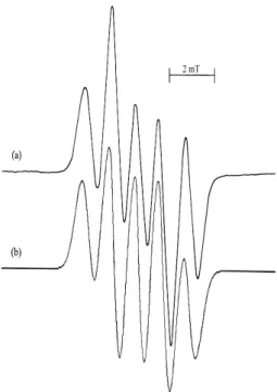
Figure 1 presents the EPR spectrum of LAMHCl powder, gamma-irradiated and recorded at room temperature. This spectrum has approximately 1:2:2:2:1 intensity ratio, and can be attributed to the NH2(NH)NH ˙CHCH2CH2CH(NH2)COOH radical. It can be seen that it consists of a doublet with a spacing of 2.10 mT. Then each line of doublet is further subdivided into three lines of spacing with 1.12 mT, with an intensity distribution of 1:2:1. A spectrum for a larger number of inequivalent protons can be found by graphic construction, which is a mathematical record of an EPR spectrum [15]. The binomial expansion for this spectrum is given as:
1 : 2 : 1+1 : 2 : 1=1 : 2 : 2 : 2 : 1.
Similar EPR spectrum and intensity distribution were ob-served for the gamma-irradiated L-arginine powders at room temperature [16]. In this case, the same radical produced by the abstraction of one hydrogen atom form the same carbon atom. A simulation of the LAMHCl spectrum is shown fig-ure 1b, using the hyperfine coupling constantsaα=2.10 mT,
aβ=1.12 mT andaN=0.28 mT. The linewidth of the
spec-trum is somewhat larger than the hyperfine coupling constant of the nitrogen nuclei, and therefore the hyperfine splitting of the nitrogen nuclei is not observed in the spectrum. The measuredgvalue of the radical isg= 2.0028±0.0005. It is known that the value of thegfactor in aliphatic aminoalkyl radicals vary between 2.0027 - 2.0031 [17]. Thegvalue in this study is in this range.
430 Murat AYDIN
FIG. 1: (a) The EPR spectrum of gamma-irradiated LAMHCL pow-der at room temperature, (b) simulation form of the spectrum using
aα=2.10 mT,aβ=1.12 mT,aN=0.28 mT and linewidth 0.32 mT.
0.0005. Thegvalue of the radical is similar to those where the unpaired electron is placed on the carbon atoms [18]. A spectrum simulated with the above given values agrees with the experiment (Figure 2b).
FIG. 2: (a) The EPR spectrum of gamma-irradiated ala-ala pow-der at room temperature, (b) simulation form of the spectrum using
aα=2.02 mT,aβ=2.24 mT and linewidth 0.46 mT.
The EPR parameters of the radical discussed here agree with the values measured in the irradiated alanine and their
derivatives [11, 16, 19 - 21]. X-irradiated L-alanine single crystal has been investigated by EPR technique, and the re-ported values of the hyperfine coupling constants areaα=
1.51 mT,aβ=1.88 mT at low temperature andaα=2.14
mT,aβ=2.38 mT at room temperature [22]. The measured
values at room temperature are in agreement with our results. The amine group abstraction is one of the most common mechanisms of producing free radicals in alanine compounds [23 - 25].
The EPR spectrum of DLBL in figure 3 exhibits an inten-sity distribution as 1:6:15:20:15:6:1. This spectrum belongs to the (CH3)2CCH(NH˙ 2)CH2COOH radical due to the hy-perfine interactions of the unpaired electron with six equiva-lent protons of the two methyl groups, one CH proton and 14N nucleus. A simulation of the spectrum is shown fig-ure 3b, using the hyperfine coupling constantsa(CH3)2=2.10
mT,aCH=0.63 mT,aN =0.52 mT and linewidth 0.46 mT.
The g value of the radical is measured as g = 2.0028 ± 0.0005. Since the linewidth of the spectrum is fairly large, the hyperfine splitting of the CH proton and the nitrogen nu-clei are not observed in the spectrum. It can be state that this radical is obtained by the removal of hydrogen atom from the tertiary carbon atom of the DLBL.
FIG. 3: (a) The EPR spectrum of gamma-irradiated DLBL pow-der at room temperature, (b) simulation form of the spectrum using
a(CH3)2=2.10mT,aCH=0.63mT,aN=0.52 mT and linewidth 0.46
mT.
The measured hyperfine coupling constants of the methyl protons and thegvalue for this radical are in agreement with those of the radicals which are derived from leucine and va-line [26 - 29]. In gamma-irradiated L-vava-line methyl ester hydrochloride powder, at room temperature, the free radi-cal has been attributed to the (CH3)2CCH(NH˙ 3Cl)COOCH3 radical [30]. This is similar to our proposed radical and the reported values ofa(CH
3)2 =2.39 mT,aCH=0.61 mT and
aN=0.61 mT are in good agreement with our resuls. In the
Brazilian Journal of Physics, vol. 40, no. 4, December, 2010 431
unpaired electron is localized at the carbon atom binding the two methyl groups.
4. CONCLUSION
In this study, we have been indicated that the structure and the identity of the free radicals trapped in the LAHCl, ala-ala and DLBL have been investigated using an EPR technique. The EPR parameters of these radicals could be determined
and these values were found to be consistent with previous results in the literature. The measurements of magnetic prop-erties of these radicals can be helpful in the study of similar radicals found in biological systems.
Acknowledgments
This work was supported by Grant No. EFBAP2009-0005 of the Research Fund of Adiyaman University (ADYAP).
[1] N. D. Yordanov and R. Mladenova, Int. J. Food Sceince Tech.,
42, 1384 (2007).
[2] T. Kiljunen, E. Popov, H. Kunttu, J. Eloranta, J. Chem. Phys.,
130, 164504 (2009).
[3] U. Sayin, E. T¨urkan, ¨O. Dereli, H. Y¨uksel, M. Birey, Radiat. Phys. Chem.,79, 863 (2010).
[4] [4] B. Karabulut, R. Tapramaz, Radiat. Phys.,55, 331 (1999). [5] N. D. Yordanov and B. Mladenova, Radiat. Phys. Chem.,60,
191 (2001).
[6] N. D. Yordanov, R. Mladenow, Spectrochim. Acta A,60,1395 (2004).
[7] M. Tabak, D. de Sousa Neto and C.E.G. Salmon, Braz. J. Phys.,36, 83 (2006).
[8] Kinoshita, L. Figuty and O. Baffa, Braz. J. Phys., 36, 93 (2006).
[9] M. Aydin, M.H. Basksan, Y. E. Osmanoglu, Braz. J. Phys.,39, 583 (2009).
[10] M. Aydin, S. Osmanoðlu, M.H. Baskan, I.Y. Dicle, J. Mol. Struct.,975, 30 (2010).
[11] S.B. Zincircioglu, N. Canoruc, S. Osmanoglu, M.H. Baskan, I.Y. Dicle, M. Aydin, Z. Naturforsch.,61a, 577 (2006). [12] ] G.A. Almanov, G.A. Bogdanchikov, O.M. Usov, Radiat.
Phys. Chem.,29, 75 (1987).
[13] N. D. Yordanov, Appl. Mag. Res.,10, 339 (1996). [14] R.D. McKelvey, J. Chem. Educ.,64, 497 (1987). [15] M. Birey, Z. Naturforsch.,57a, 36 (2002).
[16] M. Aydin, M.H. Baskan, S. Yakar, S.F. Ulak, M. Aydınol, B. Aydınol, M. B¨uy¨um, Radiat. Eff. Defect. Solids,163, 41 (2008).
[17] R. K¨oseoglu, E. K¨oseoglu, F. K¨oksal, App. Radiat. Isot.,58, 63 (2003).
[18] L. Horvath, V.H. Laslo, H. Bilinsky, Radiat. Phys. Chem.,32, 801 (1988).
[19] A. Horsfield, J.R. Morton, D.H. Whiffen, Mol. Phys.,4, 425 (1961).
[20] J.R. Morton, A. Horsfield, J. Chem. Phys.,35, 1142 (1961). [21] A. Horsfield, J.R. Morton, D.H. Whiffen, Mol. Phys.,5, 115
(1962).
[22] J.W. Sinclair, M.W. Hanna, J.Phys. Chem.,71, 84 (1967). [23] I. Miyagawa, W. Gordy, J. Chem. Phys.,32, 255 (1960). [24] W.C. Lin, C.A. Mcdowell, Can. J. Chem.,40, 1062 (1962). [25] E. Sagstuen, E. O. Hole, S. R. Haugedal, W. H. Nelson, J.
Phys. Chem. A,101, 9763 (1997).
[26] G.H. Schnepel, Int. J. Rad. Biol.,25, 555 (1974).
[27] G.A. Almanov, G.A. Bogdanchikov, O.M. Usov, J. Structural Chem.,36, 912 (1989).
[28] V . N. Laslo, T. A. Himdan, H. Bilinski, Radiat. Res.,132, 1 (1992).
[29] M. Aydin, Braz. J. Phys.,40, 306 (2010).