DOI: http://dx.doi.org/10.1590/2446-4740.04116 Volume 33, Number 2, p. 91-96, 2017
Introduction
Osteoporosis is deined as a metabolic bone disorder and skeletal disease characterized by low bone mass and microarchitectural deterioration. It generally occurs in women, at the menopause age (usually over 50) attributed
to declining levels of estrogen and in men (usually over age 70) by testosterone or estradiol deiciency (Ebeling, 2008; Society..., 2014). Osteoporosis is characterized
by a progressive loss of bone mass, increased porosity, deterioration of microarchitecture, cracks, degradation of collagen ibers and unbalance in reabsorption and bone formation rates. Speciically in trabeculae there is a reduction of number, thickness and connectivity and shape deterioration from plate to rod format (National..., 2014; Parfitt et al., 1987). These factors, combined or
isolated, causes lower resistance which entails higher fracture risk with little or without trauma, mainly in trabecular bone from regions of femoral neck and vertebrae (Hernlund et al., 2013), increasing morbidity and mortality (Kanis et al., 2013).
Trabecular bone, from Latin trabeculae which means “small beam”, is considered a composite, viscoelastic, heterogeneous and anisotropic material (Cauley et al., 2010; Doblaré et al., 2004). It presents a complex structure
Axial compressive strength of human vertebrae trabecular bones
classiied as normal, osteopenic and osteoporotic by quantitative
ultrasonometry of calcaneus
Reinaldo Cesar
1*, Tomaz Puga Leivas
2, Cesar Augusto Martins Pereira
2, Ricardo Simionato Boffa
3,
Roberto Guarniero
2,4, Rodrigo Bezerra de Menezes Reiff
5, Antônio Mandeli Netto
1,
Carlos Alberto Fortulan
1, João Manuel Domingos de Almeida Rollo
61 Department of Mechanical Engineering, São Carlos School of Engineering, University of São Paulo, São Carlos, SP, Brazil.
2 Institute of Orthopedics and Traumatology, General Hospital, Faculty of Medicine, University of São Paulo, São Carlos, SP, Brazil.
3 Interunits Bioengineering Post-graduate Program, University of São Paulo, São Carlos, SP, Brazil.
4 National Institute of Science and Technology in Biofabrication, São Paulo, SP, Brazil. 5 Departament of Medicine, Federal University of São Carlos, São Carlos, SP, Brazil.
6 Departament of Materials Engineering, São Carlos School of Engineering, University of São Paulo, São Carlos, SP, Brazil.
Abstract Introduction: Biomechanical assessment of trabecular bone microarchitecture contributes to the evaluation of fractures risk associated with osteoporosis and plays a crucial role in planning preventive strategies. One of the most widely clinical technics used for osteoporosis diagnosis by health professionals is bone dual-energy X-ray absorptiometry (DEXA). However, doubts about its accuracy motivate the introduction of congruent technical analysis such as calcaneal ultrasonometry (Quantitative Ultrasonometry - QUS). Methods: Correlations between Bone Quality Index (BQI), determined by calcaneal ultrasonometry of thirty (30) individuals classiied as normal, osteopenic and osteoporotic, and elastic modulus (E) and ultimate compressive strength (UCS) from axial compression tests of ninety (90) proof bodies from human vertebrae trabecular bone, which were extracted from cadavers in the twelfth thoracic region (T12), irst and fourth lumbar (L1 and L4). Results: Analysis of variance (ANOVA) showed signiicant differences for E (p = 0.001), for UCS (p = 0.0001) and BQI. Spearman’s rank correlation coeficient (rho) between BQI and E (r = 0.499) and BQI and UCS (r = 0.508) were moderate. Discussion: Calcaneal ultrasonometry technique allowed a moderate estimate of bone mechanical strength and fracture risk associated with osteoporosis in human vertebrae.
Keywords Osteoporosis, Calcaneal ultrasonometry, Trabecular bone, Human vertebra, Axial compression test, Correlation.
This is an Open Access article distributed under the terms of the Creative Commons Attribution License, which permits unrestricted use, distribution, and reproduction in any medium, provided the original work is properly cited.
How to cite this article: Cesar R, Leivas TP, Pereira CAM, Boffa RS, Guarniero R, Reiff RBM, Mandeli A No, Fortulan CA, Rollo JMDA. Axial compressive strength of human vertebrae trabecular bones classified as normal, osteopenic and osteoporotic by quantitative ultrasonometry of calcaneus. Res Biomed Eng.2017; 33(2):91-96. DOI: 10.1590/2446-4740.04116.
with irregular organization, which is dependent on the applied mechanical stress (Cesar et al., 2013).
Bone mineral density (BMD) (measured in g/cm2)
evaluated by dual-energy X-ray absorptiometry (DEXA) is the main clinical method (gold standard) for predicting morphometric vertebral fracture in postmenopausal women, however it cannot predict osteoporosis accurately because it evaluates density as the only relevant parameter (Adams, 2013; Watts et al., 2005).
Quantitative ultrasound (QUS) of calcaneal bone is considered a low-cost technique and a fast exam with absence of ionizing radiation. This technique allows clinical assessment of bone quality from the elastic properties, which vary as function of bone loss (Raum et al., 2014). The most commonly used parameters are the speed of sound (SOS), broadband ultrasound attenuation (BUA) and bone quality index or “stiffness index” (BQI) (Guglielmi and Terlizzi, 2009). BQI is a measure that is not necessarily related to the mechanical strength of bone calculated from normalized values of SOS and BUA, BQI = (0.67 x BUA + 0.28 x SOS) - 420, based on the average young adults (Liu et al., 2012) and usually expressed as “T-scores” in the same manner as those obtained by DEXA (Shenoy et al., 2014) and classiied as normal (> -1), osteopenic (between -1 and -2.5) and osteoporotic (< -2.5) (National..., 2014).
Axial compression tests allow the analysis of the biomechanical behavior of cylindrical or cubic proof bodies of trabecular bone (dry or wet) with standardized dimensions. Axial compression is the most common type of biomechanical loading in the human body due to gravity and allows the characterization of bone quality by the analysis of the elastic modulus (E) and ultimate compressive strength (UCS) associated with osteoporosis (Roux et al., 2010).
The hypothesis of this study is the existence of correlations and signiicant differences between trabecular bone analyzed by different characterization techniques, among which: BQI values, QUS and mechanical properties. Hence, 30 Brazilian individuals (human cadavers) were classiied as normal (considered healthy), osteopenic and osteoporotic by QUS from the calcaneus of which also was performed BQI values, followed respective extraction of 90 proof bodies of vertebral regions T12, L1 and L4 for axial compression to obtain E and UCS.
Methods
Vertebral segments of human cadavers were extracted from the thoracic region 10 (T10) to 5 lumbar (L5), according to the procedures approved by the Research Protocol nº 408/11 of the Research Ethics Committee of the Medical School of the University of São Paulo (CEP-USP) and regulations established by the Capital Deaths Checking Service (SVOC) of the University of São Paulo. The vertebrae of the regions T12 and L1 were chosen because they have a higher rate of fractures and
the L4 has a better distribution of trabeculae and axial alignment to the cranial-caudal axis in the vertebral body (Suzuki, 2009). The minimum number of samples (N = 8.64) was determined as a function of the quantitative variables for an ininite population (Fontelles et al., 2010) and is deined by Equation 1.
2 2 8.6 Z SD N N SE α = → = (1)
were, Zα/ 2: critical value for the desired degree of
conidence (usually: 1.96 (95%); SD: standard deviation of the average; and SE: standard error of the average (SD = 1.80 MPa, SE = 1.2 MPa, according to Rodrigues (2004). The adopted sample size was ten (N = 10) for each group: normal, osteopenic and osteoporotic, totalizing thirty (30) subjects.
Cadavers were selected with known cause of death, 12 hours after death interval, over 21 years of age and excluded if diagnosed with infectious diseases, trauma history, deformities, cancer, injuries, deformations, excessive feet fatty tissue layers or signs of surgeries on the thoracic-lumbar region.
Calcaneus QUS measurements were performed by the Aquilles Insight ultrasonometry equipment
(GE MEDICAL SYSTEMS LUNAR) three (03)
times on the left and right foot each, taking the general average between them.
Extraction of vertebral segments was made with the cadavers in the dorsal decubitus position and the vertebrae interest location (from T10 to L5) was set apart by inserting a surgical needle in the adjacent intervertebral discs. Appropriate surgical tools were used for dissection and separation of the vertebrae from the discs and immediately stored in a commercial chest freezer at -20 °C. Samples were extracted using a trephine drill, with an internal diameter of 10 mm and at low speed (bench drilling machine) (Figure 1a) in order to avoid excessive heat, crushing or shearing of trabeculae during trephine penetration in the axial direction. The upper and lower surfaces of the cylindrical samples were cut with a diamond disk orthogonally to obtain parallelism and achieve the specimen standardized dimensions of 20 mm of height (Figure 1b). Most of bone marrow was removed (Figure 1c) by physiological saline application (sodium chloride 0.9%) and stored in an amber vial (for protection against light degradation) in freezing temperature (4 °C).
The obtained specimens (20x10 mm) were immersed in saline solution and kept at room temperature (21 °C) for 1 hour before the analysis in a Universal Testing Machine equipped with a 981 N load cell (KRATOS, model K5002, Cotia-Brasil), shown in Figure 2.
for Testing and Materials (2016) for compression tests on trabecular bone, compression was performed at the lowest rate (5 mm/min) to minimize experimental errors.
Levene and Brown-Forsythe tests for Homogeneity of Variances (HOV) assumption showed no signiicance (p > 0.05) and then ANOVA analysis of variance (5% signiicance level) was performed to compare the mechanical parameters E, UCS and BQI, according to the classiication done by calcaneus ultrasonometry (normal, osteopenic and osteoporotic). When the difference was signiicant, Tukey Kramer HSD multiple comparison test was used. Spearman’s rank correlation coeficient (denoted by ρ or rho) was calculated for analysis of independent non-parametric parameters. A linear correspondence was adopted when the correlation coeficient (r) was equal or greater than the critical value (rc) (Fontelles et al., 2010), given by Equation 2.
2
2 Z + Z
N = 4 + rc 0.499 0.5 1 rc
0.5 ln 1 rc
α β
→ = ≅
+
+
(2)
were, N: number of samples (N = 30); Zα/ 2: critical
value for the desired degree of conidence (1.96
(95%)); and Zβ: critical value for adopted degree of
conidence (0.84 (20%)). The Pearson correlation (r) expresses quantitatively the relationship between two variables and is classiied as follows: r = 1 (perfect correlation); r = 0.8 → 1 (very high correlation); r = 0.6 → 0.8 (high correlation) and r = 0.4 → 0.6 (moderate correlation). Therefore, all correlation values equal or above rc = 0.499 were accepted and otherwise rejected.
Results
The results of descriptive statistics (mean - M, standard deviation - SD, minimum value - Min, maximum value – Max), ANOVA analysis, multiple comparisons by Tukey Kramer HSD of the bone quality index (BQI) of 30 individuals and mechanical properties (E) and (UCS) of 90 proof bodies of trabecular bone proportionally distributed by regions T12, L1 and L4 of 30 vertebral segments (30 human cadavers), are presented in the Tables 1, 2. Table 3 shows the values of the respective Spearman’s correlations coeficients of the parameters Age, BQI, E and UCS.
Figure 1. (a) Vertebral body trephine drill penetration; (b) Cylindrical specimens of trabecular bone; (c) Specimens (20x10) mm without bone
marrow of L1 region, classiied as normal, osteopenic and osteoporotic.
Discussion
Mean values (Table 1) of Age of individuals (human cadavers) on the vertebral segments (T12, L1 and L4) were smaller in the normal group, intermediate in osteopenic and higher in osteoporotic, while BQI, E and UCS were higher in the normal group, intermediate in osteopenic and lower in osteoporotic. Mean values of E showed a
reduction of 51.654 % of the osteoporotic group compared to the normal and UCS a reduction of 58.414 %.
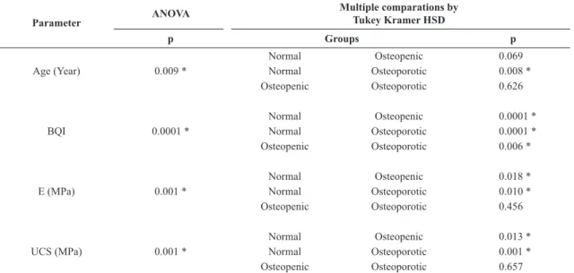
ANOVA (Table 2) showed signiicant differences in the parameters Age, E and UCS, extremely signiicant for BQI. Multiple comparisons by Tukey Kramer HSD showed signiicant differences in the E (Normal/Osteopenic and Normal/Osteoporotic) (p = 0.018 and p = 0.010) and UCS (Normal/Osteopenic), very signiicant differences in the Age (Normal/Osteoporotic), BQI (Osteopenic/Osteoporotic) and UCS (Normal/Osteoporotic) and extremely signiicant differences in the BQI (Normal/Osteopenic and Normal/Osteoporotic). However, ANOVA analysis showed the same signiicant difference between those groups. Multiple comparisons procedures by Tukey-Kramer (HSD) set out UCS as the most signiicant difference between normal and osteoporotic groups. This result can be justiied due to
Table 1. Descriptive statistics of Age, BQI, E and UCS of trabecular bone between the regions T12, L1 and L4 of 30 human vertebral segments,
determined by mechanical testing of axial compression and classiied as normal, osteopenic and osteoporotic bone by the QUS.
Parameter
Groups M ± SD (Min - Max)
Normal Osteopenic Osteoporotic
Age (Year) (47.000 - 87.000)65.000 ± 15.549 (62.00 - 93.000)77.500 ± 9.301 (68.000 - 95.000)82.500 ± 10.277
BQI 107.167 ± 18.764 72.017 ± 6.518 53.950 ± 5.364
(77.000 - 133.000) (60.333 - 80.333) (41.833 - 60.500)
E (MPa) 134.693 ± 50.999 85.475 ± 32.397 65.119 ± 23.905
(52.089 - 196.301) (45.946 - 134.882) (38.618 - 105.825)
UCS (MPa) 2.270 ± 1.142 1.239 ± 0.476 0.944 ± 0.396
(0.711 - 4.789) (0.617 - 1.889) (0.477 - 1.536)
Table 3. Correlation of BQI, E and UCS by Spearman test.
Variables r
BQI Age (Year) -0.501
BQI E (MPa) 0.499
BQI UCS (MPa) 0.508
E (MPa) Age (Year) -0.584
UCS (MPa) Age (Year) -0.684
Table 2. Analysis of comparisons of Age, BQI, E and UCS by ANOVA test and multiple comparisons by Tukey Kramer HSD.
Parameter ANOVA
Multiple comparations by Tukey Kramer HSD
p Groups p
Normal Osteopenic 0.069
Age (Year) 0.009 * Normal Osteoporotic 0.008 *
Osteopenic Osteoporotic 0.626
Normal Osteopenic 0.0001 *
BQI 0.0001 * Normal Osteoporotic 0.0001 *
Osteopenic Osteoporotic 0.006 *
Normal Osteopenic 0.018 *
E (MPa) 0.001 * Normal Osteoporotic 0.010 *
Osteopenic Osteoporotic 0.456
Normal Osteopenic 0.013 *
UCS (MPa) 0.001 * Normal Osteoporotic 0.001 *
Osteopenic Osteoporotic 0.657
the higher mineral density and trabecular bone strength of calcaneus in healthy subjects. One hypothesis for the lesser signiicant differences between osteopenics and osteoporotics groups with respect to E and UCS is the inaccuracy of QUS in deining the transition point.
BQI parameters (Table 3) showed moderate inversely proportional correlations with Age and directly proportional correlations with E and UCS. The elastic modulus (E) also presented moderate inversely proportional correlations with Age, even superior to those of BQI, yet there was no signiicant correlations with UCS, which in turn was highly inversely correlated with Age.
These results indicate that the complementary use of calcaneal ultrasonometry technique in the therapeutic evaluation of mechanical strength (trabecular bone quality) and fracture risk of human vertebrae is associated with osteoporosis. However useful, it is not a substitute method to the bone densitometry (DEXA) of human vertebrae, which is the gold standard clinical reference, but a relevant additional clinical examination.
The hypothesis was conirmed, moderate Spearman’s rank correlation coeficients were identiied between the results of bone quality index (“stiffness index” - BQI) determined by calcaneal ultrasonometry and the age of the individuals, elastic modulus (E) and the ultimate compressive strength (UCS) evaluated by axial compression tests of human vertebrae (cadavers).
Acknowledgements
This research was supported by Coordination for the Improvement of Higher Level Personnel (CAPES), National Council for Scientiic and Technological Development (CNPq) (protocol n. 149100/2010-8) and São Paulo Research Foundation (FAPESP) (protocol n. 2010/51698-0). The authors would like to thank the Institute of Orthopedics and Traumatology of the Clinical Hospital of University of São Paulo - School of Medicine, member of the National Science and Technological Institute for Biofabrication - INCT BIOFABRIS, the School of Engineering of São Carlos for the partnership, GE MEDICAL SYSTEMS LUNAR by loan of the Aquilles Insight ultrasonometry
equipment and to all people involved in the collection and testing of samples.
References
Adams JE. Advances in bone imaging for osteoporosis. Nature Reviews. Endocrinology. 2013; 9(1):28-42. PMid:23232496. http://dx.doi.org/10.1038/nrendo.2012.217.
American Society for Testing and Materials – ASTM International. West Conshohocken: ASTM [cited 2016 Jan. 1]. Available from: https://www.astm.org
Cauley JA, Blackwell T, Zmuda JM, Fullman RL, Ensrud KE, Stone KL, Barrett-Connor E, Orwoll ES. Correlates of trabecular and cortical volumetric bone mineral density at the femoral neck and lumbar spine: the osteoporotic fractures in men study (MrOS). Journal of Bone and Mineral Research. 2010; 25(9):19-58. PMid:20572023.http://dx.doi.org/10.1002/ jbmr.86.
Cesar R, Boffa RS, Fachine LT, Leivas TP, Silva AMH, Pereira CAM, Reiff RBM, Rollo JMDA. Evaluation of trabecular microarchitecture of normal osteoporotic and osteopenic human vertebrae. Procedia Engineering. 2013; 59:6-15. http://dx.doi. org/10.1016/j.proeng.2013.05.087.
Doblaré M, Garcia JM, Gomez MJ. Modeling bone tissue fracture and healing: a review. Engineering Fracture Mechanics. 2004; 71(13-14):1809-40. http://dx.doi.org/10.1016/j. engfracmech.2003.08.003.
Ebeling PR. Osteoporosis in men. The New England Journal of Medicine. 2008; 358(14):1474-82. PMid:18385499.http:// dx.doi.org/10.1056/NEJMcp0707217.
Fontelles MJ, Simões MG, Almeida JC, Fontelles RGS. Metodologia da pesquisa: diretrizes para o cálculo do tamanho da amostra. Revista Paraense de Medicina. 2010; 24:57-64. Guglielmi G, Terlizzi F. Quantitative ultrasound in the assessment of osteoporosis. European Journal of Radiology. 2009; 71(3):425-31. PMid:19651483.http://dx.doi.org/10.1016/j. ejrad.2008.04.060.
Hernlund E, Svedbom A, Ivergård M, Compston J, Cooper C, Stenmark J, Kanis JA. Osteoporosis in the European Union: medical management, epidemiology and economic burden. Archives of Osteoporosis. 2013; 8(1-2):136. PMid:24113837. http://dx.doi.org/10.1007/s11657-013-0136-1.
Kanis JA, McCloskey EV, Johansson H, Cooper C, Rizzoli R, Reginster JY. European guidance for the diagnosis and management of osteoporosis in postmenopausal women. Osteoporosis International. 2013; 24(1):23-57. PMid:23079689. http://dx.doi.org/10.1007/s00198-012-2074-y.
Liu JM, Ma LY, Bi YF, Xu Y, Zhao HY, Sun LH, Tao B, Li XY, Wang WQ, Ning G. A population-based study examining calcaneus quantitative ultrasound and its optimal cut-points to discriminate osteoporotic fractures among 9352 Chinese women and men. The Journal of Clinical Endocrinology and Metabolism. 2012; 97(3):800-9. PMid:22170722.http://dx.doi. org/10.1210/jc.2011-1654.
National Osteoporosis Foundation – NOF. Clinician’s Guide to Prevention and Treatment of Osteoporosis [Internet]. Washington: NOF; 2014 [cited 2016 Jan. 1]. Available from: http://nof.org/files/nof/public/content/file/2791/upload/919.pdf Parfitt AM, Drezner MK, Glorieux FH, Kanis JA, Malluche H, Meunier PJ, Ott SM, Recker RR. Bone histomorphometry: Standardization of nomenclature, symbols, and units. Journal of Bone and Mineral Research. 1987; 2(6):595-610. PMid:3455637. http://dx.doi.org/10.1002/jbmr.5650020617.
Raum K, Grimal Q, Varga P, Barkmann R, Glüer CC, Laugier P. Ultrasound to assess bone quality. Current Osteoporosis Reports. 2014; 12(2):154-62. PMid:24652476.http://dx.doi. org/10.1007/s11914-014-0205-4.
São Carlos: University of São Paulo; 2004. http://dx.doi. org/10.11606/D.82.2004.tde-22042004-093459.
Roux JP, Wegrzy J, Arlot ME, Guyen O, Delmas PD, Chapurlat R, Bouxsein ML. Contribution of trabecular and cortical components to biomechanical behavior of human vertebrae: an ex vivo study. Journal of Bone and Mineral Research. 2010; 25(2):356-61. PMid:19653808.http://dx.doi.org/10.1359/jbmr.090803. Shenoy S, Chawla JK, Sandhu JS. Multisite quantitative ultrasound: it’s comparison with dual energy X-ray absorptiometry in the diagnosis of osteoporosis. Journal of Orthopaedics and Allied Sciences. 2014; 2(2):40-4.