*e-mail: [email protected], [email protected]
1. Introduction
Within the past decade, there were several reports about
phase separation in the rapid solidiied Ag-Cu-Zr system.
Kundig et al.1 revealed a miscibility gap of a liquid phase
in the Ag-rich region. Phase separation was predicted due to the positive enthalpy of mixing of Cu-Ag (10.8 kJ.mol–1)
and negative enthalpies of mixing of both Ag–Zr and Cu–Zr (–20 kJ.mol–1 and –23 kJ.mol–1, respectively)2. Kang & Jung3
reported a complete prediction of the Cu–Zr–Ag system based
on thermodynamic calculation. The prediction indicates an area of miscibility gap with spinodal decomposition where
the Ag-rich nanocrystalline particles are formed in an amorphous matrix up to 90 at.% of Ag content. However,
a few evidences have been reported on the liquid phase
separation of the Ag-Cu-Zr system in the Ag-rich area. Janovszky et al.4 described microstructural evolution of
the liquid phase separation in the Ag-Cu-Zr system up to 70 at.% of Ag content. Castellero et al.5 reviewed liquid
miscibility gap in the melt- spun Ag-Cu-Zr system at 73 at.% of Ag content.
Several factors may inluence the microstructural
evolution of phase-separated glass; a critical temperature, compositional range in the miscibility gap, symmetry of the spinodal curve, glass–forming ability of the liquid, and cooling rate6,7. In addition, the microstructural evolution is
inluenced by viscosity and diffusivity of the melt8. Size of the phase-separated structure also depends on time interval between an onset of the phase separation and completion
of the solidiication9. The boundaries of the liquid ternary
miscibility gap can also be enlarged by applying high cooling rates7.
Basically, two types of structures are observed in liquid-liquid phase separation owing to different separation mechanisms: interconnected-type structure due to spinodal decomposition, and droplet-type structure due to the nucleation and growth mechanism6. Park et al.10 clearly showed that the interconnected-type or droplet-type structure depends on
the alloys composition in the miscibility gap. According to
the glass-forming ability of the separated liquid phases, an amorphous/amorphous or amorphous/crystalline structure can be obtained as composite structures6,10,11. The phase separating mechanisms provide a unique opportunity for designing composite materials with hierarchical microstructure at different length scales.
The present study focused on investigating the microstructure
and phase formation of the melt-spun Ag-rich Ag-Cu-Zr system up to 80 at.% of Ag content. Microstructural evolution of phase separation was studied using XRD, DSC, SEM and TEM including Vickers micro-hardness tester.
2. Experimental
Ag100-2xCuxZrx alloys, where x = 10, 15 and 20, were prepared with high-purity starting elements (99.99% purity or higher). Ingots were melted in a water-cooled copper crucible under a Ti-gettered argon atmosphere. The alloys were re-melted at least four times in the same furnace to ensure chemical homogeneity.
Rapidly solidiied ribbons were prepared in a single roller melt-spinner in Ar atmosphere by remelting each ingot in
Phase Separation in Rapid Solidiied Ag-rich Ag-Cu-Zr Alloys
Saisamorn Niyomsoana,b*, Piter Gargarellab,c, Natthaphol Chomsaenga, Preecha Termsuksawadd,
Utha Kühnb, Jürgen Eckertb
aJewelry Materials Research and Development Center, Faculty of Gems,
Burapha University, Chanthaburi 22170, Thailand
bIFW Dresden, Institut für Komplexe Materialien, Helmholtzstraβe 20, D–01069, Dresden, Germany cDepartamento de Engenharia de Materiais, Universidade Federal de São Carlos,
Rod. Washington Luis, Km 235, CEP 13565-905, São Carlos, SP, Brazil
dSchool of Energy, Environment and Materials, King Mongkut’s University of Technology Thonburi,
Bangkok 10140, Thailand
Received: September 30, 2014; Revised: October 6, 2015
The microstructure and phase formation of rapid solidiied Ag-rich Ag-Cu-Zr alloys were
investigated. Two types of structure; interconnected- and droplet-type structures, were obtained due to phase separation mechanisms. The former was spinodal decomposition and the later was nucleation
and growth mechanism. Depending on the alloy compositions, three crystalline phases; FCC-Ag, AgZr and Cu10Zr7 phases were observed along with an in-situ nanocrystalline/amorphous composite.
Vickers hardness testing indicated a signiicant increase of hardness in the
nanocrystalline/amorphous-composite alloy.
a quartz tube and then ejecting it through a nozzle onto a spinning Cu wheel. Melt spinning was performed at 30 ms–1 wheel speed. The ribbons then experienced high cooling rate of approximately 6.9×10⁶ K.s−1[12]. An ejection temperature
of the ribbons was 1273 K and an ejection pressure was around 50 MPa. An approximate width and thickness of the melt-spun ribbons were 5 mm and 50 μm, respectively.
The structure of the ribbons was characterized using X-ray diffraction (XRD) on a STOE STADI P diffractometer
in transmission geometry (Bragg-Brentano) and equipped
with a Co-source (monochromatic radiation, λKα = 1.789 Å).
The microstructure was analysed using FEI Nova Nano SEM 450 scanning electron microscope (SEM) in backscattered mode. The composition of different phases was conirmed using a Bruker Xflash 6/30 energy-dispersive X-ray spectrometer (EDX) attached to the SEM. An experimental detection limit of the EDX measurement was around
0.1 at.%[13]. The thermal stability was studied by differential
scanning calorimetry (DSC) at a heating rate of 30 K/min in a Perkin-Elmer differential scanning calorimeter (DSC-7).
The measurements were carried out in an argon atmosphere ranging from 298 K to 923 K.
Fine microstructural features in the Ag-Cu-Zr ribbons
were further analysed by transmission electron microscopy
(TEM). Thin foils of these samples were examined using a JEOL JEM-2010 transmission electron microscope, operating at 200 kV. The sample preparation for TEM analysis was inished by a dimple grinder. Vickers hardness
was measured on the ribbon cross section with a load of 10 g
and loading time of 10 s using a HMV-2000 Microhardness tester SHIMADZU.
3. Results and Discussion
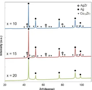
XRD patterns of the rapid solidiied Ag100-2xCuxZrx ribbons are shown in Figure 1. The analysis indicated FCC-Ag
phase in all ribbons. AgZr and Cu10Zr7 phases were clearly
detected in the ribbons with 10-15 at.% of Cu-Zr contents. Additionally, the FCC-Ag phase obtained broad XRD peaks
indicating nanocrystalline structure.
With 10-15 at.% of Cu and Zr contents, the rapid solidiied Ag100-2xCuxZrx ribbons possessed three equilibrium
phases; FCC-Ag, AgZr and Cu10Zr7, in agreement with the
prediction reported by Castellero et al.5. Phase transformation
of the Ag-rich Ag-Cu-Zr compositions went through many
isothermal reactions as follows5:
L → L1 + L2 Liquid phase separation (1)
L1 + L2 → Lʹ1 + Lʹ2 + AgZr Primary precipitation (2)
Lʹ1 (Ag-rich) → Lʹ2(Ag-poor) + AgZr + fcc-Ag
Monotectic reaction (3)
Lʹ2+ fcc-Ag → AgZr + AgCu4Zr Peritectic reaction (4)
AgZr + AgCu4Zr → Cu10Zr7 + fcc-Ag Peritectiod reaction (5)
According to the reaction sequences, phase formation of the Ag70Cu15Zr15 and Ag80Cu10Zr10 alloys started from
liquid phase separation with a precipitation of AgZr phase. Subsequently, the monotectic, peritectic and, inally, peritectiod
reactions leaded to the presence of three equilibrium phases.
Consequently, the AgCu4Zr phase might be formed by the peritectic reaction and then, in this study, completely transformed
into Cu10Zr7 phase by the peritectoid reaction above 873 K
[5].
However, the same sequence was not applicable with the phase formation in the Ag60Cu20Zr20 ribbons.
Figure 2 shows the DSC result for the Ag60Cu20Zr20 ribbon at a heating rate of 30 K/min. Three typical events of a rapid
solidiied sample could be clearly observed: relaxation, glass transition and crystallization. The relaxation occurred with
an onset relaxation temperature (Tr ) at 568 ± 2 K. The glass transition (Tg) was observed at 665 ± 2 K followed by two
exothermic crystallization peaks with an onset crystallization
temperature (Tx) at 707 ± 2 K. However, these temperatures depend on the cooling rate14. The DSC analysis proved
the presence of a glassy phase in the Ag60Cu20Zr20 ribbon.
Figure 1. XRD patterns of the melt-spun Ag100-2xCuxZrx samples,
where x = 10, 15 and 20.
Figure 2. DSC results of the melt-spun Ag60Cu20Zr20 ribbon
Combining the DSC and XRD results, it was possible to conirm the presence of the glassy phase with nanocrystalline FCC-Ag phase in the melt-spun Ag60Cu20Zr20 system.
The microstructure of the rapid-solidiied Ag100-2xCuxZrx ribbons, where x = 10, 15 and 20, was investigated by
SEM and the results are shown in Figure 3 and Figure 4.
A liquid-like phase distribution in all ribbons revealed
that phase separation performed in the liquid state. In the
ribbons with 10-15 at.% Cu and Zr contents, primary phase
separation occurred through the spinodal decomposition
mechanism. As the Cu and Zr contents reached 20 at.%, the
primary phase was separated by the nucleation and growth
mechanism. Multi-step phase separation was also observed in the Ag60Cu20Zr20 and Ag70Cu15Zr15 ribbons especially at the free surface.
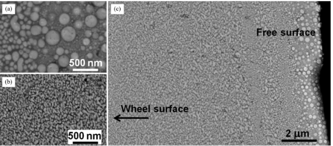
Figure 3. SEM image of the melt-spun Ag60Cu20Zr20 ribbon. (a) free surface, (b) wheel surface and (c) cross-section view of the ribbon.
The ribbon ejection temperature was 1273 K.
The microstructure of the melt-spun Ag60Cu20Zr20
ribbons consisted of a bright liquid-like phase distributed among a dark matrix (Figure 3). The bright and dark phases
corresponded with the Ag-rich phase and Cu-Zr-rich phase (or Ag-poor phase), respectively, according to the backscattered microscopy and EDX measurements. Dispersion of droplets of the Ag-rich liquid in the Cu-Zr-rich liquid indicated that volume fraction of the Ag-rich phase in the ribbon was less
than 50 vol.%[7].
Different sizes and morphologies of the melt-spun Ag60Cu20Zr20 ribbon changed across thickness of the ribbon.
Cooling rate increased as going from the free surface to the wheel surface of the ribbon. Consequently, the spherical droplet-type structure and ine irregular droplet structure
were observed as shown in Figure 3a and b. Fast heat
transfer from the melt to the spinning Cu wheel provided an extremely high driving force for nucleation. Approximately 50-100 nm-size spherical droplets were formed at the free
surface of the ribbon and there were also many droplets
below 50 nm. At the wheel side, the droplets were averagely
smaller than 50 nm, reviewing that this part of the ribbon experienced higher cooling rate.
At the free surface of the Ag60Cu20Zr20 ribbons, the multi-step phase separation was clearly observed (Figure 3a).
As a result of the second phase separation during further
cooling, the small internal droplets appeared within the large droplets as so called a marble-type structure15. From their microstructural morphology, the primary and secondary phase
separation mechanisms were identiied as the nucleation
and growth process6,9.
SEM images of the melt-spun Ag70Cu15Zr15 and Ag80Cu10Zr10 ribbons were shown in Figure 4a and b, respectively. The
interconnected structure of the bright region (Ag-rich phase) and the dark region (Cu-Zr-rich phase) was observed in both
ribbons. The experimental observation of the interconnected microstructure implied that the liquid phase separation occurred with the spinodal decomposition mechanism6.
The interconnected structure of the Ag-rich and Cu-Zr-rich
phases revealed an approximately equal volume fraction of
the two phases in the Ag70Cu15Zr15 ribbon
7. However, in the
Ag80Cu10Zr10 ribbon, a smaller scale of the interconnected
Cu-Zr-rich phase indicated a lower volume fraction of the Cu-Zr-rich phase than that of the Ag-rich phase.
The multi-step phase separation was obviously presented
at the free surface of both melt-spun Ag70Cu15Zr15 and
Ag80Cu10Zr10 ribbons (Figure 4). Small internal droplets were formed as the secondary separated phases inside the interconnected structures. The droplet morphology indicated the secondary phase separation mechanism as the nucleation and growth process6. The same multi-step phase separation
mechanism was also reported by Janovszky et al.7 in the
Ag70Cu15Zr15 alloys prepared at lower cooling rate varying from 2000 K.s–1 to 20000 K.s–1.
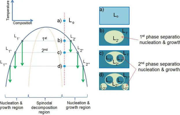
The current experimental observation showed that the
melts of the Ag100-2x CuxZrx alloys, where x = 10, 15 and 20, were rapidly cooled into the miscibility gap and then decomposed into two distinct types of structure. The purposed phase separation sequences were schematically drawn in Figure 5 and Figure 6 to describe the structural evolutions
regarding works of Ziewiec et al.9 and Chang et al.6. Sequence of the marble-type structure development in the
Ag60Cu20Zr20 ribbon due to the multi-step phase separation
during rapid cooling was sketched in Figure 5. An alloy with a starting composition locating in the nucleation region
within the miscibility gap was cooled down through the
critical temperature. At the irst separation temperature
(T1st), the primary droplets were separated from the matrix.
As the temperature was further reduced, the compositions
of each phase became supersaturated within the nucleation region, and then second phase was separated at the second separation temperature (T2nd). This marble-structure sequence is normal for the starting composition that locates in the nucleation and growth region of the symmetric spinodal curve with relatively small range of spinodal composition6.
Sequence of the multi-step structure development in
the Ag70Cu15Zr15 and Ag80Cu10Zr10 ribbons was purposed in Figure 6. The interconnected structure with internal droplets was developed from the starting composition within the spinodal region. During cooling, the primary phase separation
took place at the irst phase separation temperature (T1st).
The interconnected structure was generated with the spinodal
decomposition mechanism. Consequently, at the second
phase separation temperature (T2nd), the starting compositions located in the nucleation and growth region. Therefore,
second phases were separated in the droplet-like structure
due to the nucleation and growth mechanism.
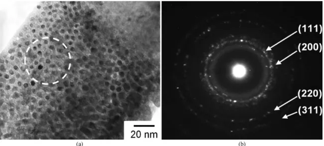
TEM results of the melt-spun Ag60Cu20Zr20 ribbons
prepared at 1273 K ejection temperature are shown in
Figure 7. The bright-ield TEM image in Figure 7a reveals spherical droplets of nanocrystalline phase with approximately 5 - 10 nanometers in diameter. Figure 7b presents the
corresponding selected area diffraction patterns (SADP) of an
area encircled in Figure 7a. The diffraction patterns consisted of a broad diffusive ring and several discrete rings which are typical for amorphous and crystalline phases, respectively. The discrete diffraction rings in Figure 7b were identiied
Figure 6. Schematic diagram showing multi-step phase separation sequence and interconnected structure with internal droplets.
as patterns from a group of crystallographic planes of FCC-Ag; (111), (200), (220) and (311) (Ref. No. 04-002-1170 of
ICDD-4+). The TEM results displayed a distribution of the FCC-Ag nanocrystalline particles among the amorphous matrix.
The in-situ nanocrystalline/amorphous composite was
clearly observed in the melt-spun Ag60Cu20Zr20 ribbons. The presence of amorphous and nanocrystalline phases
followed the prediction proposed by Kang & Jung3, which
suggests the presence of amorphous phase with the Ag-rich particles in the Ag-rich alloys containing up to 80 at.% of Ag content. The amorphous phase was a result of the Cu-Zr rich phase (Ag-poor phase) which has high glass forming ability and suppresses the crystallization in the rapid-solidiication process. In the Ag-Cu-Zr ternary system
studied by Song et al.16, the best glass-forming compositions
determined experimentally are located in the Cu-Zr rich Figure 8. Vickers hardness of Ag100-2x CuxZrx ribbons
where x = 10, 15 and 20 at.%.
area and predicted at Cu59Zr30Ag11 and Cu43Zr41Ag16 with a deviation of 6 at.%.
Vickers hardness of the Ag100-2xCuxZrx ribbons, where x = 10, 15 and 20, is shown in Figure 8. With increasing
Cu and Zr contents from 10 to 15 and 20 at.%, the hardness increased from 242 +39 to 257 + 54 and 390 +23 HV, respectively. The hardness increased with Cu-Zr contents and
dramatically increased as the content reached 20 at.% together
with the presence of both FCC-Ag nanocrystalline and Cu-Zr
rich amorphous structure. The nanocrystalline/amorphous composite exhibited excellent mechanical properties as a result of a combination of the high strength glassy phase and the ductile and tough nanocrystalline phase.
4. Conclusions
Microstructure of the rapidly solidiied Ag-rich Ag-Cu-Zr system was investigated. Liquid phase separation phenomena were experimentally observed in the melt-spun Ag100-2xCuxZrx
ribbons containing up to 80 at.% of Ag content. Depending on
the alloys composition, three crystalline phases were formed;
FCC-Ag solid solution, AgZr and Cu10Zr7 phases. Two types of phase morphology were observed; interconnected- and droplet-type structures indicating the spinodal decomposition and the nucleation and growth mechanism, respectively.
Moreover, the multi-step liquid phase separation was
observed in all alloys obviously at the free surface of the ribbons. The in-situ nanocrystalline/amorphous composite
was formed in the Ag60Cu20Zr20 ribbon which resulted in a
signiicant increase of hardness.
Acknowledgements
The authors thank S. Donath, for his technical assistance. Financial support of CNPq (Brazil), DAAD (Germany), and the Erasmus Mundus (BRIDGING THE GAP project) is
gratefully appreciated.
References
1. Kundig A, Ohnuma M, Ohkubo T, Abe T and Hono K. Glass formation and phase separation in the Ag–Cu–Zr system. Scripta Materialia. 2006; 55(5):449-452. http://dx.doi.org/10.1016/j. scriptamat.2006.05.012.
2. Boer F, Boom R, Mattens W, Miedema A and Niessen A. Cohesion in metals: transition metal alloys, cohesion and structure. Amsterdam: Elsevier; 1988.
3. Kang D and Jung I-H. Critical thermodynamic evaluation and optimization of the Ag–Zr, Cu–Zr and Ag–Cu–Zr systems and its applications to amorphous Cu–Zr–Ag alloys. Intermetallics. 2010; 18(5):815-833. http://dx.doi.org/10.1016/j.intermet.2009.12.013.
4. Janovszky D, Tomolya K, Sycheva A and Kaptay G. Stable miscibility gap in liquid Cu–Zr–Ag ternary alloy. Journal of Alloys and Compounds. 2012; 541:353-358. http://dx.doi.
org/10.1016/j.jallcom.2012.07.015.
5. Castellero A, Kang D, Jung I-H, Angella G, Vedani M and Baricco M. Rapid solidification of Ag-rich Ag–Cu–Zr alloys. Journal of Alloys and Compounds. 2012; 536S:S148-S153.
http://dx.doi.org/10.1016/j.jallcom.2011.12.089.
6. Chang H, Yook W, Park E, Kyeong J and Kim D. Synthesis of
metallic glass composites using phase separation phenomena. Acta
Materialia. 2010; 58(7):2483-2491. http://dx.doi.org/10.1016/j. actamat.2009.12.034.
7. Janovszky D, Tomolya K, Sycheva A, Pekker P and Roósz A. Liquid separation in Cu–Zr–Ag ternary alloys. Journal of Alloys and Compounds. 2014; 586:S194-S198. http://dx.doi.
org/10.1016/j.jallcom.2012.12.067.
8. Han J, Mattern N, Kim D and Eckert J. Phase separation and microstructure evolution of rapidly quenched Gd–Hf–Co–Al
alloys. Journal of Alloys and Compounds. 2011; 509S:S42-S45.
http://dx.doi.org/10.1016/j.jallcom.2010.12.210.
9. Ziewiec K, Malczewski P, Gajerski R and Ziewiec A. The
microstructure development in arc-melt and melt-spun Fe50Ni10Cu20P10Si5B5 immiscible alloy. Journal of Non-Crystalline
Solids. 2011; 357(1):73-77. http://dx.doi.org/10.1016/j.
jnoncrysol.2010.10.002.
10. Park B, Chang H, Kim D, Kim W, Chattopadhyay K, Abinandanan T, et al. Phase separating bulk metallic glass: a hierarchical
composite. Physical Review Letters. 2006; 96(24):245503. http://
dx.doi.org/10.1103/PhysRevLett.96.245503. PMid:16907253.
11. Kozieł T, Kedzierski Z, Zielinska-Lipiec A and Latuch J. The
microstructure of melt-spun alloys with liquid miscibility gap.
12. Tkatch V, Limanovskii A, Denisenko S and Rassolov S. The
effect of the melt-spinning of processing parameters on the rate cooling. Materials Science and Engineering A. 2002; 323(1-2):91-96. http://dx.doi.org/10.1016/S0921-5093(01)01346-6.
13. Kuisma-Kursula P. Accuracy, precision and detection limits of SEM–WDS, SEM–EDS and PIXE in the multi-elemental analysis
of medieval glass. X-Ray Spectrometry. 2000; 29(1):111-118. http://
dx.doi.org/10.1002/(SICI)1097-4539(200001/02)29:1<111::AID-XRS408>3.0.CO;2-W.
14. Brodowsky H and Schaller H-J. Relaxation and crystallization of metallic glasses. In: Brodowsky H and Schaller H-J, editors.
Thermochemistry of alloys: recent developments of experimental methods. Dordrecht: Springer Netherlands; 1989. p. 464-466. 15. Nagase T, Yokoyama A and Umakoshi Y. Multi-scale crystalline
Cu globule dispersed Fe-based metallic glass formation by
multi-step liquid phase separation. Journal of Alloys and
Compounds. 2010; 494(1-2):295-300. http://dx.doi.org/10.1016/j.
jallcom.2010.01.015.