MEDIAL OLIVOCOCHLEAR SYSTEM AND GENOTOXICITY
IN STUDENTS OF THE TOBACCO-PRODUCING REGION
Sistema olivococlear medial e genotoxicidade em escolares
de região fumicultora
Letícia Regina Kunst(1), Michele Vargas Garcia(1), Alencar Kolinski Machado(1),
Fernanda Barbisan(1), Aron Ferreira da Silveira(2)
(1) Universidade Federal de Santa Maria - UFSM, Santa Maria,
RS, Brasil.
(2) Departamento de Morfologia da Universidade Federal de
Santa Maria - UFSM e Programa de Pós-graduação Distúr-bios da Comunicação Humana da UFSM, Santa Maria, RS, Brasil.
Conlict of interest: non-existent
they end up exposed to several toxic substances. In tobacco culture, different types of pesticides, classes and toxicities are widely used, especially organophosphates. The indiscriminate use of these chemicals ensures greater productivity and reduces losses of the season, but the health harms in short, medium and long term are not considered¹. Besides the pesticides, the population is exposed to other highly toxic organic compounds present in the leaves of tobacco, among them, there is nicotine.
Children constitute a group with particular characteristics of exposure and special vulnerability to environmental toxicants. In addition to the fact they are in development, they differ from adults because they have relative immaturity of physi-ological and biochemical functions of the systems in
INTRODUCTION
Brazil is one of the largest producers and exporters of tobacco in the world, being tobacco farming an activity of great social and economic importance in the country. It is usually carried out by small farmers who get aid from family members, residing near the plantation. That is the reason why
ABSTRACT
Purpose: to evaluate the association between the function of the medial olivocochlear system and
the biomarkers of genotoxicity in resident students from the tobacco-producing region. Methods:
the study group was composed by 21 normal-hearing students from the tobacco-producing region and the control group by 25 normal-hearing students who did not live in the countryside. The medial olivocochlear system was assessed by the of distortion product otoacoustic emissions and genotoxic biomarkers, such as: comet assay, micronucleus test and luorimetric assay for the quantiication of DNA. The data were subjected to statistical analysis. Results: by comparing the occurrence of suppression of emissions between the groups, no signiicant association was detected. Considering the comet assay and the luorimetric assay for quantitation of DNA, the mean of the study group was considerate signiicantly higher than the mean of the control group. In the micronucleus test, it was found a signiicant difference in the sum of abnormal cells and in the frequency of binucleated cells, with the mean of the study group higher than the one found in the control group. And, in relation to the frequency of cells with micronucleus, it showed no signiicant difference between both groups. No association was found between the occurrence of suppression and the results of genotoxicity biomarkers. Conclusion: the study group had no change in medial olivocochlear system, which
was evidenced by the presence of emissions suppression, but it presented injury rates considerate signiicantly higher in relation to the genotoxic biomarkers. However, there was no association between suppression of emissions and genotoxicity.
transient or permanent, the majority of the biomoni-toring studies have used the micronucleus (MN) test and comet assay. These two techniques have been widely used to investigate DNA damage in people who were occupationally exposed and they have shown to be rapid and sensitive tests³. Currently, the use of luorescent probes, such as the luorimetric assay for quantiication of DNA with the use of the Picogreen reagent, has been highlighted. This is an objective and highly sensitive technique, which detects small amounts of DNA in solution. Moreover, it plays an increasingly important role in different studies and biological applications, being quite used in molecular biology17. It may also be used to
analyze the genotoxic or genoprotector effect of a given compound.
Based on what was described above, the aim of this study was to evaluate the association between the function of the medial olivocochlear system (MOCS) and genotoxic biomarkers in students from the tobacco-producing region.
METHODS
This is an observational, prospective, cross-sectional study. It was approved by the Ethics and Human Research Committee of the insti-tution, registered under the protocol number of 0237.0.243.000-11. It had the support of the Reference Center for Occupational Health (CEREST) of Santa Maria. All subjects agreed to participate of the research and they presented the Written Consent Form (WCF) signed by their guardians.
The study subjects were selected from public schools in two cities of the central region of Rio Grande do Sul. All schools allowed the dissemi -nation of research and they signed an Institutional Consent Term.
The students, in order to participate in this study, presented the following inclusion criteria: being aged between seven and 14 years, being normal-hearing people, presence of DPOAE, not being continuously exposed to intense noise and to cigarette smoke. As exclusion criteria, the following factors were used: presenting a history of hearing loss presenting auditory disorders, according to the criterion adopted for this study, and presenting chronic diseases and/or making use of continuous medication.
The study group (SG) was constituted by students from the tobacco-producing region, and the control group (CG) by the students from the urban area of another municipality, outside the tobacco-producing region. The students of the CG were selected in a different municipality of the SG with the aim of the proportion of body components (water, protein,
fat and minerals), the anatomical structure of the organs, in the competence of metabolizing and excreting toxic substances². They also present more speciic habits, such as “putting hand to mouth”, which increase the chances of ingesting toxic compounds present in water, soil and household dust³.
The range of adverse health effects, resulting from pesticides, includes acute and chronic damages³. Currently, several studies have demon -strated a close relationship between hearing loss and exposure to pesticides4,5. Organophosphate
pesticides induce alterations in the auditory and vestibular system, being also veriied its neurotoxic potential4, affecting the central auditory system. In
relation to the chronic health effects resulting from dermal exposure to the nicotine of the tobacco leaf, there is no much information about it. The main discussion of the toxic effects of nicotine in the smoke is related to the smoking habit. Some studies report that nicotine may have direct ototoxic effect and cause cochlear ischemia6, as well as interfering
in the neural transmission of auditory information7.
The central auditory system consists of afferent and efferent auditory pathways. The efferent auditory pathway is divided into two parts: lateral olivoco -chlear system and medial olivoco-chlear system (MOCS). MOCS consists of myelinated ibers and predominantly crossed, which will innervate the outer hair cells (OHC) 8. The normal functioning of
the MOCS can be evidenced by the abolition or reduction of OAE with the application of an ipsilateral competing noise or contralaterally9. The registration
of OAE and analysis of the suppression effect can be used for early detection of hearing impairment of cochlear and retrocochlear origin and also for the establishment of preventive actions in audiology10.
Human biomonitoring is the most eficient way to prevent and diagnose early damage from human exposure to chemicals with genotoxic potential11. The
human body is subjected to oxidative stress, which is deined as the imbalance between oxidant systems (reactive oxygen species) and antioxidants in favor of the irst ones12, causing damage in many cellular
constituents, such as unsaturated lipids, proteins and DNA13. Several pesticides have been tested and
they showed to have genotoxic potential14. Smoking
has also been reported as the cause of increased rates of genotoxicity15. Genotoxicity is the capability
that some substances have to induce alterations in the genetic material (DNA) of organisms exposed to them16. This alteration is considered a primary risk
factor for long-term effects.
presented in the intensity of 65dBSPL and F2 in L2 = 55dBSPL. For DPOAE measures, the frequencies 1500, 2000, 3000, 4000 , 5000 and 6000Hz were tested. DPOAE was considered present when the signal/noise relationship was greater than or equal to 6 dBSPL at least three frequencies.
The contralateral acoustic stimulation was a white noise, in the intensity of 60 dBHL (25), generated by the audiometer which was mentioned above (Interacoustics model AC 40 via earphone TDH -39). In order to avoid manipulation of the DPOAEs probe, the phone was attached to the ear in a contralateral way to the recording of DPOAE before the beginning of the test. In this research, it was respected the following order of tests: DPOAE in the right ear (RE) without noise, DPOAE in the RE with noise, DPOAE in the left ear (LE) without noise and DPOAE in LE with noise.
The calculation of DPOAE suppression was performed by subtracting the level of DPOAE without contralateral acoustic stimulation of the answer level of DPOAE with contralateral acoustic stimulation. The analysis of suppression effect was a response (overall response). The response is calculated from the geometric mean of the frequencies under test. This study considered the effect of this deletion with decreasing of DPOAE amplitudes of at least 0.5 dB and suppression effect absent when the difference was less than 0.5 or negative. According to Collet et al. 18, an effect of suppression from 0.5 to 1.0 dB
shows the integrity of MOCS.
Finally, the collection of biological material to perform genotoxic tests was performed, however; only 18 subjects from the SG and 18 subjects from the CG performed this procedure, reducing the sample to these evaluations.
Genotoxicity tests performed in this study were: comet assay, micronucleus test (MN) and luori -metric assay for quantiication of DNA. The collection of biological material (blood and epithelial cells of the oral mucosa), for the testing, was performed by an accomplished nursing technician. After the collection, the blood was immediately stored in a tube with anticoagulant heparin and used in the comet assay and luorimetric assay for quantifying DNA. The epithelial cells, for the MN test, were deposited in a Falcon conical tube containing 2 mL of saline solution or PBS pH 7.4 solution.
The Comet assay was carried out according to the method proposed by Singh et al.19 and modiied
by Collins Ma and Duthie20
. For each subject, slides
were prepared in duplicate . All the steps were conducted without direct light to prevent additional DNA damage. In order to apply the technique, 5μL of sample (leukocytes) with 90μL of 0.75 % agarose were mixed in an eppendorf. The solution was added ensuring that these subjects were free of exposure
to pesticide and to the nicotine derived from tobacco leaf.
In the selection of the SG, 103 students attended the inclusion criteria of the study, but only 25 showed interest in participating of the research. From these ones, 22 participated of the research. On the other hand, for the selection of the CG, three public schools were visited, being invited 250 students on mean, but only 57 subjects showed interest, and only 26 students participated of the survey.
The convenience sample of this study initially had 48 volunteers. From these ones, two presented alterations in the basic audiometric assessment, being, so, excluded from the research and they were sent to proper treatment. The inal sample consisted of 46 students - 21 belonging to the SG and 25 belonging to the CG.
The audiometric assessments and the collec-tions of biological materials, from both groups, were carried out at CEREST, preferably all of them in the same day.
Initially, parents and/or guardians and the subject herself/himself underwent a questionnaire, to identify the criteria for inclusion and exclusion. All children were subjected to visual inspection of the external auditory canal by using the Clinical Otoscope Welch-Allyn Klinic, to verify any altera-tions that could make dificult the testing and the audiometric assessment, consisting of: pure tone audiometry (PTA) and immittanciometry.
PTA was carried out in a sound-treated booth with the audiometer Interacoustics AC40 model and earphone TDH-39. In the PTA air conduction, thresholds were investigated in the frequencies of 500, 1000, 2000 and 4000 Hz. The chosen technique was the descending-ascending one. Normal-hearing subjects were considered those ones who presented tritone mean (500, 1000 and 2000 Hz) less than or equal to 25 dBHL (decibel hearing level).
The acoustic impedance measurements were carried out with the AT 235 equipment, Interacoustics and 226 Hz probe tone, for research of the tympa-nometric curve and acoustic relexes. These ones were investigated in the frequencies from 500 to 4000Hz bilaterally, in a contralateral mode. Only children with type A tympanogram and acoustic relexes were present in the sample.
ive minutes and placed in a bowl containing staining solution for 25 minutes at 37 °C. After staining, the slides were washed three times in distilled water and again left to dry in room temperature.
The slides were analyzed using the binocular optical microscope Olympus®, model CX40, with an increase of 400 times. A score was made for each sample, of 100 cells (50 per slide). The slides were analyzed by two independent observers, and, for the damage index (DI), it was considered the mean damage of the two analyzed slides. The calcu-lation of the damage index (DI) was made from the formula proposed by Cavalcanti et al.21: ID = (x 0 n0)
+ (1 x n1) + (2 x n2) + (3 x n3) + (4 x n4), where n = number of nuclei for each analyzed class.
The ive categories that were used for the classi -ication of the Comet are those ones proposed by García et al. 22 and shown in Figure 1.
to a pre-covered slide with 1 % normal agarose and covered by a coverslip, being in the fridge for ive minutes. After the coverslip was removed and the slide placed in a vat with lysis solution for one day at 4 °C. The slides were removed from the lysis solution, washed with distilled water. Then, they were placed in a horizontal vat containing electro -phoresis solution. The slides were left in this solution for 20 minutes at rest to permit the DNA unwinding, subsenquently, the electrophoresis was performed for 20 minutes at 25 Voltz (V) and 300 microam -peres (mA). Then, the slides were placed in a vat with neutralizing solution for ive 45 minutes, being washed three times with distilled water and dried until the next day at room temperature. The slides were rehydrated for ive minutes and placed in a vat containing ixative solution for ten minutes. After that, they were washed three times and left to dry at room temperature. They were rehydrated again for
Fígure 1 – Classiication of Damage to DNA. (Fronza et al., 2011)
In the MN test, the samples of the epithelial cells of the oral mucosa were centrifuged at 1000-1500 RPM during ten minutes at room temperature. Then, the supernatant was discarded using individual Pauster pipettes, being careful to not remove the cell pellet. So, it was added 1.5 ml of ixative solution and centrifuged again at 1000-1500 rpm for 1-2 minutes. Again the supernatant was discarded, keeping some of the ixative solution in the tube, and then the content was homogenized with Pauster pipettes to resuspend the cells. The contents were deposited in clean slides and directly identiied, which ones were left to dry at room temperature for 10-15 min. After, it was carried out the staining by using the
panoptic stain. Finally, the slides were washed with distilled water in a way to remove the excess stain and they were left to dry at room temperature during 20-25 min.
Fígure 2 - Classiication of cells of MN test: A (cell without alteration); B (cell with MN); C (BN cell). (FRONZA et al., 2011)
For comparison purposes, only the index results of DNA damage in comet assay were used. And, for the MN test, we considered the sum of abnormal cells, the total cells with MN and the total binucleated cells.
In the luorometric assay for quantiication of DNA, initially the total blood was centrifuged at 2000 RPM for ten min. For this technique, the blood plasma was used.
Before starting the experiment it was done the reading of the Elisa plate (empty). Then, 10μL of plasma was pipetted into the Elisa plate (black) in, at least, quadruplicates. After that, 10μL of the reagent PicoGreen ® was added. After the completion of pipetting, the plate was incubated for 5 min at room temperature and protected from light (once the Picogreen reagent is photosensitive). After the incubation period, it was carried out the veriication of the luorescence plate, with 480nm of excitation and 520 nm of emission. The interpretation of the obtained values is given so that the higher the luorescence value rises; the more DNA is veriied in the environment, which indicates cell death.
Statistical analyzes were performed with the help of the SPSS software (Statistical Package for the Social Sciences) version 17.0. To determine the normality of the variables, it was used the
Kolmogorov-Smirnov test. In all analyzes, we adopted a signiicance level of 5%.
To analyze the occurrence of suppression of DPOAE, Fisher exact test was used. Comparing the comet assay, luorimetric assay quantiication of DNA and micronucleus test (MN) results, we used the Student-t test for independent samples. And, to compare the occurrence of suppression effect and genotoxic tests, the Mann Whitney test was used.
RESULTS
All subjects, in both groups, showed DPOAE in both ears.
When comparing the occurrence of suppression between RE and LE, both SG and CG, it was veriied no signiicant difference (p <0.05). As a result, we started to consider the presence or absence of suppression of DPOAE in RE and LE simultane-ously, taking into account only the group to which the students were part of. It was considered absence of suppression of DPOAE when it was absent in both ears.
In the MN test, when the sum of abnormal cells was compared between the groups, it was veriied signiicant statistical difference (p <0.01), so that the mean of the SG (24.9 ± 9.8) showed to be higher than the mean of the CG (14.9 ± 7.8) (Table 2). Regarding the results of the comparison between groups in the frequency of binucleated cells, it was detected a statistically signiicant difference
Table 1- Comparative analysis of the occurrence of suppression effect of DPOAE between the study
and control groups (n=46)
Suppression Groups Value-pʄ
Study (n=21) Control (n=25)
Present 19 (90,5%) 25 (100,0%)
0,203 Not present 2 (9,5%) 0 (0,0%)
ʄ: Exact Fisher Test; * p<0,05
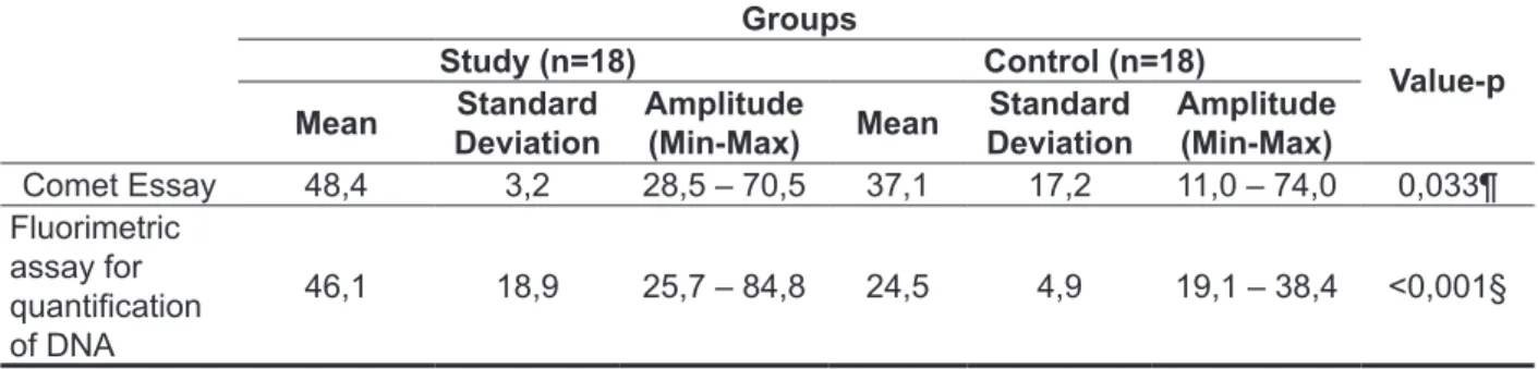
In comparison to the mean percentage of damage in comet assay, the results showed that the mean of the SG (48.4 ± 3.2) was signiicantly higher than the one found in the control group (37.1 ± 17.2). A signiicant difference is also conigured in
the comparison of the luorimetric assay test score for quantiication of DNA, where the mean was higher in SG than in the CG (46.1 ± 18.9 vs.24, 5 ± 4.9, p <0.001) (Table 2).
Table 2 - Comparative analysis of the average (mean and standard deviation) values of the damage index of the comet assay and luorimetric assay for quantiication of DNA between the study and
control groups (n=36)
Groups
Value-p
Study (n=18) Control (n=18)
Mean Standard
Deviation
Amplitude
(Min-Max) Mean
Standard Deviation
Amplitude
(Min-Max)
Comet Essay 48,4 3,2 28,5 – 70,5 37,1 17,2 11,0 – 74,0 0,033¶ Fluorimetric
assay for quantiication of DNA
46,1 18,9 25,7 – 84,8 24,5 4,9 19,1 – 38,4 <0,001§
Captions: Min-Max = minimum and maximum;
¶: Student-t Test for independent groups, assuming homogeneity of variances; §: Student-t test for independent groups, assuming
heterogeneity of variances;
*p< 0,05;
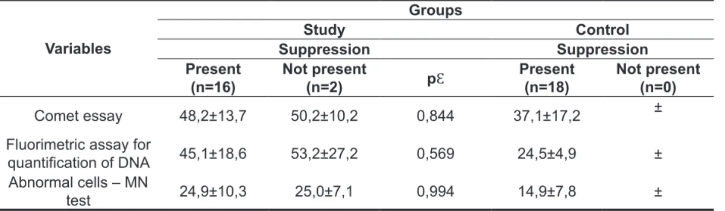
Based on the comparison of the presence and the absence of the effect of DPOAE suppression, it was compared in each group, the damage index of the Comet assay and luorimetric assay for quanti -fying DNA, and the frequency of abnormal cells in the MN test. As the results in Table 4, no statisti-cally signiicant differences (p> 0.05) were detected,
Table 3 - Comparative analysis of the average (mean and standard deviation) values of the variables of the MN test between the study and control groups (n = 36)
Variables
Groups
Value-p
Study (n=18) Control (n=18)
Mean Standard
Deviation Amplitude Mean
Standard
Deviation Amplitude
Abnormal cells 24,9 9,8 3,0 – 42,0 14,9 7,8 7,0 – 40,0 0,002§* Binucleated 12,2 6,5 2,0 – 27,0 6,3 2,2 3,0 – 11,0 0,002§ Micronuclei 11,6 7,2 0,0 – 24,0 8,2 7,2 1,0 – 32,0 0,168¶
Captions: MN = micronuclei
¶: Student-t Test for independent groups, assuming homogeneity of variances; §: Student-t test for independent groups, assuming
heterogeneity of variances;
*p< 0,05;
Table 4 - Association between DPOAE suppression effect and index of DNA damage in genotoxic tests between the study and control groups
Variables
Groups
Study Control
Suppression Suppression
Present (n=16)
Not present
(n=2) pƐ
Present
(n=18) Not present(n=0)
Comet essay 48,2±13,7 50,2±10,2 0,844 37,1±17,2 ± Fluorimetric assay for
quantiication of DNA 45,1±18,6 53,2±27,2 0,569 24,5±4,9 ± Abnormal cells – MN
test 24,9±10,3 25,0±7,1 0,994 14,9±7,8 ±
Ɛ: Mann Whitney Test; *p < 0,05;
indicating that the means of the compared variables are not related to suppression in the SG. In the infor -mation regarding to the CG, the absences of DPOAE suppression were not registered, which prevented the performance of a comparative analysis (Table 4).
DISCUSSION
The potential health effects associated to the exposure of children to pesticides are matter of constant concern. However, the chronic effects of continuous exposure to pesticides, as well to nicotine (tobacco leaf), on the human body in devel-opment are not well understood.
As previously mentioned, the pesticides can affect the peripheral and central auditory system. In this study, it was studied the effect of pesticides and nicotine on MOCS by DPOAE suppression. When
the occurrence of suppression of DPOAE between groups are compared, no statistically signiicant association was detected (p> .05), which may indicate a relative independence between groups and occurrence of DPOAE suppression. These results suggest that exposure to toxic substances (pesticides and nicotine) did not affect the functions of the MOCS in the population who was studied.
showed alterations in OHC cytoarchitecture, with greater loss of animals which received high dosage. In this research, it was also observed no functional alteration in the MOCS.
Hearing loss may be an early manifestation of the pesticide poisoning, injuring the peripheral component and also the central hearing27. The OAE
and the analysis of suppression can be used for early detection of hearing impairment with cochlear and retrocochlear origin10. Our study showed no alter
-ation in the auditory system, which was evidenced by the presence of DPOAE and suppression effect.
Another way to prevent risk regarding devel -opment of various diseases from the exposure to toxic substances is through biomonitoring. This is a useful tool to estimate the genetic risk from an integrated exposure to a complex mixture of chemic products14. In this study, genotoxic biomarkers were:
comet assay, luorimetric assay for quantiication of DNA and MN test.
In the comet assay, the group of exposed students showed a signiicantly higher damage index than the unexposed one. These indings corroborate the results of other authors28 who, when studying
genotoxic effects on tobacco growers through the comet test also showed that the treatment group presented a signiicant increase of DNA damage if compared to the CG. Da Silva et al.29 also observed
that the DNA damage by comet assay in tobacco growers had increased three times when it was compared to the unexposed group.
The possible genotoxic damages caused by nicotine are still unknown30. Other authors31
observed genotoxic effect of nicotine through the comet assay.
In luorimetric assay for the quantiication of DNA, the group of exposed children also presented higher mean regarding the CG, this difference was highly signiicant (p <0.001). The luorometric assay for the quantitation of DNA is a test that uses the ultrasensitive luorescent reagent Quant-iT™ PicoGreen® dsDNA (Invitrogen), which detects small amounts of double-stranded DNA in solution32.
The PicoGreen (PG) binds to DNA and, when that happens, its luorescence increases > 1000 times, and this is proportional to the amount of present DNA (circulating) 33,34.
In the literature, studies that used the luorimetric assay for the quantiication of DNA as a biomarker of pesticide exposure are limited. However, it is a technique that plays an increasingly important role in many studies and biological applications, such as molecular biology and diagnostic techniques as biomarker for severe diseases such as Dengue Hemorrhagic Fever Dengue Hemorrágica35.
In the absence of those ones, this research was related to studies with subjects exposed to noise and/or to other kinds of ototoxic substances and also to studies that assessed the central auditory system by means of auditory evoked potentials.
The most used organophosphate pesticides, in the tobacco plantation, present a mechanism of action based on the inhibition of acetylcholines-terase, which consequently increases the level of the neurotransmitter acetylcholine in synapses24.
According to Harkrider, Champlin, McFadden7,
nicotine also has effect on the neurotransmitter acetylcholine. The same researchers evaluated the results of OAE and BAEP of ten normal-hearing subjects and nonsmokers, after administration of nicotine, realized that it interferes in the neural trans-mission of auditory information. The effect of nicotine on high neural centers may have an inhibitory effect on the OHC. That is justiied by the acceleration of acetylcholine, which is the neurotransmitter of the efferent auditory system, implying an increase in the effect of OAE suppression.
Another study evaluated the effect of smoking on the auditory system, comparing the results of conventional and high-frequency audiometry of TOAE and the suppression effect between smokers and nonsmokers, concluding that cigarette smoking has an adverse effect on hearing. Speciically in relation to the evaluation of MOCS, the researchers have noted a reduction in the response level of otoacoustic emissions (suppression) in 100% of cases in both groups, and the group of smokers showed higher suppression values when compared to the non-smoking group25. These results disagree
with the two other already mentioned studies, once it was found no increase in DPOAE suppression effect of the students exposed to pesticides and nicotine.
A study that evaluate the condition of the MOCS of subjects exposed to organic solvents through the suppression effect of TEOA observed that the presence of TEOA suppression effect was higher in the control group (72%) if compared to the study group (58%), but this difference was not statisti -cally signiicant26. In this study, the presence of the
suppressive effect was also higher in the CG (100%) when compared to the SG (90%), but this difference was even less signiicant.
Other authors1 investigated the effect of
authors had speciically studied children exposed to pesticides and they also observed a signiicant increase in the frequency of cells with MN in the exposed group3,39. The results of our study disagree
with the indings of these authors. However, these results are consistent with the indings of Pastor et al 40 who also found no difference between subjects
exposed and not exposed to pesticides in relation to the frequency of MN .
In this study, we tried to examine the association between the MOCS functions (presence and absence of DPOAE suppression) and genotoxic damages. The results of this study indicate that the damage index which was found in genotoxic tests do not depend on the DPOAE suppression in the SG, ie, there are no positive association between the variables. Fronza et al.18 veriied that smokers have
elevated levels of genotoxicity, evidenced by the Comet assay, however, no signiicant associations between the lack of effect of DPOAE suppression and genotoxicity were observed.
CONCLUSION
In this study, no alterations in MOCS were observed, which was evidenced by DPOAE suppression in students who live in the tobacco-producing region. However, signiicantly elevated damage indexes of genotoxic biomarkers in exposed students have been observed. However, no association between DPOAE suppression and Genotoxicity was veriied.
These indings show that there is no alteration in relation to the auditory function (MOCS), but the results of genotoxic biomarkers indicate the presence of a susceptible genetic proile for the development of future pathologies resulting from the exposure to these pesticides, being the hearing loss one of them.
In this study, the luorimetric assay for quanti-ication of DNA, using the reagent Picogreen, was shown to be an effective and highly sensitive technique for analyzing the genotoxic effect of pesticides on the exposed population. These results corroborate the indings of Parra, Sanchez-Fortún and Castanõ36, although these tests had been made
in different kinds of cells (lymphocytes and tissue cells). They studied the applicability of the reagent Picogreen in the quantiication of DNA to determine the genotoxic effects induced by organophosphorus pesticides in tissue cells of ish. They concluded that this method was able to put in evidence the degree of DNA damage induced by these pesticides. Therefore, it can be used to prevent the genotoxic effects of pesticides in these kinds of cells. They also note that the necessary amount of chemicals, the cost and time required for the test is drastically reduced, so it could replace other genotoxicity tests.
On the other hand, the MN test is a widely used technique in studies of biomonitoring of populations exposed to different kinds of toxic substances. In our study, in the MN test, it was detected a statistically signiicant difference when the sum of abnormal cells and the frequency of binucleated cells between the groups are compared, with the mean of the SG higher than the one for the CG. However, in comparison of the frequency of cells with MN, the difference was not set, although the mean of the SG is higher than the one for the CG. These indings agree in parts with the results of other researchers who found signiicant differences in all types of abnormal cells when comparing the exposed group with the CG3,29,37.
7. Harkrider AW, Champlin CA, Mcfadden D. Acute effect of nicotine on non-smokers: OAEs and ABRs. Hear Res. 2011;160:73-88.
8. Durante AS. Emissões otoacústicas. In: Bevilacqua MC, Martinez MAN, Balen AS, Pupo AC, Reis ACMB, Frota S (eds). Tratado de Audiologia. São Paulo: Santos; 2011, pp. 145-58.
9. Muñiz JF, Ventura AM, Algarra JM. Estudio de la correlación existente entre el efecto supresor contralateral y la fatiga auditiva mediante otoemisiones acústicas transitórias. Acta Otorrinolaringol Esp. 2006;57(5):199-203.
10. Bernardi APA. Trabalhadores expostos simultaneamente a ruído e tolueno: estudo das emissões otoacústucas evocadas e efeito de supressão [dissertação]. São Paulo (SP): Universidade de São Paulo, Faculdade de Saúde Pública; 2000.
11. Angerer J, Ewers U, Wilhelm M. Human biomonitoring: state of the art. Int. J. Hyg. Environ – Health. 2007;210(3-4):201-28. In: Da Silva FR. Risco ocupacional em fumicultores: genotoxicidade associado à suscetibilidade genética [tese]. Porto Alegre (RS): Universidade Federal do Rio Grande do Sul; 2011.
12. Halliwell B, Whiteman M. Measuring reactive species and oxidative damage in vivo and in cell culture: how should you do it and what do the results mean? Br J Pharmacol. 2004.142(2):231-44.
REFERENCES
1. Körbes D, Silveira AF, Hyppolito MA, Munaro G. Ototoxicidade por organofosforados: descrição dos aspectos ultraestruturais do sistema vestibulococlear de cobaias. Braz J Otorhinolaryngol. 2010;76(2):238-44.
2. Perry MJ. Children’s agricultural health traumatic injuries and hazardous inorganic exposures. J Rural Health. 2003;19(3):269-78.
3. Benítez-Leite S, Machi ML, Fernandez V, Franco D, Ferro EA, Mojoli A et al. Daño celular en una población infantil potencialmente expuesta a pesticidas. Pediatr . (Asunción). 2010;37(2):97-106. 4. Hoshino ACH, Pacheco-Ferreira H, Taguchi CK, Tomita S, Miranda MF. Estudo da ototoxidade em trabalhadores expostos a organofosforados. Rev Bras Otorrinolaringol. 2008;74(6):912-8.
5. Finkler AD, Silveira AS, Munaro G, Zanrosso CD. Otoprotection in guinea pigs exposed to pesticides and ginkgo biloba. Braz J Otorhinolaryngol. 2012;78(3):122-8.
6. Cocchiorella LA, Sharp DS, Persky VW. Hearing threshold shifts white-cell count and smoking status in working men. Occup Med. 1995; 45(4):179-85. In: Oliveira DCCM, Lima MAMT. Da audiometria tonal laminar em baixa e alta freqüência: comparação dos limiares auditivos entre tabagistas e não-tabagistas. Braz J Otorhinolaryngol. 2009;75(5):738-44.
RESUMO
Objetivo: avaliar a associação entre a função do sistema olivococlear medial e biomarcadores genotó-xicos em escolares residentes de região fumicultora. Métodos: trata-se de um estudo observacional, prospectivo e transversal. O grupo estudo foi composto por 21 escolares normo-ouvintes residentes de região fumicultora e o grupo controle por 25 escolares normo-ouvintes que não residiam na zona rural. O sistema olivococlear medial foi avaliado por meio da supressão das Emissões otoacústicas produto de distorção, e os biomarcadores genotóxicos foram: ensaio cometa, teste de micronúcleos e ensaio luorimétrico de quantiicação de DNA. Os dados obtidos foram submetidos à análise esta -tística. Resultados: ao comparar a ocorrência do efeito de supressão das emissões entre os grupos,
não foi detectada associação signiicante. Tanto no ensaio cometa como no ensaio luorimétrico de quantiicação de DNA a média do grupo estudo mostrou-se signiicantemente mais elevada que a do grupo controle. No teste de micronúcleos, veriicou-se diferença signiicante quanto ao somatório de células alteradas e à frequência de células binucleadas, sendo a média do grupo estudo mais elevada que a do grupo controle. Já referente à frequência de células com micronúcleo, não se observou dife -rença signiicante entre os grupos. Não foi detectada associação entre ocorrência do efeito de supres -são e os resultados dos biomarcadores genotóxicos. Conclusão: o grupo estudo não apresentou alterações no sistema olivococlear medial, evidenciado pela presença de supressão das emissões, porém apresentou índices de dano signiicantemente mais elevados dos biomarcadores genotóxicos. Entretanto, não se veriicou associação entre supressão das emissões e genotoxicidade.
26. Quevedo LS, Tochetto TM, Siqueira MA. Condição coclear e do sistema olivococlear medial de frentistas de postos de gasolina expostos a solventes orgânicos. Arquivos Int. Otorrinolaringol. 2012;16(1):50-6.
27. Manjabosco CAW, Morata TC, Marques SJM. Peril audiométrico de trabalhadores agrícolas. Arq Int Otorrinolaringol. 2004;8(4):284-95.
28. Juffo DD, Silva FR, Rohr P, Kvitko K, Da Silva J. Avaliação do dano causado ao DNA de trabalhadores da lavoura de fumo do município de Venâncio Aires RS. In: V Jornada de Iniciação Cientíica- Meio Ambiente- FZB-RS e FEPAM, 2009, Porto Alegre. Anais da V Jornada de Iniciação Cientiica-Meio Ambiente-FZB-RS e FEPAM, 2009. p. 1.
29. Da Silva FR, Da Silva J, Dias JF, dos Santos CEI, Kahl V, Rohr P et al. Genotoxic biomonitoring of tobacco farmers: Biomarkers of exposure of early biological effects and of susceptibility. J Hazard Mater. 2012; 225-226:81-90.
30. Da Silva FR. Risco ocupacional em fumicultores: genotoxicidade associado à suscetibilidade genética [tese]. Porto Alegre (RS): Universidade Federal do Rio Grande do Sul; 2011.
31. Sobkowiak R, Lesicki A. Genotoxicty of nicotine in cell culture of Caenorhabdilis elegans evaluated by the comet assay. Drug Chem Toxicol. 2009;32(3):252-7. In: Da Silva FR. Risco ocupacional em fumicultores: genotoxicidade associado à suscetibilidade genética [tese]. Porto Alegre (RS): Universidade Federal do Rio Grande do Sul; 2011.
32. Ahn SJ, Costa J, Emanuel JR. PicoGreen quantitation of DNA: effective evaluation of samples pre- or post-PCR. Nucleic Acids Research. 1996;24(13):2623-5.
33. Ikeda Y, Iwakiri S, Yoshimori T. Development and characterization of a novel host cell DNA assay using ultra-sensitive luorescent nucleic acid stain ‘‘PicoGreen’’. J Pharm Biomed. Anal. 2009;49(4):997-1002.
34. Dragan AI, Bishop ES, Geddes CD. Metal-enhanced PicoGreen luorescence: application for double-stranded DNA quantiication. Anal Biochem. 2010;396(1):8-12.
35. Ha TTN, Huy NT, Murao LA, Lan NTP, Thuy TT et al. Elevated Levels of Cell-Free Circulating DNA in Patients with Acute Dengue Virus Infection. PLoS ONE. 2011;6(10):1-7.
36. Parra JM, Sánchez-Fortún S, Castanõ A. Assessment of genotoxic effects induced by selected pesticides on RTG-2 ish cells by means of a modiied fast micromethod assay. EnvironToxicol. 2010;27(4):238-43.
13. Findlay VJ, Tapiero H, Tolunsend DM. Suliridoxin: a potential therapeutic agent? Biomed Pharmacother. 2005;59(7):374-9.
14. Kumar LP, Panneerselvam N. Toxic effects of pesticides: a review on cytogenetic biomonitoring studies. Medicine and Biology. 2008;15(2):46-50. 15. Fronza AB, Barreto DCM, Tochetto TM, Cruz IBMC, Silveira AF. Association between auditory pathway efferent functions and genotoxicity in young adults. Braz J Otorhinolaryngol. (Impr.). 2011;77(1):107-14.
16. Kohatsu AGS, Shimabukuro F, Gattás GJF. Utilização dos testes de mutagenicidade para a avaliação de exposição ocupacional. Saúde, Ética e Justiça. 2007;12(1):15-21.
17. Dragan AI, Casas-Finet JR, Bishop ES, Strouse RJ, Schenerman MA, Geddes CD. Characterization of PicoGreen Interaction with dsDNA and the Origin of Its Fluorescence Enhancement upon Binding. Biophys J. 2010;99(9):3010-9.
18. Collet L, Veuillet E, Bene J, Morgon A. Effects of contralateral white noise on click evoked emissions in normal and sensorineural ears: towards an exploration of the olivocochlear system. Audiology.1992;31(1):1-7.
19. Singh NP, McCoy MT, Tice RR, Schneider EL. A simple technique for quantitation of low levels of DNA damage in individual cells. Exp Cell Res. 1988;175:184-91.
20. Collins AR, Ma AG, Duthie SJ. The kinetics of repair of oxidative DNA damage (strand breaks and oxidised pyrimidines) in human cells. Mutat Res. 1995;336:69-77.
21. Cavalcanti BC, Sombra CML, Oliveira JHHL, Berlinck RGS, Moraes MO, Pessoa C. Citotoxicity and genotoxicity of ingenamine G isolated from the Brazilian marine schponge Pachychalina alcaloidifera. Comp Biochem Physiol A. 2008;147(4):409-15.
22. Garcia O, Mandina T, Lamadrid AI, Diaz A, Remigio A, Gonzalez Y et al. Sensitivity and variability of visual scoring in the comet assay: Results of an interlaboratory scoring exercise with the use of silver staining. Mutat Res. 2004;556:25-34.
23. Quant-iT™ PicoGreen ® dsDNA Reagent and Kits. Molecular Probes Invitrogen detection technologies. 2008.
24. Oliveira-Silva JJ, Alves SR, Meyer A, Perez F, Sarcinelli PN, Mattos RCOC et al . Inluência de fatores socioeconômicos na contaminação por agrotóxicos, Brasil. Rev Saúde Pública. 2001;35(2):130-5.
39. Neri M, Bonassi S, Knudsen LE, Sram RJ, Holland N, Ugolini D et al. Children’s exposure to environmental pollutants and biomarkers of genetic damage. I. Overview and critical issues. Mutat Res. 2006;612(1):1-13.
40. Pastor S, Creus A, Parrón T, Cebulska-Wasilewska A, Siffel C, Piperakis S et al. Biomonitoring of four european populations occupationally exposed to pesticides: use of micronuclei as biomarkers. Mutagenesis. 2003;18:249-58.
37. Martínez-Valenzuela C, Gómez-Arroyo S, Villalobos-Pietrini R, Waliszewski S, Calderón-Segura ME, Félix-Gastélum R et al. Genotoxic biomonitoring of agricultural workers exposed to pesticides in the north of Sinaloa State, Mexico. Environ Int. 2009;35(8):1155-9.
38. Bortoli GM, Azevedo MB, Silva LB. Cytogenetic biomonitoring of Brazilian workers exposed to pesticides: micronucleus analysis in buccal epithelial cells of soybean growers. Mutat Res. 2009;675(1-2):1-4.
Received on: April 16, 2013 Accepted on: December 17, 2013
Mailing address: Letícia Regina Kunst
Av. Itaimbé, 655, apto 307 – Centro Santa Maria – RS – Brasil
CEP: 97050-331