Reproductive Aspects of the Mosquito
Culex quinquefasciatus
(Diptera:Culicidae) Infected with
Wuchereria bancrofti
(Spirurida: Onchocercidae)
Catarina A Lima, Walkiria R Almeida, Hilary Hurd*, Cleide MR Albuquerque/
+Departamento de Zoologia, Universidade Federal de Pernambuco, Av. Moraes Rego s/no, 50670-420 Recife PE, Brasil *Centre for Applied Entomology and Parasitology, School of Life Sciences, Keele University, Staffordshire, UK
This study reports on the relationship between Wuchereria bancrofti infection and female body size, intake of blood and fecundity in the mosquito Culex quinquefasciatus, vector of this filarial parasite in Recife (Brazil). Adults from field collected larvae were infected via a membrane feeding procedure, using blood with parasitaemia ranging from 724-6,000 mf/ml. A positive correlation was observed between mosquito size (measured by wing length) and egg production in uninfected females. However, this relationship did not exist in W. bancrofti infected mosquitoes. This change is unlikely to be the result of changes in blood ingestion as no significant difference was found when infected and uninfected females were compared. Variation in egg production observed between trials could not be associated with parasite density in the blood. These results suggest infection with W. bancrofti may disrupt the relationship between mosquito size and egg production during the first gonotrophic cycle of C. quinquefasciatus
such that fecundity is sometimes reduced. However, this overall affect is variable and many groups of mosquitoes do not respond in this way.
Key words: Wuchereria bancrofti - Culex quinquefasciatus - mosquito - fecundity - body size - blood feeding
The parasite Wuchereria bancrofti is distributed throughout tropical regions (Central and South America, Africa, Asia, Pacific areas) and also in subtropical coun-tries in the Middle East (Service 1996). Current estimates (Ottesen & Ramachandran 1995) suggest that approxi-mately 120 million people are infected with all types of lymphatic filariae, particularly bancroftian filariasis. In Recife, a Brazilian coastal city (on 8°4’3’’ south latitude) bancroftian filariasis is endemic in many areas and is trans-mitted by Culex quinquefasciatus. The high fecundity of this mosquito species, together with the increase in num-ber of breeding places due to uncontrolled urbanization, may have contributed to the recent increase in prevalence rates of W. bancrofti from 9.7% (Azevedo & Dobbin 1952) up to 14.9% (Maciel et al. 1996).
Previous work in our laboratory has shown that C. quinquefasciatus (Recife strain) is able to control the number of developing W. bancrofti (1-2/mosquito) when ingesting variable densities of parasite up to 2,600 mi-crofilariae (mf) per ml (Albuquerque et al. 1999). These observations suggest that there is a regulation mecha-nism that controls the number of infective larvae that can successfully mature in C. quinquefasciatus. This mecha-nism is likely to involve a defence response although
en-Financial support: Wellcome Trust via a Research Develop-ment Award in Tropical Medicine (047085 to CRMA) and CNPq (process no. 521126/98-3).
+Corresponding author. Fax: +55-81-271.8359. E-mail:
cleide@ufpe.br Received 18 June 2002 Accepted 14 November 2002
capsulated and/or melanized mf are rarely observed. What-ever the method of parasite elimination, we expect it to be costly to the mosquito in terms of resources (Sheldon & Verhulst 1996), as will repair to the damage to the midgut caused by mf invasion.
Observations from several parasite/insect systems have shown that infection costs can manifest as a reduc-tion in host reproductive fitness or longevity and that fecundity reduction may occur very early in the infection, before the developing parasites are likely to cause a drain on host metabolic resources (reviewed by Hurd & Webb 1997).
Of particular relevance to this study are examples of reduction in the reproductive output of mosquitoes resulting from protozoan (for example, Hogg & Hurd 1995a, b, 1997) and nematode infections (Javadian & Macdonald 1974, Christensen 1981, Ferdig et al. 1993). In Anopheles gambiae, a reduction of 41.2% in fecundity and 61.8% in fertility was observed in mosquitoes infected with rodent malaria parasite Plasmodium yoelii nigeriensis (Ahmed et al. 1999). Parasite-induced fecundity reduction in Aedes trivittatus was related to the number of Dirofilaria immitis
it harboured, with a severe reduction in egg production occurring in females infected with more than 15 filarial parasites (Christensen 1981).
fitness of C. quinquefasciatus. In addition, factors known to affect the fecundity, such as body size (Colless & Chellapah 1960, Briegel 1990) and bloodmeal size (Lea et al. 1978), were investigated to assess the possibility that they might contribute to any fecundity reduction observed in infected females.
MATERIALS AND METHODS
Mosquito collection - C. quinquefasciatus larvae were collected from the filariasis endemic area of Azeitona, lo-cated in the district of the Metropolitan Zone in Recife, PE, Brazil. Collections were made in the same habitat, a moderately shaded drainage ditch 40 m long and 0.5 m wide. Samples were collected and transported to the labo-ratory where last stage larvae (L4) were separated from the associated fauna and were preliminary screened for species identification based on external morphological characters (Forattini 1996). Male and female mosquitoes emerging within a 24 h period were kept together for mat-ing and were provided with sugar lumps and cotton pads soaked in water. Mosquito adults were maintained at 27 ± 3oC, 85 ± 10% humidity in a 12:12 h light/dark cycle. Be-fore adult dissection, species confirmation was carried out based on diagnose from Consoli and Oliveira (1994).
Infection via artificial blood feeding - Artificial feed-ing was carried out as previously described by Albuquer-que et al. (1999) using three to four day old female mos-quitoes. Blood with different densities of circulating para-sites, ranging between 724-6000 mf/ml,was used in this work. Blood was obtained from human lymphatic filari-asis carriers, who participated in a survey carried out by Centro de Pesquisa Aggeu Magalhães-Fiocruz, Recife, after written informed consent. Venous blood was col-lected between 23.00 and 24.00 h in vacutainers contain-ing EDTA (1 mg/ml) as an anticoagulant. At the end of the work, donors were treated with diethylcarbamazine ac-cording to the therapeutic recommendation of the World Health Organization (WHO 1987). Microfilarial density was estimated by counting the number in 60 mm3 (3x20 mm3 aliquots) of blood and expressed as mf/ml. Groups of mosquitoes were allowed to feed for 30 min on the in-fected blood, heated to 37oC, using glass feeders cov-ered with cow gut membrane. To ensure mf homogeneity the blood was kept agitated using a stirrer at 1 red/sec. After feeding, individual females were transferred to uni-versal tubes and maintained for 3 days for the total excre-tion of haematin, used to estimate the blood meal size. Only females that were fully engorged were used in the experiments. A control group of mosquitoes was fed on uninfected human blood.
All mosquitoes were dissected to check for the pres-ence of mf in the thoracic muscles at the time of ovary dissection (4-7 days post blood meal). A subsample of infected mosquitoes from all groups was dissected imme-diately post infection to confirm that microfilariae had been ingested.
Haematin assay - Assessment of mosquito bloodmeal size by haemoglobinometry (Briegel et al. 1979) was not feasible, as this technique requires killing the insect be-fore egg production occurs. Haemoglobin intake was thus measured indirectly by making a spectrophotometric
de-termination of the amount of haematin excreted by each mosquito. Haematin analysis was performed by dissolv-ing the excretory products, deposited in the tubes, in 1 ml of 1% lithium carbonate. The absorbance was measured at 395 nm and the amount of haematin present determined by comparison with bovine haematin standards (Briegel 1980, Hogg & Hurd 1997).
Assessment of egg production and fertility - Follow-ing blood meal digestion mosquitoes were moved from the first vial to oviposition tubes contain 1.5 ml of water previously used to culture larvae. The number of eggs laid by each surviving female was recorded from 4 to 7 days post blood meal. Retention of fully developed eggs was determined following ovary dissection after oviposi-tion, to observe the presence of any retained eggs. Total egg production was considered as the sum of laid and retained eggs from the first gonotrophic cycle. Fertility was measured as number of larvae hatched from each raft.
Mosquito size - Mosquito body-size was expressed in terms of wing length. One wing was selected from each mosquito, mounted on a slide, and its length measured from the tip, excluding fringe, to the distal end of the allula (Lyimo & Koella 1992) using a calibrated dissecting mi-croscope with an ocular micrometer.
Statistical analysis - Data collected on wing length, haematin excretion and egg production were tested for normality. A comparison of egg production, blood meal size and wing length between different parasitaemia lev-els and between infected and uninfected groups within the same parasitaemia level was made by multiple analy-sis of variance (MANOVA) and the LSD post hoc test. A
p value of < 0.05% was accepted as significant. Compari-sons of percentage survivorship and fertility between uninfected and infected groups were assessed by Chi-square analysis. The correlation of blood meal size and wing length on egg production within species was deter-mined by regression analysis. All statistical tests were performed using Statistic 5.0 and Bio Estatistic 6.0 ver-sions. All mean values were expressed ± standard devia-tions.
RESULTS
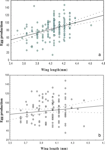
Mean wing size of each group was assessed in five of the trials (see Table II). Body size ranged from 3.69-4.25 mm and 3.72-4.22 mm in infected and uninfected females respectively. Some variation in size occurred throughout the collection period (one year), probably as a result of changes in environmental conditions at the collection site. However, when data from uninfected and infected mos-quitoes were analyzed via pair wise comparisons, using the Scheffé test at 0.05%, no significant differences were detected between the mosquitoes assigned to infected or control groups in any individual trial (Table II). Thus size differences could not account for differences in egg pro-duction. Likewise, statistically significant differences in blood meal size (as measured by the amount of haematin excreted) were not observed when infected and control females were compared in any of the individual trials (Table II). Blood meal sizes were not affected by the density of mf in the blood meal used for feeding in these five trials. As expected, mosquito size showed a significant over-all positive correlation with egg production by linear re-gression when mosquitoes fed on uninfected blood (R =
0.38, P = 0.0001). However, the presence of mf destroyed this relationship and no positive correlation between mosquito size and egg production was observed in the infected group (R = 0.12, P = 0.06) (Fig. 1 a, b).
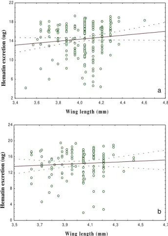
Considerable variation in haematin excretion occurred in all 5 trials and although, overall, a positive relationship was observed between haematin excretion and egg pro-duction in mosquitoes fed on uninfected blood, this was not a significant correlation (at p = 0.05 level) (R = 0.12; P = 0.06; N = 236). No relationship was demonstrated for TABLE I
Eggs production by Culex quinquefasciatus uninfected and infected with Wuchereria bancrofti at different microfilariae
(mf) rate in the blood
Mf density in Mean no. of eggs produced/female ± SD the blood/ml Infected N Uninfected N
724 96.40 ± 20.90 a (25) 79.50 ± 27.60 (28) 800 c 72.31 ± 18.61 (19) 79.95 ± 18.49 (49) 1150 97.20 ± 22.90 a (27) 86.80 ± 18.40 (47) 1200 66.50 ± 14.70 (20) 66.60 ± 17.90 (46) 1500 72.40 ± 16.50 b (30) 85.50 ± 19.60 (45) 1750 68.07 ± 20.60 (14) 69.26 ± 24.47 (15 ) 2900 82.50 ± 17.90 b (32) 97.10 ± 16.40 (71) 6000 c 72.32 ± 17.80 (40) 79.95 ± 18.40 (49)
Total (207) (350)
a: significantly greater than the respective control (P < 0.05); b: significantly less than the respective control (P < 0.05); c: infected mosquitoes in these trials were from the same generation but fed on different blood sources from subjects with different parasitaemias; N: sample size
TABLE II
Wing size and haematin excretion of Culex quinquefasciatus infected with Wuchereria bancrofti. Females were fed on blood with different parasitaemia levels
Wing size (mm) Haematin (µg)
X ± DP (N) X ± DP (N)
Mf/ml Infected Controls p Infected Controls p
724 3.89 ± 0.12 (18) 3.94 ± 0.21 (21) 0.99 15.10 ± 2.37 (25) 14.70 ± 2.80 (28) 0.40
1150 4.00 ± 0.16 (25) 4.06 ± 0.12 (30) 0.98 15.80 ± 1.52 (27) 14.60 ± 0.97 (47) 0.99
1200 3.82 ± 0.13 (20) 3.90 ± 0.18 (45) 0.93 14.70 ± 1.94 (20) 15.10 ± 0.97 (46) 0.99
1500 4.07 ± 0.15 (28) 4.08 ± 0.19 (44) 1.00 14.60 ± 2.98 (30) 15.70 ± 2.21 (45) 0.98
2900 4.14 ± 0.11 (32) 4.10 ± 0.12 (65) 0.99 15.80 ± 0.65 (32) 14.90 ± 1.86 (70) 0.99
Total 123 205 134 236
Wing length(mm)
Egg production
0 20 40 60 80 100 120 140 160
3,4 3,6 3,8 4,0 4,2 4,4 4,6 4,8
Wing length (mm)
Egg production
20 40 60 80 100 120 140 160
3,5 3,7 3,9 4,1 4,3 4,5 4,7
Fig. 1: linear regression of body size against egg production, for
Culex quinquefasciatus. a: uninfected group (y = -104 + 46.83 x; N = 205); b: infected group (y = 21.01 + 15.61 x; N = 123)
a
mosquitoes feeding on infected blood (R = 0.04; P = 0.63; N = 134) (data not shown) Similarly, although haematin production increased with mosquito size, this relation-ship was not significant in this study (Fig. 2 a, b).
Although analysis of the total data set from 5 trials demonstrated that infection disrupted the usual relation-ship between size and reproductive output, comparison of mean egg production by uninfected and infected fe-males in all eight individual trials did not produce a con-sistent picture (see Table I). Comparisons between infected groups and their respective controls showed that signifi-cantly fewer eggs were produced by infected females fed on blood containing 1,500 and 2,900 mf/ml. In contrast, a significant increase in the number of eggs produced oc-curred in infected females fed on blood with parasitaemia levels of 724 and 1,150 mf/ml and no statistically signifi-cant difference between infected and uninfected mosqui-toes was observed in the other four experiments.
Similar results were observed when the number of eggs produced by infected females and those submitted to an infected blood meal but remaining uninfected were com-pared. At the time of dissection, it was impossible to de-termine whether mosquitoes in the latter group had been infected and eliminated the parasite or whether no
para-site had been ingested during bloodfeeding. Data from these uninfected mosquitoes were therefore removed from the study when the effect of the parasite on egg produc-tion was examined.
Our comparison of egg production by mosquitoes from the same generation, but feeding on blood with different mf densities (800 and 6,000 mf/ml) revealed that a similar number of eggs was produced by infected females in each group (Table I), confirming that parasite density in the blood meal did not affect egg production
The presence of the parasite in the blood had no ef-fect on egg retention in the oviduct, which was minimal and only observed at parasitaemia of 724 mf/ml(0.4% of eggs) and 1,500 mf/ml (0.5% of eggs). In mosquitoes fed on uninfected blood, 0.4% of eggs produced were not laid within the 7 days post blood meal.
Data for fertility was only obtained for the experiment with 2,900 mf/ml. Although total egg production was sig-nificantly reduced by infection in this trial, ingestion of an infected blood meal did not significantly reduce egg fertility, as measured by the hatching rate of eggs laid by infected (94.2%, N = 1,799 eggs) and uninfected females (93%, N = 1,373 eggs), respectively (χ2 = 0.064, df = 1, P = 0.823).
The prevalence of infection was strongly affected by mosquito size (Fig. 3). The greatest proportion of mosqui-toes with one or more worms fell within the mid size range of our sample and the largest mosquitoes were the least likely to be infected with W. bancrofti microfilariae.
An overall increase of 0.9% in mortality rate was ob-served in mosquitoes fed on infected blood compared to controls up to 3-4 days post feeding. However, no statis-tically significant difference was found at any parasitaemia levels (χ2 = 1.68, df = 1, P = 0.23).
Wing length (mm)
Hematin excretion (ug)
2 6 10 14 18 22
3,4 3,6 3,8 4,0 4,2 4,4 4,6 4,8
Wing length (mm)
Hematin excretion (ug)
0 4 8 12 16 20 24
3,5 3,7 3,9 4,1 4,3 4,5 4,7
Fig. 2. linear regression of body size against hematin excretion
from Culex quinquefasciatus. a: untreated group (R = 0.11, p = 0.093, N = 205; y = 6.15 + 2.05 x); b: infected group (R = 0.06, p
= 0.45, N = 123; y = 8.89 + 1.34 x).
0 10 20 30 40 50
3.7 3.8 3.9 4.0 4.1 4.2 4.3 4.4 Wing length (mm)
% infected females
Fig. 3: frequency distribution of wing lengths of adult females of
Culex quinquefasciatus infected with Wuchereria bancrofti in classes with intervals of 0.1 mm.
DISCUSSION
Parasitic infections are generally costly to their host and this cost is often manifested as a reduction in repro-ductive fitness. This is so for most nematode/insect as-sociations that have been examined (Hurd 1990). How-ever the cost of filarial infection have been shown to be complex. A positive relationship between parasite den-sity and degree of reproductive repression have been a
described in A. aegypti infected with Brugia pahangi
(Javadian & Macdonald 1974) and A. trivittatus infected with D. imittis (Christensen 1981) during the second gonotrophic cycle post infection. In contrast, reduction on reproductive fitness was obtained during the first gonotrophic cycle in Simulium ornatum infected with only one Onchocerca lienalis worm(Renshaw & Hurd 1994) and no significant dose effect was observed.
Most previous studies were performed with labora-tory strains of vector and may not reflect interactions that occur between a natural vector and the strain of patho-gen that it transmits in the field. We thus used adults originated from field collected last stage larvae (Scorza 1972) as the source of our experimental mosquitoes and infected blood collected from subjects living in the same local. Many factors affect the number of eggs produced by mosquitoes including larval rearing conditions, adult size and age, the quality and quantity of blood ingested (Hurd et al. 1995). Although the larvae used in this study were collected from the same field site, we observed a broad range in the body size of adults that emerged and considerable variation both within and between experi-ments. Evidence from other studies demonstrated that, in the field, mosquito size is affected by changes in tempera-ture, larval density, food quality and availability, which vary seasonally (Landry et al. 1988).
Overall we demonstrated a positive correlation be-tween mosquito size and egg production in uninfected females, confirming that body size is one of the factors influencing egg production in this species, as suggested by Scorza (1972) in C. pipiens fatigans Wied. (= quin-quefasciatus) from Venezuela. Highly significant positive correlation between egg production and mosquito blood meal size have been described many times in the litera-ture (e.g. Clements & Boocock 1984, Briegel 1990, Hurd et al. 1995). As with previous studies of vector reproduction (Hogg & Hurd 1997) we found that this relationship was disrupted in C. quinquefasciatus infected with W. bancrofti microfilaria. However, no relationship between parasite density could be demonstrated and fertility (egg hatch) was not affected. This latter result is in agreement with Hammad (1997) who found no effect of W. bancrofti
on C. quinquefasciatus (from Egypt) fertility. Unfertilized females were not investigated in this work and the ab-sence of sperm in the spermateca must be considered.
In this study, our experimental protocol dictated that we used haematin excretion to assess the size of blood meal even though it is not as reliable a measure as haemoglobinometry (Hurd personal observations). Haematin production was highly variable and although we were not able to demonstrate a statistically significant correlation between blood meal size and egg production, our data suggests the infection may have altered this re-lationship. Blood sources used for feeding infected mos-quitoes in this study were from various subjects. Donors may have differed with respect to age and state of health and some may have been anaemic. These parameters were not monitored but, as with differences in mosquito size, are likely to have contributed to variability between our infected groups.
The presence of malaria parasites in a bloodmeal was not found to affect the size of meal taken by anopheline mosquitoes (Hogg & Hurd 1997, Jahan & Hurd 1997, Ahmed et al. 1999). We have now shown that, indepen-dent of the parasitaemia levels, the bloodmeal size, taken by C. quinquefasciatus was not significantly affected by the presence of W. bancrofti. This suggests that the pres-ence of mf in a bloodmeal does not inhibit feeding behaviour and that they pass through the mosquito feed-ing apparatus without interruptfeed-ing feedfeed-ing.
The variation in the effect of infection upon mosquito fecundity observed among our individual trials is difficult to explain. Haemoglobin (measured by haematin excre-tion) provides the major source of protein for egg produc-tion and intake of this component did not differ between infected and uninfected groups within a trial and thus is unlikely to have given rise to differences in egg produc-tion. Similarly, mosquito size did not differ between groups and is thus unlikely to have contributed to these differ-ences. We may however be observing differences in mos-quito response to a diversity of parasite population within the locality in which blood samples were collected. Alter-natively, mosquito populations may vary over time with respect to their response to filarial worm infection.
Ferdig et al. (1993) took a mechanistic approach to the examination of Brugia malayi infections in Armigeres subalbatus, which is partially refractory to infection. They suggested that costs imposed by mounting an immune response, including melanization of worms, were reflected in the low tyrosine and protein levels in the ovary, the abnormal development of follicles and the retardation in oviposition time. Our previous study of this symbiosis demonstrated that there was a positive correlation be-tween mf density in the donor blood and number of mf ingested by the female. However, the number of parasite that achieved the infective stage (L3) appeared to be in-dependent of mf density in the blood, at parasitaemias ranging from 550-2,600 mf/ml(Albuquerque et al. 1999). Such difference cannot be attributed to melanization since melanized mf was only occasionally seen. We can assume that the efforts of the mosquito to eliminate the parasite and to repair tissue damage must have incurred some costs but these may not have been so great as a melanization response. Reproductive costs associated with mounting an antibacterial peptide immune response have recently been demonstrated in lipopolysaccharide injected A. gambiae (Ahmed et al., in press).
In the field, Lyimo and Koella (1992) found that large
An. gambiae supported fewer malaria sporozoite infec-tions than did medium sized-females. They attributed their observation to enhanced mortality rates caused by heavier parasite loads in large mosquitoes. We also found that the proportion of females infected with the filarial parasite was higher among medium sized mosquitoes than large ones. However we found no significant difference in para-site burdens of dead mosquitoes that could indicate in-crease in mortality of large females nor could we detect a significant effect of infection on survivorship.
C. quinquefasciatus is a natural vector of W. bancrofti
relationship. In this mosquito species the majority of eggs laid by the females are produced during the first gonotrophic cycle (Suleman & Reisen 1979). Therefore any metabolic benefits of shifting resources away from egg production and into storage may be available when the parasite is developing. We hypothesise that parasites that cause such an alteration of nutrient partitioning in their host may have a selective advantage. Clearly further studies need to be performed with this natural parasite/ vector association to determine whether this is so.
REFERENCES
Ahmed AM, Baggott SR, Maingon M, HurdH. Anopheles
gambiae: The fitness costs of mounting an immune re-sponse. Oikos (in press).
Ahmed AM, Maingon RD, Taylor PJ, Hurd H 1999. The ef-fects of infection with Plasmodium yoelii nigeriensis on the reproductive fitness of the mosquito Anopheles gambiae.
Invertebr Reprod Devel36: 217-222.
Albuquerque CMR, Cavalcanti VMS, Melo MAV, Verçosa P, Regis LN, Hurd H 1999. Bloodmeal microfilariae density and the uptake and establishment of Wuchereria bancrofti
infections in Culex quinquefasciatus and Aedes aegypti.
Mem Inst Oswaldo Cruz94: 591-596.
Azevedo R, Dobbin JE 1952. Filariose (Wuchereria bancrofti) no grupo residencial do IAPB no bairro de Afogados (Recife).
Publ Avulsas Inst Aggeu Magalhães 1: 157-162.
Briegel H 1990. Metabolic relationship between female body size, reserves, and fecundity of Aedes aegypti. J Insect Physiol 36: 165-172.
Briegel H, Lea AO, Klowden MJ 1979. Hemoglobinometry as a method for measuring blood meal size of mosquitoes (Diptera:Culicidae). Med Entomol 5: 235-238.
Clements NA, Boocock MR 1984. Ovarian development in mosquitoes: stages of growth and arrest, and follicular re-sorption. Physiol Entomol9: 1-8.
Christensen BM 1981. Effect of Dirofilaria immitis on the fecundity of Aedes trivittatus. Mosq News 41: 78-81. Colless DH, Chellapah WT 1960. Effects of body weight and
size of blood-meal upon egg production in Aedes aegypti
(Linnaeus) (Diptera:Culicidae). Ann Trop Med Parasitol 54: 475-482.
Consoli RAGB, Lourenço-de-Oliveira R 1994. Principais Mos-quitos de Importância Sanitária no Brasil, Fiocruz, Rio de Janeiro, 228 pp.
Ferdig MT, Beerntsen BT, Spray FJ, Li J, Christensen B 1993. Reproductive costs associated with resistance in a mos-quito-filarial worm system. Am J Trop Med Hyg49: 756-762.
Forattini OP 1996. Culicidologia Médica. I Princípios Gerais Morfologia, Glossário Taxonômico, Universidade de São Paulo, São Paulo, 548 pp.
Hammad REI 1997. Effect of Wuchereria bancrofti Cobbold infection on the biological activities of Culex pipiens L. (Diptera: Culicidae) in Egypt. J Egypt Soc Parasitol 27: 863-870.
Hogg JC, Hurd H 1995a. Plasmodium yoelii nigeriensis: the effect of high and low intensity of infection upon the egg production and bloodmeal size of Anopheles stephensi dur-ingthreegonotrophiccycles. Parasitology111: 555-562.
Hogg JC, Hurd H 1995b. Malaria-induced reduction of fecun-dity during the first gonotrophic cycle of Anopheles stephensi mosquitoes. Med Vet Entomol9: 176-180. Hogg JC, Hurd H 1997. The effects of natural Plasmodium
falciparum infection on the fecundity and mortality of
Anopheles gambiae s.l. in north east Tanzania. Parasitol-ogy 114: 325-331.
Hurd H 1990. Physiological and behavioural interactions be-tween parasites and invertebrate hosts. Adv Parasitol29: 271-318.
Hurd H, Webb TJ 1997. The role of endocrinologically active substances in mediating changes in insect hosts and insect vectors. In NEBeckage, Parasites: Effects on Host Hor-mones and Behaviour, Chapman and Hall, London, p. 179-201.
Hurd H, Hogg JC, Renshaw M 1995. Interactions between bloodfeeding, fecundity and infection in mosquitoes.
Parasitol Today 11: 411-416.
Jahan N, Hurd H 1997. The effects of infection with Plasmo-dium yoelii nigeriensis on the reproductive fitness of Anoph-eles stephensi. Ann Trop Med Parasitol91: 365-369. Javadian E, Macdonald WW 1974. The effect of infection with
Brugia pahangi and Dirofilaria repens on the egg produc-tion of Aedes aegypti.Ann Trop Med Parasitol 68: 477-481. Landry SV, Defoliart GR, Hogg DB 1988. Adult body size and survivorship in a field population of Aedes triseriatus.
J Am Mosq Control Assoc4: 121-128.
Lea AO, Briegel H, Lea HM 1978. Arrest, resorption or matu-ration of oocytes in Aedes aegypti: dependence upon the quality of the blood meal and the interval between blood meals. Physiol Entomol3: 309-316.
Lyimo EO, Koella JC 1992. Relationship between body size of adult Anophelesgambiae s.l. and infection with the malaria parasite Plasmodiumfalciparum. Parasitology104: 233-237.
Maciel A, Rocha A, Marzochi KB, Medeiros Z, Carvalho AB, Regis L, Souza W, Lapa T, Furtado A 1996. Epidemiologi-cal study of bancroftian filariasis in Recife, Northeastern Brazil. Mem Inst Oswaldo Cruz 91: 449-455.
Ottesen EA, Ramachandran CP 1995. Lymphatic filariasis in-fection and disease: control strategies. Parasitol Today11: 129-131.
Renshaw M, Hurd H 1994. The effect of Onchocerca infection on the reproductive physiology of the British blackfly,
Simulium ornatum. Parasitology109: 337-345.
Scorza JV 1972. Observaciones Bionómicas sobre Culex pipiens fatigans Wied., 1829 de Venezuela, Universidad de los Andes, Mérida, 198 pp.
Service MW 1996. Medical Entomology for Students, Chapman and Hall, London, 278 pp.
Sheldon BS, Verhulst S 1996. Ecological immunology: costly parasite defences and trade-offs in evolutionary ecology.
Trends Ecol Evol11: 317-321.
Suleman M, Reisen WK 1979. Culex quinquefasciatus Say: life table characteristics of adults reared from wild-caught pu-pae from North West Frontier province, Pakistan. Mosq News 39: 756-762.