online | memorias.ioc.fiocruz.br
Antifungal activity of extracts from Atacama Desert fungi against
Paracoccidioides brasiliensis and identification of Aspergillus felis as a
promising source of natural bioactive compounds
Graziele Mendes1,2, Vívian N Gonçalves1, Elaine M Souza-Fagundes3, Markus Kohlhoff2,
Carlos A Rosa1, Carlos L Zani2, Betania B Cota2, Luiz H Rosa1, Susana Johann1/+
1Universidade Federal de Minas Gerais, Instituto de Ciências Biológicas, Departamento de Microbiologia, Belo Horizonte, MG, Brasil 2Fundação Oswaldo Cruz, Centro de Pesquisa René Rachou, Laboratório de Química de Produtos Naturais, Belo Horizonte, MG, Brasil
3Universidade Federal de Minas Gerais, Departamento de Fisiologia e Biofísica, Belo Horizonte, MG, Brasil
Fungi of the genus Paracoccidioides are responsible for paracoccidioidomycosis. The occurrence of drug toxic-ity and relapse in this disease justify the development of new antifungal agents. Compounds extracted from fungal extract have showing antifungal activity. Extracts of 78 fungi isolated from rocks of the Atacama Desert were tested in a microdilution assay against Paracoccidioides brasiliensis Pb18. Approximately 18% (5) of the extracts showed minimum inhibitory concentration (MIC) values ≤ 125.0 µg/mL. Among these, extract from the fungus UFMGCB 8030 demonstrated the best results, with an MIC of 15.6 µg/mL. This isolate was identified as Aspergillus felis
(by macro and micromorphologies, and internal transcribed spacer, β-tubulin, and ribosomal polymerase II gene
analyses) and was grown in five different culture media and extracted with various solvents to optimise its antifungal activity. Potato dextrose agar culture and dichloromethane extraction resulted in an MIC of 1.9 µg/mL against P. brasiliensis and did not show cytotoxicity at the concentrations tested in normal mammalian cell (Vero). This extract was subjected to bioassay-guided fractionation using analytical C18RP-high-performance liquid chromatography (HPLC) and an antifungal assay using P. brasiliensis. Analysis of the active fractions by HPLC-high resolution mass spectrometry allowed us to identify the antifungal agents present in the A. felis extracts cytochalasins. These results reveal the potential of A. felis as a producer of bioactive compounds with antifungal activity.
Key words: rock-inhabiting fungi - Atacama Desert - Paracoccidioides brasiliensis - antifungal - Aspergillus felis
doi: 10.1590/0074-02760150451
Financial support: CNPq, FAPEMIG (CBB-APQ-02587-14), CAPES, UFMG
+ Corresponding author: sjohann@icb.ufmg.br Received 11 December 2015
Accepted 15 February 2016
Paracoccidioidomycosis (PCM) is a human sys-temic mycosis endemic in Latin America (Tavares et al. 2005). Approximately 10 million people in this re-gion are infected (Stürme et al. 2011), with 85% of cases occurring in Brazil (Andrade et al. 2005), and specific social groups, such as rural workers, being particularly affected (Shikanai-Yasuda et al. 2006). PCM is acquired by inhaling airborne propagules derived from the myce-lial form of Paracoccidioides brasiliensis (Tavares et al. 2005) and Paracoccidioides lutzii (Teixeira et al. 2009). They adhere to the alveolar epithelium, where they transform into pathogenic yeasts (Torres et al. 2010).
Despite the effectiveness of treatments with current-ly available drugs (amphotericin B, azoles, and sulfon-amides), they require long term administration protocols capable of causing toxic effects (Borges-Walmsley et al. 2002, Palmeiro et al. 2005, Shikanai-Yasuda et al. 2006, Visbal et al. 2011).
In addition, antifungal chemotherapy does not en-sure the complete elimination of the fungus from the
pa-tient (Travassos & Taborda 2012). The discovery of new antifungal agents with higher efficacies and fewer side effects is needed in order to increase treatment options for this infection.
Abadio et al. (2015) using the rational combination of molecular modelling simulations and virtual screen-ing identified compounds against thioredoxin reduc-tase of P. lutzii,which is a promising target for drugs. Transcriptome is another potential experimental strat-egy to elucidate the mechanism of action of bioactive compounds using the change in gene expression. Argen-tilactone, for example, appears to be capable of modu-lating cellular targets by inducing oxidative stress and interfere with cell wall biosynthesis in P. lutzii (Araújo et al. 2016). Proteomic profile of this fungus indicated a global metabolic adaptation in the presence of argenti-lactone. Enzymes of important pathways were repressed in P. lutzii, while proteins involved in cell rescue, de-fense, and stress response were induced in the presence of argentilactone (Prado et al. 2015).
Rock-inhabiting fungi are among the most stress-tolerant organisms on Earth, able to cope with the va-riety of stressors associated with bare rocks in envi-ronments of hot and cold extremes (Tesei et al. 2012). These surfaces are unique habitats where rapid changes in radiation, temperature, water and nutrient availability represent a challenge to microbial survival in different environments across the world (Gueidan et al. 2008).
long-stand-ing aridity adds value to the study of biological adapta-tions, since that, organisms have been exposed to chal-lenging environmental conditions for sufficiently long to bear witness to evolution and natural selection processes (Wierzchos et al. 2013). It is believed that species adapted to live in such environments constitute potential sources of enzymes with special characteristics and novel genes with possible industrial applications (Dalmaso et al. 2015).
The present study aimed to evaluate the activity of crude extracts from a collection of fungi isolated from the Atacama Desert against the human pathogenic fun-gus P. brasiliensis. Extract of the strain UFMGCB 8030 showed outstanding antifungal activity against this fun-gus of medical importance, and thus it was selected for further investigation.
MATERIALS AND METHODS
Fungal material - The 78 fungal isolates used in this study were obtained from rocks collected in the Atacama Desert (Gonçalves et al. 2015). These fungi have been deposited in the Collection of Microorganisms and Cells of the Federal University of Minas Gerais (UFMG), Bra-zil, under codes UFMGCB 8010-8090 (Table I).
Fungal cultivation and preparation of extracts for biological assays - All fungal isolates were cultivated and extracts prepared according to protocols established by Rosa et al. (2013). A stock solution of each extract was prepared in dimethyl sulfoxide (DMSO) (Merck, USA) at a concentration of 100 mg/mL and stored at -20ºC. Extract of sterile yeast mold medium (YM) (0.3% yeast extract, 0.3% malt extract, 0.5% peptone, 2% glucose, and 2% agar), generated using the same extraction pro-tocol, was used as a control in the screening procedure.
Antifungal assay - Fungal isolate and inoculum - Antifungal activity of the extracts was evaluated using
P. brasiliensis Pb18 (Fungi Collection of the Faculty of Medicine of São Paulo University, Brazil). Isolate Pb18 belongs to the cryptic phylogenetic species S1 (Matute et al. 2006) and was maintained at the Microbiology Depart-ment of the UFMG by weekly transfer onto solid yeast peptone dextrose medium (1% yeast extract, 0.1% peptone, 1% dextrose, and 2% agar) at 37ºC. Isolated Pb18 cells were suspended in sterile saline and the transmittance of the resulting suspension at a wavelength of 530 nm was adjusted to 70% (1-5 × 10⁶ cells/mL) using a
spectropho-tometer (SP-22; Biospectro, Brazil). The yeast-cell stock suspension was diluted in a 1:10 solution of RPMI-1640 medium (Sigma-Aldrich, USA) plus 3-(N -morpholino)-propanesulfonic acid broth (Sigma-Aldrich) for a final inoculum of 1-5 × 10⁵ cells/mL (Cruz et al. 2012).
Antifungal activity screen - Extracts were diluted in RPMI medium for final concentrations of 500 µg/ mL with DMSO at 0.5% v/v. RPMI medium with in-oculum was used as a growth control, while the former was used on its own as a sterility control. DMSO (0.5% v/v) was used as a control for toxicity and itraconazole (0.05-0.0005 µg/mL) (Sigma-Aldrich) as a susceptibility control. The 96-well plates were prepared in duplicate and incubated at 37ºC for 10 days. After this period, the plates were visually assessed and 10 µL of 5 mg/
mL thiazolyl blue tetrazolium bromide (MTT) (Sigma-Aldrich) was added to each well prior to 4-h incubation. Following MTT metabolism, 100 µl of 5% v/v sodium dodecyl sulfate/isopropanol was added per well. The ab-sorbance of test wells was measured at 530 nm using a microtitre plate spectrophotometer (VersaMax; Molecu-lar Devices, USA) and compared with that of the growth control well. The inhibition of yeast growth (% inhib.) was calculated as a percentage according to the follow-ing equation where OD signifies optical density: % inhib. = (OD of negative control well - OD of sample tested) × 100 _________________________________________OD of negative control well
Extracts demonstrating 70% inhibition of isolate
Pb18 growth were considered active and subjected to a minimum inhibitory concentration (MIC) assay.
Determination of MIC - Microdilution assays were performed using the same conditions as those described for the antifungal activity screen (CLSI 2008, Johann et al. 2010). By dilution in RPMI-1640 broth, 10 two-fold serial dilutions of the selected extracts, ranging from 500.0-0.9 µg/mL, were tested. DMSO (0.5% v/v) was used as a control for toxicity and itraconazole (0.05-0.0005 µg/mL) as a susceptibility control. The MIC was considered to be the lowest concentration completely in-hibiting Pb18 growth compared to the growth control, expressed in µg/mL. All tests were performed in dupli-cate in three independent experiments.
Molecular identification - The DNA extraction pro-tocol and amplification of the internal transcribed spac-er (ITS) region, achieved using the univspac-ersal primspac-ers ITS1 and ITS4 (White et al. 1990), have been described
by Rosa et al. (2009). Amplification of β-tubulin (Glass
& Donaldson 1995) and ribosomal polymerase II genes (RPB2) (Houbraken et al. 2012) was performed with Bt2a/Bt2b and RPB2-5F-Pc/RPB2-7CR-Pc 7CR primers, respectively, according to protocols established by God-inho et al. (2013). To achieve species-rank identification
based on ITS, β-tubulin, and RPB2 data, consensus se -quences were aligned using all se-quences of related spe-cies retrieved from the National Center for Biotechnology Information GenBank database using the Basic Local Alignment Search Tool (Altschul et al. 1997). The
se-quences obtained were subjected to ITS, β-tubulin, and
RPB2-based phylogenetic analyses using comparisons with sequences of type species deposited in GenBank, with estimations calculated by MEGA v.5.0 (Tamura et al. 2011). The maximum composite likelihood method was employed to estimate evolutionary distances, with boot-strap values calculated from 1,000 replicate runs. Infor-mation concerning fungal classification generally follows Kirk et al. (2008) and the MycoBank (mycobank.org) and Index Fungorum (indexfungorum.org) databases.
TABLE I
Minimum inhibitory concentrations (MIC) of extracts of fungi isolated from Atacama Desert rocks against
Paracoccidioides brasiliensis Pb18
Fungal species UFMGCBa
MIC (µg/mL)
Alternaria cf. arborescens 8010 500.0 Aspergillus felis 8011 250.0
Alternaria sp. 1 8012 500.0
Alternaria sp. 2 8013 500.0
Cladosporium halotolerans 8014
-Neosartorya cf. udagawae 8015 125.0
Cladosporium cf. cladosporioides 8017 500.0
A. cf. arborescens 8018
-A. felis 8019 500.0
Hypoxylon cf. trugodes 8020 500.0
N. cf. udagawae 8021 62.5
Fusarium oxysporum 8023
-A. felis 8024 31.2
A. felis 8025
-A. felis 8026 31.2
Eupenicillium javanicum 8027 500.0
Cladosporium cf. oxysporum 8028
-Aspergillus sp. 8029
-A. felis 8030 15.6
Aspergillus lentulus 8031 500.0
Neosartorya sp. 2 8032 500.0
F. oxysporum 8033 500.0
E. javanicum 8034 500.0
A. lentulus 8035
-Penicillium cf. puvillorum 8036
-Neosartorya sp. 2 8037 500.0
E. javanicum 8038
-Neosartorya sp. 2 8039 500.0
A. felis 8040
-C. halotolerans 8041
-C. halotolerans 8042
-Penicillium crysogenum 8043 500.0
Didymellaceae sp. 8044
-P. crysogenum 8045 500.0
Aspergillus persii 8046
-Aspergillus westerdijkiae 8047 500.0
Cladosporium cf. gossypiicola 8048
-P. crysogenum 8049 500.0
Macroventuria cf. anomachaeta 8050
-Fungal species UFMGCBa
MIC (µg/mL)
Penicillium cf. citrinum 8051
-P. cf. citrinum 8052
-P. crysogenum 8053
-P. crysogenum 8054
-P. crysogenum 8055 500.0
C. halotolerans 8056
-P. crysogenum 8057 500.0
Aspergillus sydowii 8058
-P. cf. citrinum 8059
-P. cf. citrinum 8060
-Devriesia sp. 8061
-P. cf. citrinum 8062
-Neosartorya sp. 2 8063
-Neosartorya sp. 2 8064
-Neosartorya sp. 2 8065
-Neosartorya sp. 1 8066
-Neosartorya sp. 1 8067
-Neosartorya sp. 2 8068
-Neosartorya sp. 2 8069
-C. halotolerans 8070
-Neosartorya sp. 2 8071
-Neosartorya sp. 2 8072
-P. cf. citrinum 8073
-P. crysogenum 8074 500.0
C. halotolerans 8075
-C. cf. gossypiicola 8076
-C. halotolerans 8077
-P. cf. citrinum 8078
-Neosartorya cf. udagave 8079
-P. cf. citrinum 8080
-P. crysogenum 8081
-Pseudogymnoascus cf. 8082
-Cladosporium cf. 8083
-Cladosporium cf. 8084
-Cladosporium cf. 8085
-A. cf. arborescens 8086
-C. halotolerans 8087
-P. cf. citrinum 8089 500.0
P. cf. citrinum 8090
of the fungus UFMGCB 8030 were incubated for seven days in the dark at 25ºC. Fungal reproductive structures were produced by microculture technique, stained with lactophenol cotton blue (0.05% w/v), and evaluated un-der an optical microscope (DM750; Leica, Germany) at 40X magnification (Klich 2002).
Cultivation and extraction of UFMGCB 8030 using different culture media - The fungus UFMGCB 8030 was grown on the following five culture media in order to evaluate the antifungal activity of its extracts: potato dextrose (PDA) [2% w/v glucose, 30% w/v potato infu-sion (HiMedia)], YM, MEA, corn meal (HiMedia), and minimal medium containing 6.98 g/L K2HPO4, 5.44 g/L KH2PO, and 4.1 g/L (NH4)2SO4, and supplemented with 5, 10, 15, 20, and 30 g/L glucose. The cultures were incubated at 25 ± 2ºC for 15 days and extracted with ethanol (Vetec, Brazil) for 24 h at ambient temperature. After filtration, the organic phase was concentrated on a rotary evaporator. Residual solvent was removed with a SpeedVac system (Savant SPD 121P; Thermo Scientific, USA) at 40ºC to yield crude extracts.
Production of UFMGCB 8030 extracts using differ-ent solvdiffer-ents - UFMGCB 8030 was grown on PDA medi-um at 25 ± 2ºC for 15 days, with cultures being subjected to extraction three times at 48 h intervals using 20 mL of hexane, dichloromethane (DCM), ethyl acetate or etha-nol (all Vetec). The extracts were obtained by the proce-dure described above.
Cytotoxicity assay - The VERO (African green mon-key kidney cells) lineage was used as a model of normal cells. This lineage was maintained in the logarithmic phase of growth in Dulbecco’s modified Eagle’s medium
supplemented with 100 IU/mL penicillin and 100.0 μg/
mL streptomycin enriched with 5% foetal bovine serum. VERO cells were maintained at 37ºC in a humidified incubator with 5% CO2 and 95% air. The medium was changed twice weekly and the cells were regularly exam-ined and used until 20 passages. Vero cells were seeded at a density of 1 × 10⁴ cells before being pre-incubated
for 24 h at 37ºC to allow for their adaptation prior to ad-dition of the test sample. The extract was dissolved in DMSO (0.5% v/v) before dilution and tested over a range of concentrations (8 nonserial dilutions from 100-1.5 µg/ mL). All cell cultures were incubated in a humidified 5% CO2/95% air atmosphere at 37ºC for 48 h. The negative control comprised treatment with 0.5% v/v DMSO. Con-trols included drug-containing medium (background) and drug-free complete medium. Drug-free complete medium was used as a control (blank) and was treated in the same way as the drug-containing media. Results were expressed as a percentage of inhibition of cell viability compared to the 0.5% DMSO control and were calculat-ed as follows: % inhibition of cell viability (%) = 100 - (mean OD treated - mean OD background)/(mean OD un-treated culture, i.e., 0.5% DMSO - mean OD blank wells) x 100. Interactions between compounds and media were estimated on the basis of variations between drug-con-taining media and drug-free media to avoid
false-posi-tives or false-negafalse-posi-tives (Monks et al. 1991). All samples were tested in triplicate in two independent experiments.
Chromatographic separation of UFMGCB 8030 DCM extract and identification of active compounds - Analyti-cal chromatography was performed on a reversed-phase high-performance liquid chromatography (RP-HPLC) system (Shimadzu, Japan) equipped with a manual in-jector, two pumps (LC-10A), and a diode array detector (SPD-M10A). DCM extract (500 µg) was injected into an analytical HPLC column [Shim-pack ODS, 4 µm, 3.9 × 150 mm (Shimadzu)] and eluted at a flow rate of 1 mL/ min using a gradient of 15-100% acetonitrile (ACN) in water for 16 min, followed by 100% ACN for 8 min. The effluent was collected in a 96-well plate (300 µL per well in 80 wells) using a fraction collector (SF2120; Advantec MFS, USA). The experiment was repeated four times and the plates obtained were dried in a SpeedVac vacuum cen-trifuge at 40ºC. Fractions from two plates were dissolved in 100 µL RPMI medium containing 0.5% v/v DMSO before being transferred to fresh plates for the P. brasi- liensis Pb18 bioassay. Fractions showing 70% inhibition of isolate Pb18 growth were considered active.
Active compounds were dissolved by addition of ACN to the appropriate wells prior to liquid chromatography-mass spectrometry (LC-MS) [tandem MS (MS/MS)] analysis. This was performed on a Nexera UHPLC system (Shimadzu) coupled to a maXis ETD high-resolution ESI-QTOF mass spectrometer (Bruker, USA) and controlled by the Compass 1.5 software package (Bruker). Fractions (20 µL) were injected into a Shim-Pack XR-ODS III col-umn [C18, 2.2 µm, 2.2 × 200 mm (Shimadzu)] at 40ºC using a flow rate of 200 µL/min. The components of the mobile phase, A and B (0.1% formic acid in water and ACN, respectively), formed an eluent gradient as follows: 5% B for the initial 0.5 min, then a linear gradient to 100% B over 12.5 min, and a final hold for 1 min of 100% B. Ultraviolet chromatograms were recorded at wavelengths of 214 and 254 nm. The mass spectra were acquired in positive mode at a spectra rate of 2 Hz. Ion-source param-eters were set to 500 V end plate offset, 4,500 V capil-lary voltage, 2.0 bar nebuliser pressure, and 8.0 L/min and 200ºC dry gas flow and temperature, respectively. Data-dependent of precursor fragmentation was performed at collision energies of 40 eV. Ion cooler settings were opti-mised within an m/z range of 40-1,000 using a solution of 10 mM sodium formate in 50% 2-propanol for calibration. Mass calibration was achieved by an initial ion-source in-fusion of 20 µL calibration solution and post-acquisition recalibration of the raw data.
Compound detection was performed by chromato-graphic peak analysis with subsequent formula determi-nation according to exact mass and isotope pattern (MS1) and database comparison of compound fragment spectra (MS2). An in-house database of standard compounds and the public spectra database MassBank (Horai et al. 2010) served as sources of reference ESI fragment spectra.
RESULTS
When the 78 extracts of fungi obtained from rocks in the Atacama Desert were tested at a single concen-tration (500.0 µg/mL) against P. brasiliensisPb18 35% were found to inhibit 70% of growth. These were con-sidered to be active and their MICs were determined. Approximately 18% (5) of these active extracts
exhib-ited MICs ≤ 125.0 µg/mL (Table I). UFMGCB 8030 ex -tract demonstrated the lowest MIC with a value of 15.6 µg/mL followed by those of isolates UFMGCB 8024 and UFMGCB 8026, with values of 31.2 µg/mL, and UFMGCB 8021, with an MIC of 62.5µg/mL. The fun-gal isolates have been identified based on ITS sequence analysis by Gonçalves et al. (2015), being grouped into 30 species belonging to 13 genera. The fungi providing the most active extracts in the present work were Neo-sartorya cf. udagawae (UFMGCB 8015 and 8021, with MICs of 125.0 and 62.5 µg/mL, respectively) and Asper-gillus felis (UFMGCB 8024, 8026, and 8030, with MICs of 31.2-15.6 µg/mL) (Table I). Among the most active extracts, A. felis UFMGCB8030 was of particular inter-est, showing promising activity against P. brasiliensis Pb18 (MIC = 15.6 µg/mL). Thus, a more detailed study to identify both this fungus and the active compounds in its extract was performed.
Although the ITS-based identification of A. felis
UFMGCB 8030 gave satisfactory results (Gonçalves et
al. 2015), we also sequenced its β-tubulin and RPB2 (Fig.
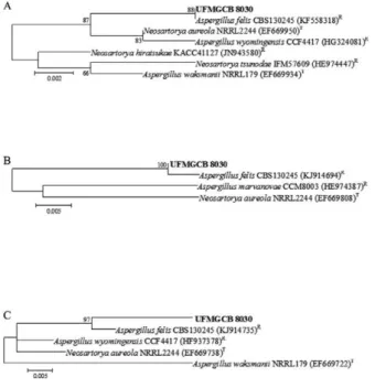
1). A combination of phylogenetic evaluation (Fig. 1) and analysis of micro and macro-morphological features (Fig. 2) increased the degree of confidence in this identification.
The ITS nucleotide sequence showed 100% query coverage and 100% similarity with that of A. felis
(Gen-Bank accession KF558318). In addition, the β-tubulin
and RPB2 sequences of this isolate shared 84% and 100% query coverage and 99% and 98% of similarity, respectively, with the corresponding A. felis sequences (GenBank accessions KJ914694 and KJ914735,
respec-tively). ITS, β-tubulin, and RPB2 references or type spe -cies sequences were retrieved from GenBank and used in a neighbour-joining phylogenetic analysis with 1,000 bootstrap replicates (Fig. 1). This approach revealed dis-tinct clustering of the organism of interest in this study with A. felis, confirming it to be the species most geneti-cally similar to isolate UFMGCB 8030.
The following characteristics of the Aspergillus iso-late were observed, as shown in Fig. 2: colony diameters of 5.0 and 5.5 cm after seven days at 25ºC on CYA and MEA media, respectively, and sporulation on MEA at 25ºC on the 14th day of culture. On CYA medium, colo-ny texture is mostly floccose; colonies are usually white, with a cream-to-light-brown reverse, and often sporu-late poorly. Furthermore, yellow soluble pigments are diffused into the agar. On MEA, colonies are somewhat velvety with greenish sporulation occurring after seven days. Colonies have a cream reverse. Conidiophores are uniseriate with greenish stipes (12 × 5.0 µm) and green globose conidia 1.5-2.5 µm in length. Phialides are 6.0 × 2.0 µm and vesicles are pyriform with a diameter of
13 mm. After taxonomic analysis using molecular and morphological methods, fungus UFMGCB 8030 was confirmed to be A. felis (Barrs et al. 2013).
In the present study, the production of bioactive com-pounds was assessed by varying certain culture conditions of A. felis UFMGCB 8030 and testing the resulting extracts with a P. brasiliensis bioassay. In regard to culture media, Fig. 1: phylogenetic analysis of nucleotide sequences obtained from fungus UFMGCB 8030 (in bold) associated with rocks from the Ata-cama Desert in comparison with type (T) and reference (R) sequences deposited in GenBank. Trees were constructed based on
ITS1-5.8S-ITS2 (A), β-tubulin (B), and ribosomal polymerase II gene (C) se -quences using the maximum composite likelihood model.
the most striking results were obtained with extracts from fungi cultivated on PDA (MIC = 7.8 µg/mL) followed by those from YM and corn meal cultures (MIC = 15.6 µg/ mL). On MEA medium, the MIC of the ethanol extract was 62.5 µg/mL (Table II). Extracts obtained after cultivation of this isolate on minimal medium supplemented with glucose showed no antifungal activity against Pb18.
As the ethanolic extract of A. felis UFMGCB 8030 grown on PDA demonstrated the lowest MIC, this me-dium was used to identify the optimal solvent for the production of extracts with the highest antifungal activ-ity. The extract obtained using DCM was found to be the most active against P. brasiliensisPb18 (MIC = 1.9 µg/ mL), followed by that produced with ethanol (MIC = 7.8 µg/mL). Extracts prepared with ethyl acetate and
hex-ane were only minimally active (with MICs of 500.0 and 250.0 µg/mL, respectively) (Table III). The PDA/DCM
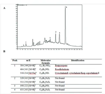
A. felis UFMGCB 8030 extract did not show cytotoxic-ity at the concentrations tested when assayed with Vero cells, demonstrating that this extract exhibits some selec-tivity towards fungal cells compared to mammalian cells. The PDA/DCM extract was then subjected to bioas-say-guided fractionation using RP-HPLC and a P. brasi- liensis assay (Fig. 3). The active fractions were analysed by HPLC-high resolution mass spectrometry (HRMS) with electrospray ionisation in positive-ion mode to ob-tain accurate mass measurements. A tentative identifica-tion based on the resulting mass spectra was achieved by manual verification using SciFinder and KNApSAcK data. The HRMS data corresponding to active fraction 1 consisted of m/z signals at 584.249 [M+H]+, 464.243
[M+H]+, and 518.214 [M+Na]+ that were tentatively
iden-tified as known compounds pyripyropene A (Omura et al. 1993), rosellichalasin (Kimura et al. 1989), and cy-tochalasin E (Aldridge et al. 1972), cycy-tochalasin Kasp (Kimura et al. 1989), or aspochalasin E (Steyn et al. 1982), respectively. Active fraction 1 comprised mul-tiple compounds, but the effective identification of these based on patterns of substitution was not possible due to a lack of information in the literature. The resulting formulas obtained from the fractions 2-4 did not match against SciFinder and KNApSAcK database to search for known metabolites. It could be hypothesised that these fractions can contain metabolites that were not previously isolated from Aspergillus species.
TABLE II
Minimum inhibitory concentrations (MIC) against Paracoccidioides brasiliensisPb18 of ethanol extracts from Aspergillus felis (UFMGCB 8030)
grown on different culture media
Culture medium (g/L)
MIC (µg/mL)
MM (5)
-MM (10)
-MM (15)
-MM (20)
-MM (30)
-PDA 7.8
YM 15.6
MEA 62.5
Corn meal 15.6
Itraconazole 0.001
MEA: malt extract agar; MM: minimal medium supplemented with 5-30 g/L glucose; PDA: potato dextrose agar; YM: yeast mold; -: no activity.
TABLE III
Minimum inhibitory concentrations (MIC) against Paracoccidioides brasiliensisPb18 of various solvent
extracts from Aspergillus felis (UFMGCB 8030) cultures grown on potato dextrose agar
Solvent
MIC (µg/mL)
Hexane 250.0
Dichloromethane 1.9
Ethyl acetate 500.0
Ethanol 7.8
Fig. 3: identification of secondary metabolites in dichloromethane (DCM) extract of Aspergillus felis UFMGCB 8030 grown for 15 days on potato dextrose agar medium. A: high-performance liquid chroma-tography chromatogram of A. felis DCM extract (ultraviolet detection at
DISCUSSION
In the present work, the DCM extract of A. felis
UFMGCB 8030 displayed promising activity against
P. brasiliensis Pb18, although in a previous screen with
Candida albicans, Candida krusei, and Cladosporium sphaerospermum it was shown to be inactive (Gonçalves et al. 2015). Although this fungus has previously been identified using ITS sequences (Gonçalves et al. 2015), in this work the identity of isolate UFMGCB 8030 was confirmed using molecular, morphological, and phylo-genetic methodologies. According to Barrs et al. (2013), species belonging to the Aspergillus,section Fumigati, cannot be identified only on the basis of morphologi-cal aspects only, therefore the use of other approaches for the identification of such organisms is key. Barrs et al. (2013) recently described the identification of A. fe-lis in human and animal hosts (dogs and cats) with in-vasive aspergillosis. The isolation of this fungus from environmental samples was first reported by our group, as a result of an investigation of Atacama Desert’s rock samples (Gonçalves et al. 2015).
In fungi, the biosynthesis of secondary metabolites is regulated in response to nutrient availability or as a result of changes in the environment or developmental phase (Sanchez & Demain 2002, Zain et al. 2011). Altering the media used to culture microorganisms can enhance the production of bioactive compounds (Abdel-Fattah & Ol-ama 2002). A good understanding of the role of culture conditions in the biosynthesis of metabolites may lead to improved exploitation of microorganisms-derived com-pounds (Miao et al. 2006). The ethanol extract obtained by cultivation of A. felis UFMGCB 8030 on PDA resulted in the strongest effect, i.e., the lowest MIC, in an evaluation of culture media, while minimal medium failed to pro-vide conditions suitable for the production of antifungal compounds against Pb18. Bhattacharyya and Jha (2011) showed that in salt-rich media such as Czapek-Dox, the growth and antimicrobial activity of an Aspergillus strain was lower than that observed using a complex medium such as PDA. In addition, Mathan et al. (2013) demon-strated that low-nutrient medium has a detrimental effect on mycelial growth and metabolite profile in Aspergillus terreus. This suggests that in salt-rich or nutrient-poor media, mycelial growth interferes with the production of antifungal metabolites by Aspergillus spp.
As A. felis was described only very recently, we were unable to find any records in the literature concerning investigation of its secondary metabolites, a fact that en-couraged us to determine the compounds in the UFMGCB 8030 DCM extract responsible for its antifungal activity.
Concerning the compounds identified in this extract, no reports of antifungal activity exist for pyripyropene, rosellichalasin, cytochalasin Kasp, or aspochalasin E. However, cytochalasin E has been tested against Fu-sarium solani (MIC > 100 µM), Gibberella saubinetti
(MIC = 100 µM), Botrytis cinerea (MIC = 100 µM), and
Alternaria solani (MIC = 50 µM), showing weak anti-fungal activity with MIC values generally greater than 50 µM (Zhang et al. 2014). Although antifungal activity against organisms of agricultural importance has thus
been documented, no investigations into the effect of the compounds identified in this work against fungi of medical interest have been carried out.
The fast tentative identification of natural products using the dereplication process can be very efficient to detect promising source of new bioactive compounds (Kildgaard et al. 2014, Petersen et al. 2014, Boruta & Bi-zukojc 2015). In one of the fractions displaying antifungal activity, cytochalasins were identified as the active me-tabolites. Cytochalasins are a group of fungal secondary metabolites with a 10-phenylperhydroisoindol-1-one skel-eton and a macrocyclic ring and are capable of various biological activities (Qiao et al. 2011). They have been described not only in the genus Aspergillus (Demain et al. 1976, Udagawa et al. 2000, Lin et al. 2009, Zheng et al. 2013), but also in Xylaria (Silva et al. 2010), Cladospo-rium (Cafêu et al. 2005), Arthrinium (Wang 2015), and
Phomopsis (Shen et al. 2014). According to Guerra et al. (2014), cytochalasins inhibit actin polimerisation and act preventing actin interaction with host cells in the fungal pathogen Cryptococcus neoformans. C. neoformans is internalised by receptor-mediated or “triggered” phago-cytosis, dependent on actin recruitment. Additionally, they can act as microfilament-disrupting agents, alter cell motility, adherence, secretion, drug efflux, deformability, morphology, and size, among many other cell properties critical to neoplastic cell pathology (Van Goietsenoven et al. 2011). Rosellichalasin and cytochalasin E isolated from
Aspergillus sp. exhibit potent cytotoxic activity against human tumour cell lines (Xiao et al. 2013). Besides these compounds, aspochalasin E shows potent activity against murine melanoma B16-F10 and human colon carcinoma HCT-116 cells (Naruse et al. 1993). Pyripyropene A acts in decrease of intestinal cholesterol absorption and cho-lesteryl oleate levels, resulting in protection of atheroscle-rosis development (Ohshiro et al. 2011).
The literature contains few reports on the isolation of compounds from fungi exhibiting activity against P. brasiliensis. However, among these are altenusin, iso-lated from an Alternaria sp. (Johann et al. 2012), and trichothecene mycotoxins (T-2 toxin and a mixture of 8-n-isobutyrylsolaniol and 8-n-butyrylneosolaniol (Campos et al. 2011).
This study indicated that fungi isolated from Ata-cama Desert rocks may constitute potential sources of novel bioactive compounds. A. felis UFMGCB 8030 pro-duced the most active extract among those studied and its antifungal activity was enhanced by changes in cul-ture conditions. The DCM extract of this fungus showed low cytotoxicity in preliminary tests and outstanding ac-tivity against one of the fungi responsible for PCM. Our results demonstrate the importance of further studies into the fungus A. felis, since the analyses presented here suggest that previously unknown bioactive compounds can be produced by this species.
REFERENCES
Abdel-Fattah YR, Olama ZA 2002. L-asparaginase production by
Pseudomonas aeruginosa in solid-state culture: evaluation and optimization of culture conditions using factorial designs. Pro-cess Biochem38: 115-122.
Aldridge DC, Burrows BF, Turner WB 1972. The structures of the fungal metabolites cytochalasins E and F. J Chem Soc Chem Commun3: 148-149.
Altschul SF, Madden TL, Schaffer AA, Zhang JH, Zhang Z, Mill-er W, Lipman DJ 1997. Gapped BLAST and PSI-BLAST: a new generation of protein database search programs. Nucleic Acids Res 25: 3389-3402.
Andrade RV, Silva SP, Torres FAG, Poças-Fonseca MJ, Silva-Pereira I, Maranhão AQ, Campos EG, Moraes LMP, Jesuíno RSA, Perei-ra M, Soares CMA, Walter ME, Carvalho MJA, Almeida NF, Brígido MM, Felipe MSS 2005. Overview and perspectives on the transcriptome of Paracoccidioides brasiliensis. Rev Iberoam Micol22: 203-212.
Araújo FS, Coelho LM, Silva LC, da Silva Neto BR, Parente-Rocha JA, Bailão AM, de Oliveira CM, Fernandes GR, Hérnandez O, Ochoa JG, Soares CM, Pereira M 2016. Effects of argentillactone on the transcriptional profile, cell wall, and oxidative stress of
Paracoccidioides spp. Plos Negl Trop Dis 10: e0004309. Azua-Bustos A, Urrejola C, Vicuña R 2012. Life at the dry edge:
mi-croorganisms of the Atacama Desert. Febs Letters586: 2939-2945. Barrs VR, Tineke MD, Houbraken J, Kidd SE, Martin P, Pinheiro
MD, Richardson M, Varga J, Samson RA 2013. Aspergillus felis
sp. nov., an emerging agent of invasive Aspergillosis in humans, cats, and dogs. PLoS ONE 8: 1-11.
Bhattacharyya PN, Jha DK 2011. Optimization of cultural conditions affecting growth and improved bioactive metabolite production by a subsurface Aspergillus strain tsf 146. Int J Appl Biol Pharm 2: 133-145.
Borges-Walmsley MI, Chen D, Shu X, Walmsley AR 2002. The pathobiology of Paracoccidioides brasiliensis. Trends Microbiol 10: 80-87.
Boruta T, Bizukojc M 2015. Induction of secondary metabolism of
Aspergillus terreus ATCC 20542 in the batch bioreactor cultures.
Appl Microbiol Biotechnol PMID: 26603760.
Cafêu MC, Silva GH, Teles HL, Bolzani VS, Araújo AR, Young MCM, Pfenning LH 2005. Antifungal compounds of Xylaria sp., an endophytic fungus isolated from Palicourea marcgravii (Ru-biaceae). Quim Nova28: 991-995.
Campos FF, Johann S, Cota BB, Alves TM, Rosa LH, Caligiorne RB, Cisalpino OS, Rosa CA, Zani CL 2011. Antifungal activity of tri-chothecenes from Fusarium sp. against clinical isolates of Para-coccidioides brasiliensis. Mycoses54: 122-129.
CLSI - Clinical Laboratory Standards Institute 2008. Reference method for broth dilution antifungal susceptibility testing of yeasts, CLSI document M27-A3, Approved standard, 3rd ed., Wayne, 25 pp. Cruz RC, Werneck SMC, Oliveira CS, Santos PC, Soares BM,
San-tos DA, Cisalpino PS 2012. Conditions for determining the min-imal inhibitory concentration (MIC) of seven antifungal agents against Paracoccidioides brasiliensis by microdilution: influ-ence of different media, incubation times, and temperatures. J Clin Microbiol50: 3415-3819.
Dalmaso GZL, Ferreira D, Vermelho AB 2015. Marine extremo-philes: a source of hydrolases for biotechnological applications.
Mar Drugs13: 1925-1965.
Demain AL, Hunt NA, Malik V, Kobbe B, Hawkins H, Matsuo K, Wogan GN 1976. Improved procedure for production of cytoch-alasin E and tremorgenic mycotoxins by Aspergillus clavatus. Appl Environ Microbiol31: 138-140.
Glass NL, Donaldson GC 1995. Development of primer sets designed for use with the PCR to amplify conserved genes from filamen-tous ascomycetes. Appl EnvironMicrobiol61: 1323-1330. Godinho VM, Furbino LE, Santiago IF, Pellizzari FM, Yokoya N,
Pupo D, Alves TMA, Júnior PAS, Romanha AJ, Zani CL, Cantrell CL, Rosa CA, Rosa LH2013. Diversity and bioprospecting of fungal communities associated with endemic and cold-adapted macroalgae in Antarctica. ISME J7: 1434-1451.
Gonçalves VN, Cantrell CL, Wedge DE, Ferreira MC, Soares MA, Jacob MR, Oliveira FS, Galante D, Rodrigues F, Alves TMA, Zani CL, Júnior PAS, Murta S, Romanha AJ,Barbosa EC, Kroon EG, Oliveira JG, Gómez-Silva B, Galetovic A, Rosa CA, Rosa LH 2015. Fungi associated with rocks of the Atacama Desert: taxonomy, distribution, diversity, ecology, and bioprospection for bioactive compounds. Environ Microbiol18: 232-245.
Gueidan C, Villaseñor CR, Hoog GS, Gorbushina AA, Untereiner WA, Lutzoni F 2008. A rock-inhabiting ancestor for mutualistic and pathogen-rich fungal lineages. Stud Mycol 61: 111-119. Guerra CR, Seabra SH, de Souza W, Rozental S 2014. Cryptococcus
neoformans is internalized by receptor-mediated or “triggered” phagocytosis, dependent on actin recruitment. PLoS ONE 9: 1-10. Horai H, Arita M, Kanaya S, Nihei Y, Ikeda T, Suwa K, Ojima Y,
Tanaka K, Tanaka S, Aoshima K, Oda Y, Kakazu Y, Kusano M, Tohge T, Matsuda F, Sawada Y, Hirai MY, Nakanishi H, Ikeda K, Akimoto N, Maoka T, Takahashi H, Ara T, Sakurai N, Suzuki H, Shibata D, Neumann S, Iida T, Tanaka K, Funatsu K, Matsuura F, Soga T, Taguchi R, Saito K, Nishioka T 2010. MassBank: a public repository for sharing mass spectral data for life sciences. J Mass Spectrom45: 703-714.
Houbraken J, Frisvad JC, Seifert KA, Overy DP, Tuthill DM, Valdez JG, Samson RA 2012. New penicillin-producing Penicillium spe-cies and an overview of section Chrysogena. Persoonia29: 78-100. Johann S, Cisalpino OS, Watanabe GA, Cota BB, Siqueira EP, Piz-zolati MG, Zani CL, Resende MA 2010. Antifungal activity of extracts of some plants used in the Brazilian traditional medicine against the pathogenic fungus Paracoccidioides brasiliensis. Pharm Biol48: 388-396.
Johann S, Rosa LH, Rosa CA, Perez P, Cisalpino PS, Zani CL, Cota BC 2012. Antifungal activity of altenusin isolated from the en-dophytic fungus Alternaria sp. against the pathogenic fungus
Paracoccidioides brasiliensis. Rev Iberoam Micol29: 205-209. Kildgaard S, Mansson M, Dosen I, Klitgaard A, Frisvad JC, Larsen
TO, Nielsen KF 2014. Accurate dereplication of bioactive second-ary metabolites from marine-derived fungi by UHPLC-DAD-QTOFMS and a MS/HRMS library. Mar Drugs12: 3681-3705. Kimura Y, Nakajima H, Hamasaki T 1989. Structure of
rosellichala-sin, a new metabolite produced by Rosellinia necatrix. Agric Biol Chem53: 1699-1701.
Kirk PM, Cannon PF, Minter DW, Stalpers JA 2008. Dictionary of the fungi, 10th ed., CAB International, Wallingford, 771 pp. Klich MA 2002. Identification of common Aspergillus species,
Cen-traalbureau voor Schimmelautures, Utrecht, 116 pp.
LinZ, Zhang G, Zhu T, Liu R, Wei H, Gu Q 2009. Bioactive cyto-chalasins from Aspergillus flavipes, an endophytic fungus asso-ciated with the mangrove plant Acanthus ilicifolius. Helv Chim Acta92: 1538-1544.
Mathan S, Subramanian V, Nagamony S 2013. Optimization and anti-microbial metabolite production from endophytic fungi Aspergil-lus terreus KC 582297. Euro J Exp Bio3: 138-144.
Para-coccidioides brasiliensis as revealed by gene genealogies. Mol Biol Evol 23: 65-73.
Miao Li, Kwong TFN, Qian P 2006. Effect of culture conditions on mycelial growth, antibacterial activity, and metabolite profiles of the marine-derived fungus Arthrinium c.f. saccharicola.Appl Microbiol Biotechnol72: 1063-1073.
Monks A, Scudiero D, Skehan P, Shoemaker R, Paull K, Vistica D, Hose C, Langley J, Cronise P, Vaigro-Wolff A 1991. Feasibility of a high-flux anticancer drug screen using a diverse panel of cultured human tumor cell lines. J Natl Cancer Inst 83: 757-766. Naruse N, Yamamoto H, Murata S, Sawsa Y, Fukagawa Y, Oki T
1993. Aspochalasin E, a new antibiotic isolated from a fungus. J Antibiot (Tokyo) 46: 679-681.
Ohshiro T, Matsuda D, Sakai K, Degirolamo C, Yagyu H, Rudel LL, Omura S, Ishibashi S, Tomoda H 2011. Pyripyropene A, an acyl-coenzyme A: cholesterol acyltransferase 2-selective inhibitor, at-tenuates hypercholesterolemia and atherosclerosis in murine models of hyperlipidemia. Arterioscler Thromb Vasc Biol31: 1108-1115. Omura S, Tomoda H, Kim YK, Nishida H 1993. Pyripyropenes, high
potent inhibitors of acyl-CoA cholesterol acyltransferase pro-duced by Aspergillus fumigates. J Antibiot (Tokyo) 46: 1168-1169. Palmeiro M, Cherubini K, Yurgel LS 2005. Paracoccidioidomicose -
Revisão da literatura. Sci Med 15: 274-278.
Petersen LM, Hoeck C, Frisvad JC, Gotfredsen CH, Larsen TO 2014. Dereplication guided discovery of secondary metabolites of mixed biosynthetic origin from Aspergillus aculeatus. Molecules 19: 10898-10921.
Prado RS, Bailão AM, Silva LC, de Oliveira CMA, Marques MF, Silva LP, Silveira-Lacerda EP, Lima AP, Soares CM, Pereira M 2015. Proteomic profile response of Paracoccidioides lutzii to the antifungal argentilactone. Front Microbiol 6: 616.
Qiao k, Chooi YH, Tang Y 2011. Identification and engineering of the cytochalasin gene cluster from Aspergillus clavatus NRRL 1.
Metab Eng 136: 723-732.
Rosa LH, Queiroz SCN, Moraes RM, Wang X, Techen N, Pan Z, Charles L, Cantrell CL, Wedge DE 2013. Coniochaeta ligniaria: antifungal activity of the cryptic endophytic fungus associated with autotrophic tissue cultures of the medicinal plant Smallan-thus sonchifolius (Asteraceae). Symbiosis60: 133-142.
Rosa LH, Vaz ABM, Caligiorne RB, Campolina S, Rosa CA 2009. Endophytic fungi associated with the Antartic grass Deschamp-sia antarctica Desv. (Poaceae). Polar Biol32: 161-167.
Sanchez S, Demain AL 2002. Regulation of fermentation processes.
Enzyme Microb Technol31: 895-906.
Shen l, Luo Q, Shen Z, Li L, Zhang X, Wei Z, Fu Y, Tan R, Song Y 2014. A new cytochalasin from endophytic Phomopsis sp. IFB-E060. Chin J Nat Med12: 512-516.
Shikanai-Yasuda MA, Telles Filho FQ, Mendes RP, Colombo AI, Moretti MI 2006. Guidelines in paracoccidioidomycosis. Rev Soc Bras Med Trop39: 297-310.
Silva GH, Oliveira CM, Teles HL, Bolzani VS, Araújo AR, Pfenning LH, Young MCM, Costa-Neto CM, Haddad R, Eberlin MN 2010. Citocalasinas produzidas por Xylaria sp., um fungo endofítico de
Piper aduncum (Piperaceae). Quim Nova 33: 2038-2041. Steyn PS, van Heerden FR, Rabie CJ 1982. Cytochalasins E and K,
tox-ic metabolites from Aspergillus clavatus. J Chem Soc1: 541-544. Stürme MHJ, Puccia R, Goldman GH, Rodrigues F 2011.
Molecu-lar biology of the dimorphic fungi Paracoccidioides spp. Fungal Biol Rev25: 89-97.
Tamura K, Peterson D, Peterson N, Stecher G, Nei M, Kumar S 2011. MEGA5: molecular evolutionary genetics analysis using maxi-mum likelihood, evolutionary distance, and maximaxi-mum parsimo-ny methods. Mol Biol Evol28: 2731-2739.
Tavares AH, Silva SS, Bernardes VV, Maranhão AQ, Kyaw CM, Poças-Fonseca M, Silva-Pereira I 2005. Virulence insights from the Para-coccidioides brasiliensis transcriptome. Genet Mol Res4: 372-389. Teixeira MM, Theodoro RC, Carvalho MJA, Fernandes L, Paes HC,
Hahn RC, Mendoza L, Bagagli E, San-Blas G, Felipe MS 2009. Phylogenetic analysis reveals a high level of speciation in the
Paracoccidioides genus. Mol Phylogenet Evol52: 273-283. Tesei D, Marzban G, Zakharova K, Isola D, Selbmann L, Sterflinger K
2012. Alteration of protein patterns in black rock inhabiting fungi as a response to different temperatures. Fungal Biol116: 932-940. Torres I, Garcia AM, Hernández O, González A, McEwen JG,
Re-strepo A, Arango M 2010. Presence and expression of the mat-ing type locus in Paracoccidioides brasiliensis isolates. Fungal Genet Biol47: 373-380.
Travassos LR, Taborda CP 2012. New advances in the development of a vaccine against paracoccidioidomycosis. Front Microbiol3: 1-6. Udagawa T, Yuan J, Panigrahy D, Chang YH, Shah J, D’Amato RJ
2000. Cytochalasin E, an epoxide containing Aspergillus-derived fungal metabolite, inhibits angiogenesis and tumor growth. J Pharmacol Exp Ther294: 421-427.
Van Goietsenoven G, Mathieu V, Andolfi A, Cimmino A, Lefranc F, Kiss R, Evidente A 2011. In vitro growth inhibitory effects of cy-tochalasins and derivatives in cancer cells. Planta Med 77: 711-717. Visbal G, San-Blas G, Maldonado A, Alvarez-Aular A, Capparelli MV,
Murgich J 2011. Synthesis, in vitro antifungal activity and mecha-nism of action of four sterol hydrazone analogues against the dimor-phic fungus Paracoccidioides brasiliensis.Steroids76: 1069-1081. Wang J, Wang Z, Ju Z, Wan J, Liao S, Lin X, Zhang T, Zhou X, Chen
H, Tu Z, Liu Y 2015. Cytotoxic cytochalasins from marine-de-rived fungus Arthrinium arundinis. Planta Med 81: 160-166. White TJ, Bruns TD, Lee SB, Taylor J 1990. Amplification and direct
sequencing of fungal ribosomal RNA genes for phylogenetics. In MA Innis, DH Gelfand, JJ Shinsky, TJ White (eds.), PCR proto-cols: a guide to methods and applications, Academic Press, San Diego, p. 315-322.
Wierzchos J, Davila AF, Artieda O, Cámara-Gallego B, Los Ríos A, Nealson KH, Valea S, García-González MT, Ascaso C 2013. Ignim-brite as a substrate for endolithic life in the hyper-arid Atacama Des-ert: implications for the search for life on Mars. Icarus224: 334-346. Xiao L, Liu H, Wu N, Liu M, Wei J, Zhang Y, Lin X 2013. Charac-terization of the high cytochalasin E and rosellichalasin produc-ing-Aspergillus sp. nov. F1 isolated from marine solar saltern in China. World J Microbiol Biotechnol29: 11-17.
Zain ME, Razak AA, El-Sheikh HH, Soliman HG, Khalil AM 2011. Influence of growth medium on diagnostic characters of Asper-gillus and Penicillium species. Afr J Microbiol Res 3: 280-286. Zhang Q, Xiao J, Sun Q, Qin J, Pescitelli G, Gao J 2014.
Characteriza-tion of cytochalasins from the endophytic Xylaria sp. and their biological functions. J Agric Food Chem 62: 10962-10969. Zheng C, Shao C, Wu Lu, Chen M, Wang K, Zhao D, Sun X, Chen G,