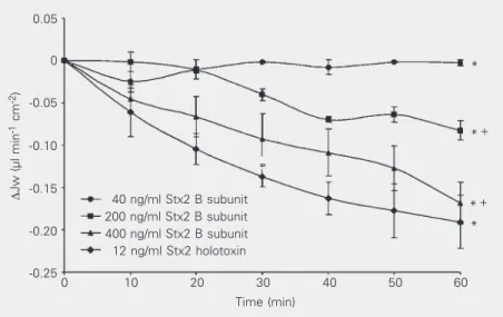
The Shiga toxin 2 B subunit inhibits net fluid absorption in human colon and elicits fluid accumulation in rat colon loops
Texto
Imagem


Documentos relacionados
Atlas: MATRIX emerged, at first, as a space of dialogue and confronta- tion where our individual research paths—a reflective practice origi- nating from the creation of ceramic
The best way to achieve this goal is to apply a solvent-free, pH-neutral, hydrophobic adhesive resin layer in a separate step, as confirmed by inferior in vitro and in vivo
Among all Opuntia ficus-indica samples, the juices from Tramagal and Sines fruits have the highest antiproliferative effect in human colon cancer cells, and this
Aberrant expression and activation of the thrombin receptor protease-activated receptor-1 induces cell proliferation and motility in human colon cancer cells.. Initiation of human
Expression of mRNA from the colon of adults and children but not from other gastrointestinal regions in Xenopus oocytes enhanced the osmotic water permeability, and the urea
Serial collection of the peritoneal fluid was conside- red to be safe and effective and analysis of total protein and LDH concentrations may be useful for early detection of
In the group undergoing surgery, the descending colon was the most affected; this location was noted in 7 cases (19.4%), followed by the entire colon also in 7 cases (19.4%),
the colon, a higher incidence of cancer in esophagus and altered distribution of the interstitial cells of Cajal in the megaesophagus and megacolon have also
