Studie s o n ATP-dipho spho hydro lase
nucle o tide -binding site s by intrinsic
fluo re sce nce
Departamento de Bioquímica y Biologia Molecular, Universidade de Chile, Santiago, Chile
A.M. Kettlun, V. Espinosa, A. Zanocco and M.A. Valenzuela
Abstract
Potato apyrase, a soluble ATP-diphosphohydrolase, was purified to homogeneity from several clonal varieties of Solanum tuberosum. Depending on the source of the enzyme, differences in kinetic and physicochemical properties have been described, which cannot be explained by the amino acid residues present in the active site. In order to understand the different kinetic behavior of the Pimpernel (ATPase/ ADPase = 10) and Desirée (ATPase/ADPase = 1) isoenzymes, the nucleotide-binding site of these apyrases was explored using the intrinsic fluorescence of tryptophan. The intrinsic fluorescence of the two apyrases was slightly different. The maximum emission wave-lengths of the Desirée and Pimpernel enzymes were 336 and 340 nm, respectively, suggesting small differences in the microenvironment of Trp residues. The Pimpernel enzyme emitted more fluorescence than the Desirée apyrase at the same concentration although both enzymes have the same number of Trp residues. The binding of the nonhydro-lyzable substrate analogs decreased the fluorescence emission of both apyrases, indicating the presence of conformational changes in the neighborhood of Trp residues. Experiments with quenchers of differ-ent polarities, such as acrylamide, Cs+ and I- indicated the existence of differences in the nucleotide-binding site, as further shown by quench-ing experiments in the presence of nonhydrolyzable substrate analogs. Differences in the nucleotide-binding site may explain, at least in part, the kinetic differences of the Pimpernel and Desirée isoapyrases. Co rre spo nde nce
A.M. Kettlun
Departamento de Bioquímica y Biologia Molecular
Universidade de Chile Casilla 174, Correo 22 Santiago
Chile
Presented at the XXVIII Annual Meeting of the Brazilian Society of Biochemistry and Molecular Biology, Caxambu, MG, Brasil, May 22-25, 1999.
Research supported by Fondecyt (No. 2970041) and the Universidad de Chile (Dirección Postgrado y Postítulo PG/068/97).
Received January 5, 2000 Accepted February 8, 2000
Ke y wo rds
·Potato apyrase
·ATP-diphosphohydrolase
·Intrinsic fluorescence
·Q uenching
·Desirée and Pimpernel isoapyrases
Intro ductio n
Apyrase (ATP-diphosphohydrolase, EC 3.6.1.5) has been described both as a soluble enzyme in plants (1) and as a membrane-bound protein in animal tissues (2,3) and parasites (4). Apyrase catalyzes the hydroly-sis of pyrophosphate bonds of nucleosides
di- and triphosphate in the presence of bivalent metal ions (5). The best effect is obtained in the
presence of Ca2+, although other ions such as
Mn2+
, Co2+
, Mg2+
and Zn2+
of the enzyme (7). Because of their extreme ATPase/ADPase ratio, the Pimpernel and
Desirée varieties of Solanumtuberosum were
selected for the present study.
Pimpernel apyrase hydrolyzes ATP ten times faster than ADP, while the Desirée enzyme splits both nucleotides at the same rate. We purified and characterized both isoapyrases and found that they differ in pI. Although the sum of the acid amino acid residues and the total number of basic resi-dues is identical for the two isoenzymes, Pimpernel apyrase is two pH units more alkaline than Desirée apyrase (8). The en-zymes also differ in the optimum pH of ADPase activity (5) and are very similar in molecular mass, approximately 49 kDa (6), in amino acid composition (8) and in the residues involved in the catalytic activity studied by chemical modification. This ap-proach has permitted us to propose that Arg,
Tyr, COO
and Trp residues may be in-volved in apyrase activities (9), while SH groups are unimportant for the catalytic ac-tivities of both apyrases (6). Since Koshland reagent inactivated both isoapyrases, and substrates protected them from inactivation, tryptophyl residues are probably involved in the nucleotide-binding site (6). Intrinsic flu-orescence experiments on the Desirée en-zyme (10) also suggest the presence of Trp near the nucleotide-binding site.
In order to investigate the environment of these residues in both isoapyrases, we stud-ied the quenching of the Trp fluorescence of Pimpernel and Desirée isoenzymes using quenchers of different polarities.
Mate rial and Me tho ds
ATP, ADP, the nonhydrolyzable substrate analogs ATP and ADP phosphonates (ADP-PCP and AMP-(ADP-PCP), Sepharose 4B-C1 and Cibacron blue were purchased from Sigma Co. (St. Louis, MO, USA). MES and ammo-nium sulfate were obtained from J.T. Baker (Phillipsburg, NJ, USA).
Apyrase purificatio n
The apyrases were purified from
homo-geneous strains of S.tuberosum cv
Pimper-nel and Desirée obtained by clonal selection, and generously supplied by the Instituto de Investigaciones Agropecuarias Remehue, Osorno, Chile. The enzymes were prepared as previously reported (10). The homogene-ity of the proteins was checked both by gel isoelectrofocusing (6) and SDS-PAGE (11).
Pro te in de te rminatio n
Protein concentration was determined by UV absorption at 280 nm.
Spe ctro sco pic me asure me nts
The emission spectra were determined with a SPEX FL2-Z2 spectrofluorometer and a 0.5-cm quartz semi-microcuvette. The ex-citation wavelength was 286 nm and
meas-urements were performed at 20oC. Protein
concentrations were 0.071 mg/ml for the Desirée enzyme and 0.064 mg/ml for the Pimpernel apyrase.
Q ue nching studie s
An aliquot of 0.06 mg/ml of each apyrase was titrated in its native conformation in 100 mM MES, pH 6.0, or denatured with 4.8 M guanidine hydrochloride (GndHCl) in 100 mM MES, pH 7.4. The intrinsic fluorescence of the proteins was quenched with 5-M solutions of acrylamide, cesium chloride, and sodium iodide prepared in 100 mM MES, pH 6.0. Titration was performed using a concentration range of quenchers from 0 to 0.35 M. Protein titration with sodium iodide was performed in the presence of 5 M sodium chloride to keep the ionic strength constant, and in 0.1%
Na2S2O3 to avoid I3
Re sults
Intrinsic fluo re sce nce o f Pimpe rne l and D e siré e apyrase s
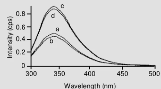
The two enzymes differed slightly in the maximum emission wavelength, which was 340 nm for Pimpernel and 336 nm for Desi-rée. The effect of ADP phosphonate on the fluorescence spectra of both isoenzymes is shown in Figure 1. The addition of the non-hydrolyzable ADP derivative produced a con-siderable decrease in the Trp fluorescence intensity of both isoapyrases. The results with the ATP derivative were very similar (data not shown). Although both enzymes contain the same number of Trp residues (Espinosa V, Kettlun AM, Zanocco A, Enci-nas V, Cardemil E and Valenzuela MA, unpublished data), the fluorescence emis-sion of the Pimpernel enzyme was greater than that of the Desirée apyrase at the same protein concentration.
Q ue nching o f Trp fluo re sce nce with acryla-mide , Cs+ and I
-Three quenchers of different polarity were used to analyze the microenvironment of Trp residues in both apyrases. Figure 2 shows the Stern-Volmer plots for Pimpernel and Desirée enzymes both in the native and de-natured form (4.8 M GndHCl) using acryla-mide, a polar uncharged quencher. Both con-formations showed an upward curvature for the Desirée apyrase, whereas only the dena-tured form presented the upward curvature for the Pimpernel enzyme. The native en-zyme had a linear Stern-Volmer plot with a Ksv = 3.77.
Figure 3 provides the Stern-Volmer plots
for both isoapyrases using Cs+
as a quencher. A downward curvature was observed for the native Pimpernel enzyme, with a fraction of exposed residues of 0.25. On the other hand, a straight line was obtained for the denatured form with a Ksv = 0.32. In contrast, for the
Figure 1 - Effect of ADP phos-phonate on the emission spec-tra of Pimpernel and Desirée apyrases. The excitation w ave-length w as 286 nm for the four spectra. The added protein w as 60 µg and 54 µg for the Desirée and Pim pernel enzym es, re-spectively, and 1.4 µM ADP phosphonate w as added in 100 mM M ES. Spectrum a: Desirée apyrase; spectrum b: Desirée apyrase + ADP phosphonate; spectrum c: Pimpernel apyrase, and spectrum d: Pimpernel apy-rase + ADP phosphonate.
Desirée enzyme linear Stern-Volmer plots were obtained for both forms of enzymes with Ksv = 0.494 and 0.976 for the native and denatured forms, respectively.
I- is a highly hydrated quencher. Figure 4
presents the Stern-Volmer plots of I
quench-ing. The denatured Pimpernel enzyme showed an upward curvature. The native Pimpernel apyrase exhibited a downward curvature with a fraction of exposed Trp of 0.5, whereas both forms of the Desirée en-zyme exhibited linear Stern-Volmer plots with a Ksv of 3.02 and 1.15 for the denatured and the native proteins, respectively.
D iscussio n
The difference in maximum emission wavelength, 340 nm for Pimpernel and 336 nm for Desirée, indicates that the Trp resi-dues are slightly more exposed in the Pim-pernel apyrase. Another relevant difference between the enzymes is the higher Trp emis-sion observed for the Pimpernel enzyme when compared with the Desirée apyrase, although both of them contain the same number of Trp residues (Espinosa V, Kettlun AM, Zanocco A, Encinas V, Cardemil E and Valenzuela MA, unpublished data). These data suggest that the tryptophan residues in the Desirée enzyme are more efficiently quenched by the solvent (water), by neighboring peptide bonds and/or by the lateral chains of amino acids such as Tyr, Cys and His (12). The decrease in fluorescence intensity of both apyrases induced by the nonhydrolyzable substrate analog (AMP-PCP) indicates that upon the nucleotide binding the local environment of
In
te
n
s
it
y
(
c
p
s
) 0.8
0.6
0.4
0.2
0
300 350 400 450 500
Wavelength (nm) c
d
a
Trp varies. These results indicate conforma-tional changes in the neighborhood of Trp in terms of nucleotide binding in both the Pim-pernel and Desirée enzymes. Since acryla-mide is a polar but uncharged quencher, it can probably collide with both exposed and nonexposed residues (13). The quenching experiments show the contribution of static quenching (upward curvature) for both the native and denatured forms of Desirée apy-rase and for the denatured form of the Pim-pernel enzyme, while the native PimPim-pernel apyrase has no contribution of static quench-ing and there is only one population of Trp equally quenched by acrylamide, thus with a linear Stern-Volmer plot.
Although the efficiency of Cs+ in
quench-ing indole groups is low, it may collide with the exposed or with the Trp in a negative environment. A downward curvature in the Stern-Volmer plot indicates that more than one population of fluorophores are present and that they have different accessibility to the quencher (14). On the basis of the Stern-Volmer plots, we conclude that the native Pimpernel enzyme has Trp populations with
different accessibility to Cs+
, whereas dena-turation of the Pimpernel enzyme induces a conformational change that renders all Trp
equally accessible to Cs+
. On the other hand, both Desirée apyrase conformations have one population of Trp residues accessible to
Cs+
. According to the Ksv values, Trp resi-dues are more exposed to the quencher in the denatured form.
I
can only interact with the exposed Trp residues. The denatured Pimpernel confor-mation exhibits contributions of static quenching (Stern-Volmer plot with upward curvature). The native Pimpernel apyrase
has half of its Trp residues accessible to I
-and the other half does not collide with this quencher. In contrast, both forms of the Desirée enzyme show one population of Trp
residues equally exposed to I
-. Since the Ksv value is higher (3.02) than the one obtained for the native form (1.15), the Trp residues
F
o
/F
2.0 1.8 1.6 1.4 1.2
F
o
/F 1.81.6 1.4 1.2 1.0
0 0.1 0.2 0.3 0.4 0 0.1 0.2 0.3 0.4
I- concentration (M ) I- concentration (M )
Pimpernel apyrase Desirée apyrase
Fo/F nat Fo/F denat 1.0
2.0 2.2 2.2
2.4
Figure 4 - Stern-Volmer plots of I- quenching of Pimpernel and Desirée native (nat) and denatured (denat) apyrases. The emission intensity w as measured at 340 and 336 nm for Pimpernel and Desirée, respectively. The protein concentration w as 0.06 mg/ml in 100 mM M ES for both enzymes. The addition of 4.8 M GndHCl produced the denatured forms. Corrections w ere made for dilution upon I- addition. Fo: Fluorescence emission in the absence of quencher; F: fluorescence emission in the presence of quencher.
F
o
/F
1.5 1.4 1.3 1.2 1.1
F
o
/F
1.30
1.20
1.10
0 0.1 0.2 0.3 0.4 1.000 0.1 0.2 0.3 0.4
CS+ concentration (M ) CS+ concentration (M )
Pimpernel apyrase Desirée apyrase
Fo/F nat Fo/F denat
Figure 3 - Stern-Volmer plots of Cs+ quenching of Pimpernel and Desirée native (nat) and denatured (denat) apyrases. The emission intensity w as measured at 340 and 336 nm for Pimpernel and Desirée, respectively. The protein concentration w as 0.06 mg/ml in 100 mM M ES for both enzymes. The addition of 4.8 M GndHCl produced the denatured forms. Corrections w ere made for dilution upon Cs+ addition. Fo: Fluorescence emission in the absence of quencher; F: fluorescence emission in the presence of quencher.
1.0
1.40
F
o
/F
6 5 4 3 2
F
o
/F
4.0 3.5 3.0 2.5 2.0
0 0.1 0.2 0.3 0.4 0 0.1 0.2 0.3 0.4
1.5 1.0
Acrylamide concentration (M ) Acrylamide concentration (M )
Pimpernel apyrase Desirée apyrase
Fo/F nat Fo/F denat
Figure 2 - Stern-Volmer plots of acrylamide quenching of Pimpernel and Desirée native (nat) and denatured (denat) apyrases. The emission intensity w as measured at 340 and 336 nm for Pimpernel and Desirée, respectively. Protein concentration w as 0.06 mg/ml in 100 mM M ES for both enzymes. The addition of 4.8 M GndHCl produced the denatured forms. Corrections w ere made for dilution upon acrylamide addition. Fo: Fluorescence emission in the absence of quencher; F: fluorescence emission in the presence of quencher.
of the denatured form are more exposed to
I-. All the quenching experiments led us to
conclude that the microenvironment of the Trp residues of the two isoapyrases is differ-ent. The changes in the Stern-Volmer plot of the native forms of both Pimpernel and Desirée isoenzymes upon nucleotide bind-ing (ADP-PCP and AMP-PCP) induce con-formational changes that alter the microen-vironments of the Trp residues in/near the nucleotide-binding site. The present results indicate that the nucleotide-binding site of
Pimpernel apyrase is different from the nucle-otide-binding site of Desirée apyrase, possi-bly explaining the kinetic variations.
Ackno wle dgme nts
We are indebted to Drs. José Santos and Julio Kalasich, Instituto de Investigaciones Agropecuarias, Remehue, Osorno, for gen-erously supplying the potato varieties. We also thank Mr. Claudio Telha for a critical reading of the manuscript.
Re fe re nce s
1. Thomas C, Sun Y, Naus K, Lloyd A & Roux S (1999). Apyrase functions in plant phos-phate nutrition and mobilizes phosphos-phate from extracellular ATP. Plant Physiology, 119: 543-552.
2. Battastini AM O, Emanuelli T, Koester M R, Wink M R, Bonan CD, Dias RD & Sarkis JJF (1998). Studies on the anchorage of ATP diphosphohydrolase in synaptic plas-ma membranes from rat brain. Interna-tional Journal of Biochemistry and Cell Biology, 30: 669-678.
3. Valenzuela M A, Kettlun AM , Sandoval S, García L, M ancilla M , Neckelmann G, Chayet L, Alvarez A, Cuevas F, Collados L, Espinosa V, Traverso-Cori A, Bravo I, Acevedo CG & Aranda E (1996). Compari-son of the biochemical properties, regula-tion and funcregula-tion of ATP-diphosphohydro-lase from human placenta and rat kidney. BrazilianJournal of M edical and Biological Research, 29: 589-597.
4. Vasconcelos EG, Ferreira ST, de Carvalho TM U, de Souza W, Kettlun AM , M ancilla M , Valenzuela M A & Verjovski-Almeida S (1996). Partial purification and
immuno-histochemical localization of ATP diphos-phohydrolase from Schistosoma mansoni. Journal of Biological Chem istry, 271: 22139-22145.
5. Valenzuela M A, Kettlun AM , M ancilla M , Calvo V, Fanta N & Traverso-Cori A (1988). The effect of bivalent m etal ions on ATPase-ADPase activities of apyrase from Solanum tuberosum. Phytochemistry, 27: 1981-1985.
6. Kettlun AM , Uribe L, Calvo V, Silva S, Rivera J, M ancilla M , Valenzuela M A & Traverso-Cori A (1982). Properties of tw o apyrases from Solanum tuberosum. Phy-tochemistry, 21: 551-558.
7. Kettlun AM , Leyton M , Valenzuela M A, M ancilla M & Traverso-Cori A (1992). Iden-tification and subcellular localization of tw o isoenzymes of apyrase from Solanum tuberosum. Phytochemistry, 31: 1889-1894.
8. M ancilla M , Kettlun AM , Valenzuela M A & Traverso-Cori A (1984). Structural studies of tw o apyrases from Solanum tubero-sum. Phytochemistry, 23: 1397-1400. 9. Kettlun AM , Uribe L, Silva S, Rivera J,
Valenzuela M A & Traverso-Cori A (1981). Chemical modification of aromatic, acid and basic amino acids of tw o isoenzymes of apyrase from Solanum tuberosum. Archivos de Biologia y M edicina Experi-mentales, 14: 171-175.
10. Espinosa V, Kettlun AM , Zanocco A, Cardemil E & Valenzuela M A (1999). Fluo-rescence studies of ATP diphosphohydro-lase from Solanum tuberosumvar. Desi-rée. Phytochemistry (in press).
11. Laemmli UK (1970). Cleavage of struc-tural proteins during the assembly of the head of bacteriophage T4. Nature, 227: 680-685.
12. Chen Y & Barkley M D (1998). Tow ard un-derstanding tryptophan fluorescence in proteins. Biochemistry, 37: 9976-9982. 13. Efnick M R & Ghiron CA (1981).
Fluores-cence quenching studies w ith proteins. Analytical Chemistry, 114: 199-227. 14. Lacow itcz JR (1983). Principle of