T
ABSTRACT
ULTRASTRUCTURAL EVALUATION OF THE
RADIOPROTECTIVE EFFECT OF SODIUM SELENITE ON
SUBMANDIBULAR GLANDS IN RATS
Maria Luiza dos Anjos PONTUAL1, Fabrício Mesquita TUJI2, Silvana Pereira BARROS3, Frab Norberto BÓSCOLO4, Pedro Duarte NOVAES3, Solange Maria de ALMEIDA4
1- PhD, Adjunt Professor, Department of Clinic and Social Dentistry, School of Dentistry, FederalUniversity of Paraíba. João Pessoa, PB,Brazil. 2- PhD, Associate Professor, Department of Oral Diagnosis, Federal University of Pará, Belém, PA, Brazil.
3- PhD, Associate Professor, Department of Morphology, Dental School of Piracicaba, State University of Campinas, Piracicaba, SP, Brazil. 4- PhD, Associate Professor, Department of Oral Diagnosis, Dental School of Piracicaba, State University of Campinas, Piracicaba, SP, Brazil.
Corresponding address: Maria Luiza dos Anjos Pontual - Rua Guerra de Holanda, 79, 52061-010, Bairro: Casa forte - Recife-PE Phone: 55 81 3268-6201 - Fax: 55 81 3446-3947 - e.mail: [email protected]
Received: November 16, 2005 - Modification: November 23, 2006 - Accepted: March 22, 2007
he aim of this study was to evaluate the radioprotector effect of sodium selenite on the ultrastructure of submandibular glands in rats. Fifty-seven male albino Wistar rats were randomized to 4 groups: control, irradiated, sodium selenite and irradiated/sodium selenite. The animals in the sodium selenite and irradiated/sodium selenite groups received intraperitoneal injections of sodium selenite (0.5 mg/kg body weight) 24 h before irradiation. The animals belonging to the irradiated and irradiated/sodium selenite groups were submitted to 15 Gy of gamma radiation in the head and neck region. The submandibular glands were removed at 4, 8, 12, 24, 48 and 72 h after irradiation. The ionizing radiation induced damage to the secretory cells, especially the serous cells, right from the first period. Vacuolization, lysis of cytoplasmic inclusions and nuclear alterations occurred. The sodium selenite group also presented cellular alterations in the study periods, but with less damage compared to that caused by radiation. There was greater similarity between the irradiated/sodium selenite group and the control group than with the other groups treated in all study periods. Despite the alterations observed in the sodium selenite group, sodium selenite presented a radioprotective action on the secretory cells of submandibular glands.
Uniterms: Submandibular gland; Sodium selenite; Selenium; Ultrastructure; Radiotherapy; Ionizing radiation; Rat.
INTRODUCTION
The inevitable exposure of salivary glands to radiation occurs frequently during radiotherapy of the head and neck region, which results in decreased saliva secretion, called xerostomia, shortly after a few radiation fractions. This may persist for the rest of the patient’s life, contributing to oral infections, caries and reduction in taste, and has been shown to be very prejudicial to the quality of life4.
Of the cells that comprise the salivary glands, the secretory cells are the most radiosensitive, especially the serous secretors4,7. The submandibular gland has two types
of secretory cells, serous and mucous cells, and it is frequently exposed during radiotherapy of the head and neck. In rodents, the submandibular serous cells are confined to the convoluted granular tubules, and the mucous cells are found in the acini1.
Different methods have been used to estimate the impact of various ionizing radiation doses on secretory cells, such
as qualitative descriptions of acute light and electron microscopic alterations7,9,15,22,23. In order to overcome the
influence of fibrosis and different degrees of atrophy in different cells, morphometric determinations of different cell types have been applied at the level of light microscopy. However, such measurements do not reflect subtle alterations in the morphology of individual cell types7.
Unfortunately, there is no adequate treatment for the deleterious effects of radiation on the salivary glands. Therefore, research has been undertaken on the administration of substances called radioprotectors that may inhibit or attenuate these effects. In a series of experiments demonstrating the radioprotecting effects of WR-2721, isoproterenol18 and cAMP17, the weight of the salivary glands was the only factor used to determine the relative radiation injuries under different experimental conditions.
and has been shown to have a radioprotective action in rat intestines10, to increase survival in animals24 and cell
cultures14,20,25.
Therefore, the aim of this study was to perform an ultrastructural evaluation of the radioprotective effect of sodium selenite on the damage caused by gamma radiation on the submandibular gland secretory cells in rats.
MATERIAL AND METHODS
Fifty-seven 3-month-old male albino Wistar rats, weighing 250-300 g were used. The rats were housed in polycarbonate cages (5-6 rats per cage) under a light/dark (14/10 h) cycle. Food (a standard pellet diet) and water were givenad libitum. The entire experiment was performed in agreement with the Ethical Principles for Animal Research established by the Brazilian College for Animal Experimentation (COBEA). The project was approved by the institutional Committee for Ethics in Animal Research (State University of Campinas, UNICAMP) on March 11, 2004.
The rats were randomly assigned to 4 groups: control group, irradiated group, sodium selenite group and sodium selenite/irradiated group. The control group was composed of 3 animals. The other groups comprised 18 rats each. Except for the control group, the other groups were divided into 6 sub-groups in accordance with the time of removal of the submandibular gland after irradiation: 4, 8, 12, 24, 48 and 72 h.
The animals belonging to the sodium selenite and sodium selenite/irradiated groups received 0.5 mg/kg body weight of sodium selenite (Merck KgaA, Darmstadt, Germany) intraperitoneally and saline was administered to the others. Twenty-four hours after administration of sodium selenite, all rats were anesthetized by intraperitoneal injection of sodium pentobarbital (Nembutal, 30 mg/kg
body weight). The animals in the irradiated and sodium selenite/irradiated groups had only the head and neck region irradiated with a single, fixed nominal dose of 15 Gy of gamma radiation Co60. Limitation of the exposed area was obtained
by collimating the apparatus. The treatment distance to the focal point on the skin was 80 cm, and the apparatus used was an Alcion CGR II model with a yield of 1.07 Gy/min, with an average of 1.25 MV.
At the previously established times, the animals were anesthetized by intraperitoneal injection of sodium pentobarbital (Nembutal, 40 mg/kg body weight), for
surgical removal of the right submandibular gland. Subsequently, the rats were sacrificed under general anesthetic with sodium pentobarbital (Nembutal).
The glands were sectioned into fragments of approximately by 1 mm and fixed by immersion in 2.5% glutaraldehyde at pH 7.3, 0.1 M sodium cacodylate buffer and 0.1 M sucrose for 24 h at 4°C. The specimens were post-fixed by immersion for 1 h in 1% osmium tetroxide, 0.1 M buffered in a 0.1 M phosphate buffer (pH 7.3) at 25°C. They were then dehydrated in a graded acetone series (50%, 70%,
80%, 90%, 100%) and embedded in Araldite resin8. For light
microscopy, 1-µm–thick sections were cut on an MT2B Sorvall Porter Blum ultramicrotome and stained with toluidine blue. After light microscopy field selection (area with terminal secretory portions and convoluted ducts), ultrathin sections (60 nm) were cut with an MT2C ultramicrotome for transmission electron microscopy. These sections were stained with uranyl acetate and lead citrate and examined with a Zeiss EM-10 transmission electron microscope (Zeiss,. Oberkochen, Germany), operated at 60 kV. The alterations in the serous and mucous cells were evaluated by qualitative descriptions. Only alterations that were observed in 3 selected rats of each sub-group were considered. The figures presented in the Results section are representative of these 3 rats per sub-group.
RESULTS
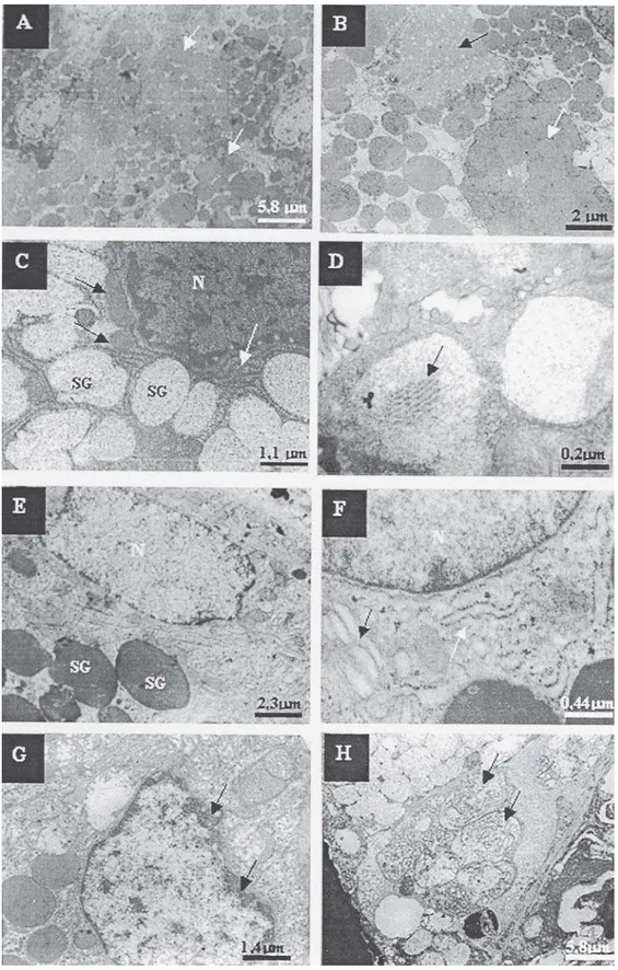
Four hours after irradiation, vascularization, cytoplasmic and nuclear alterations were observed in both types of secretory cells in the irradiated group. Diminished concentration, union, undefined limits and alterations in the electrodensity of serous granule components (Figure 1B) and the presence of electrodense content inside some mucous granules (Figure 1D) were found in the secretion granules. With regard to the nuclei, pleomorphism, chromatin condensation, thickening and rupture of the membrane were observed. Around these, altered and destroyed organelles, such as endoplasmic reticulum and mitochondria, were found (Figure 1G). Due to the rupture of the rough endoplasmic reticulum, there was an increase in free ribosomes. In addition to the rupture, the spaces between the mitochondrial crests were larger. The vacuoles showed amorphous material, organelles and/or nuclear debris (Figure 1H).
In all the studied groups, the mucous cells showed greater integrity than the serous cells in the first time period studied (Figure 2A, B). However, the behavior among the groups and times was similar for the two types of cells. In the mucous secretory cells, the sodium selenite and sodium selenite/irradiated groups presented the same alterations, but with less intensity than that found in the irradiated group, mainly in the sodium selenite/irradiated group. In the serous secretory cells, the intensity of the alterations was similar to that found in the irradiated group, except for the greater integrity of the secretion granules in the sodium selenite/ irradiated group (Figure 2C, D).
FIGURE 1- Transmission electron microscopy of the control group and irradiated submandibular salivary gland tissue 4 h
After 48 and 72 h, the alterations diminished in all groups; the sodium selenite group, and especially the sodium selenite/irradiated group presented greater similarity to the control group than the irradiated group (Figure 4). These latter times were characterized by an increase in the size of the nuclei and the number of organelles, mainly the rough endoplasmic reticulum.
DISCUSSION
Gamma radiation (15 Gy) caused degenerative processes in both types of secretory cells, but with greater destruction of the serous cells, in agreement with the findings of Stern, et al.19 (1976) and Vissink, et al.22 (1991). The greater
radiosensitivity of serous cells is explained by the hypothesis put forward by Abok, et al.1(1984) according to
which, the serous secretion granules have proteolytic and metallic transmission enzymes, while the mucous secretion granules mainly contain glycoproteins. The transmission materials are known to promote the induction of oxidative stress, potentiating the damage to the serous granule membranes. As a result, the proteolytic enzymes may infiltrate and damage the cytoplasm, causing autolysis and cellular death. Later research has confirmed the involvement of the serous secretion granules in the increased damage caused by ionizing radiation4,2,11.
The alterations found in the present study are consistent with those observed in similarly investigations15,21. The
rupture of the rough endoplasmic reticulum increased the
amount of free polyribosomes in the cytoplasm, as previously reported elsewhere15,19,22. The radiosensitivity
of the mitochondria has also been observed in other studies involving submandibular glands9,15,19,22,23.
The destruction and decrease in the number of secretion granules observed right from the first assessment after irradiation have been previously observed2,15,19,22. In the
mucous granules, in addition to these alterations, electrodense fibrils were also found, corroborating the findings of Vissink, et al.22 (1991), who suggested they were
a sign of repair and regeneration.
Nuclear alterations were noticed in the present study and in other ultrastructural studies15,19,21 on the secretory
cells of submandibular glands. Among these alterations, chromatin condensation is outstanding, indicating apoptosis20. Nuclear rupture and less electrodensity of the
euchromatin were found as well, characterizing cellular necrosis according to Rafferty, et al.13 (2003).
In both types of secretory cells, alterations were observed right from the earliest assessment. The alterations found in the present research after 4 h were similar to those observed in other investigations15,22,23 but were in
disagreement with the evaluation by Stern, et al.19 (1976),
who observed the first alterations as from the fourth day after exposure to 2 Gy of neutron radiation.
In relation to the time of greatest destruction, there was greater intensity after 8 and 12 h for both cells, but Stern, et al.19 (1976),found that the first great alterations occurred on
the fourth day. Reade and Steidler15(1985), using an 8 Gy
dose of X radiation, reported greater alterations after 48 h,
FIGURE 2- Transmission electron microscopy of submandibular gland tissues for each groups at 4 h. (A) Mucous cells from
while Vissink, et al.22 (1991) observed the greatest changes
by light and electron microscopy at 72 h. With respect to the period at which destruction diminished, indications of recovery were observed from 24 h, with decrease in damage at the last time assessed. This result differed from those of other studies, in which the indications of recovery appeared from the sixth day15,22.
With the aim of avoiding or attenuating the effects of ionizing radiation, radioprotective substances are used, which act by means of antioxidant action18,11. According to
Aruoma3 (1996) animals have lines of defense like antioxidant
enzymes that are able to inhibit the production of free radicals. Manganese superoxide dismutase, and copper and zinc superoxide dismutase enzymes are considered to be agents in the first line of defense against free radicals, removing the hydrogen superoxide and peroxide3. But the
most important hydrogen peroxide removal is done by the peroxidase glutathione enzyme, which acts in the presence of selenium3,21.
Selenium is a mineral essential to the organism and has antioxidant properties6,5,16. Its probable mechanism of action
occurs by means of its covalent binding to proteins, forming selenoproteins, with emphasis on peroxidase glutathione3,5,13,14,20,21 and thioredoxin reductase, which have
similar antioxidant properties5,19,21. Among the selenium
based components, inorganic selenium components, such as sodium selenite, are the most effective antioxidants14.
In the present study, sodium selenite diminished the effects of radiation at all the times studied in both secretory cells, but did not prevent the sodium selenite/irradiated group from undergoing alterations similar to those of the irradiated group. Four hours after irradiation, the sodium selenite/
FIGURE 3- (A) Serous cells from the irradiated group 12 h (A,B) and 24 h (C,D) after irradiation. (B) Serous cells from the
irradiated group differed from the irradiated group only by the larger number of whole secretory granules. At the following times, the sodium selenite/irradiated group presented significant and progressive tissue integrity and organization in relation to the irradiated group. In view of these observations, sodium selenite probably helped to maintain the integrity of the secretion granules initially, preventing leakage of their contents into the cellular cytoplasm, with the consequent rupture of the organelles and cell destruction found with greater frequency in the irradiated group. A similar effect of sodium selenite was found by Tuji, et al.21 (2005) in wound healing in irradiated rats.
In spite of having antioxidant action, selenium in excess has toxic activity, as demonstrated by studies on cell cultures14,20,25 and in vivo studies5,6. Its toxicity is
characterized by promotion of oxidative lesions in DNA14,
cell death14, inhibition of cellular proliferation6 and
morphologic alterations5. The cytotoxicity of selenium is
associated with the oxidation of glutathione and other thiols, giving rise to selenodiglutathione and selenopersulfide glutathione which promote the formation of hydrogen superoxide and peroxide radicals14,24,25.
The sodium selenite dose used in the present study (0.5 mg/kg body weight) was chosen because it promoted a radioprotector effect on wound healing in rats in a previous study21, without causing histomorphologic damage in the
rats treated only with sodium selenite. Although this dose promoted radioprotection in the present study, this same dose in the sodium selenite group caused degenerative processes similar to those caused by the radiation in the irradiated group. This difference in the results can be
explained by the fact that the work of Tuji, et al.21 (2005) was
a histomorphologic analysis and the present study is an ultrastructural analysis. However, it is worth pointing out that during the course of the present study, no animal treated with sodium selenite died.
Ionizing radiation results in a decrease in the accumulation of selenium by breaking its bond to proteins, involving the union of sulfur groups24. In addition, this may
also explain the decreased concentration of selenium in the cell, used to diminish the concentration of hydrogen peroxide as a result of the ionizing radiation, as opposed to its greater accumulation in the sodium selenite group and the resultant oxidation of thiols. Thus, it is suggested that the difference in the results between the sodium selenite and the sodium selenite/irradiated groups is due to the changes in protein metabolism after irradiation, leading to the reduced accumulation of selenium in the cells.
Nordman, et al.12 (1976) stated that selenite uptake is
around 5-7 times greater in malignant cells than in normal tissues. Therefore, in view of the dichotomy of sodium selenite acting as a radioprotective substance, and as a toxic substance at greater concentrations, in vivo studies about the toxicity of sodium selenite in various organs are suggested, in order to find doses that may simultaneously cause toxicity in neoplastic cells, accentuate the deleterious effects of ionizing radiation, and lastly, provide radioprotection to normal cells.
FIGURE 4- (A) Serous cells from the irradiated group at 72 h after irradiation. (B) Serous cells from the sodium selenite group
CONCLUSIONS
Under this experimental conditions it was concluded that despite the alterations observed in the sodium selenite group, sodium selenite has a radioprotective action on the secretory cells of submandibular glands.
ACKNOWLEDGMENTS
The authors wish to thank Dr. Eliene Novaes for her help and advice with this experiment. This study was supported by grants from the National Council for Scientific and Technological Development (CNPq) -, Process Number 301185/95-6.
REFERENCES
1- Abok K, Brunk U, Jung B, Ericsson J. Morphologic and histochemical studies on the differing radiosensitivity of ductular and acinar cells of the rat submandibular gland. Virchows Arch B Cell Pathol Incl Mol Pathol. 1984;45:443-60.
2- Ahlner BH, Hagelqvist E, Lind MG. Influence on rabbit submandibular gland injury by stimulation or inhibition of gland function during irradiation- histology and morphometry after 15 gray. Ann Otol Rhinol Laryngol. 1994;103:125-34.
3- Aruoma OI. Characterization of drugs as antioxidant prophylactics. Free Radic Biol Med. 1996;20:675-705.
4- Coppes RP, Vissink A, Konings AWT. Comparison of radiosensitivity of rat parotid and submandibular glands after different radiation schedules. Radiother Oncol. 2002;63:321-8.
5- Delilbasi C, Demiralp S, Turan B. Effects of selenium on the structure of the mandible in experimental diabetics. J Oral Sci. 2002;44:85-90.
6- Gøenback H, Frystyk J, Ørskov H, Flyvbjerg A. Effect of sodium selenite on growth, insulin-like growth factor-binding proteins and insulin-like growth factor-I in rats. 1995;145:105-12.
7- Grehn AL, Gustafsson H, Franzén L, Thornell LE, Henriksson R. Ultrastructural morphometry of parotid acinar cells following fractionated irradiation. Oral Oncol. 1997;33(1):23-8.
8- Luft JH. Improvements in epoxy resin embedding methods. J Biophys Biochem Cytol. 1961;9:409-14.
9- Messelt EB, Dahl E. Influence of X-ray irradiation on the ultrastructure of rat submandibular gland striated-duct cells. Acta Odontol Scand. 1983;41:277-82.
10- Mutlu-Turkoglu U, Erbil Y, Öztezcan S, Olgaç V, Toker G, Uysal M. The effect of selenium and/ or vitamin E treatments on radiation-induced intestinal injury in rats. Life Sci. 2000;66:1905-13.
11- Nagler RM, Mammary Y, Fox PC, Baum BJ, Har-El R, Chevion M. Irradiation- induced damage to the salivary glands: the role of redox-ative iron a copper. Radiat Res. 1997;147:468-75.
12- Nordman E, Jászsági-Nagy E, Rekonen A. Changes in tumour cell selenite (75 Se) affinity due to irradiation. Ann Clin Res. 1976;8:43-7.
13- Rafferty TS, Beckett GJ, Walker C, Bisset YC, McKenzie RC. Selenium protects primary human keratinocytes from apoptosis induced by exposure to ultraviolet radiation. Clin Exp Dermatol. 2003;28:294-300.
14- Rafferty TS, McKenzie RC, Hunter JAA, Howie F, Arthur J R, Nicol F, et al. Differential expression of selenoproteins by human skin and protection by selenium from UVB-radiation-induced cell death. Biochem J. 1998;332:231-6.
15- Reade PC, Steidler NE. X- irradiation induced degranulation of cells of convoluted granular tubules of murine submandibular glands. J Biol Buccale. 1985;13:307-15.
16- Saito Y, Yoshida Y, Akazawa T, Takahashi K, Niki E. Cell death caused by selenium deficiency and protective effect. J Biol Chem. 2003;278:39428-34.
17- Sodicoff M, Conger AD. Radioprotection of the rat parotid gland by cAMP. Radiat Res. 1983;96:90-4.
18- Sodicoff M, Conger AD. Radioprotection of the rat parotid gland by WR-2721 and isoproterenol and its modification by propanolol. Radiat Res. 1983;94:97-104.
19- Stern MH, Turner JE, Jett LS, Mincer H, McGinnis JP. Electron microscopic changes in rat parotid and submandibular glands subsequent to total body irradiation with fast nêutrons. Oral Surg Oral Med Oral Pathol. 1976;42:620-30.
20- Stewart MS, Spaallholz JE, Neldner KH, Pence BC. Selenium compounds have disparate abilities to impose oxidative stress and induce apoptosis. Free Radic Biol Med. 1999;26:42-8.
21- Tuji FM, Almeida SM, Bóscolo FN, Manzi FR. Avaliação do efeito radioprotetor do selenito de sódio no processo de reparação tecidual em ratos. Radiol Bras. 2005;38(5):359-64.
22- Vissink A, Kalicharan D, S-Gravenmade EJ, Jongebloed WL, Ligeon EE, Nieuwenhuis P, et al. Acute irradiation effects on morphology and function of rat submandibular glands. J Oral Pathol Med. 1991;20:449-56.
23- Vissink A, S-Gravenmade EJ, Ligeonne E, Konings AWT. Effects of split-dose X irradiation on rat salivary gland function. Radiat Res. 1991;127:52-7.
24- Weiss JF, Srinivasan V, Kumar KS, Landauer MR. Radioprotection by metals: selenium. Adv Space Res. 1992;12:223-31.