Gabriella Lopes de Rezende BARBOSA(a)
Luiz André PIMenTA(b)
Solange Maria de ALMeIdA(c)
(a)Universidade de Campinas - Unicamp, Piracicaba Dental School, Department of Oral Diagnosis, Piracicaba, SP, Brazil.
(b)University of North Carolina at Chapel Hill, School of Dentistry, Department of Dental Ecology, Chapel Hill, NC, USA.
(c)Universidade de Campinas - Unicamp, Piracicaba Dental School, Department of Oral Diagnosis, Piracicaba, SP, Brazil.
Micro-CT evaluation of the
radioprotective effect of resveratrol on
the mandibular incisors of irradiated rats
Abstract:The purpose of this study was to perform a microcomputed tomographic evaluation of the radioprotective effect of resveratrol on the volume of mandibular incisors of irradiated rats. A second aim was to make a quantitative assessment of the effect of x-ray exposure on these dental tissues. Twenty adult male rats were divided into four groups: control, irradiated control, resveratrol, and irradiated resveratrol. The resveratrol groups received 100 mg/kg of resveratrol, whereas the irradiated groups were exposed to 15 Gy of irradiation.
The animals were sacriiced 30 days after the irradiation procedure,
and their mandibles were removed and scanned in a microcomputed tomography unit. The images were loaded into Mimics software to allow segmentation of the mandibular incisor and assessment of its volume. The results were compared by One-way ANOVA and Tukey’s post hoc
test, considering a 5% signiicance level. The irradiated groups showed signiicantly diminished volumes of the evaluated teeth, as compared
with the control group (p < 0.05). The resveratrol group presented higher values than those of the irradiated groups, and volumes similar to those
of the control group. High radiation doses signiicantly affected tooth
formation, resulting in alterations in the dental structure, and thus lower volumes. Moreover, resveratrol showed no effective radioprotective impact on dental tissues. Future studies are needed to evaluate different concentrations of this substance, in an endeavor to verify its potential as a radioprotector for these dental tissues.
Keywords: Odontogenesis; Radiation-Protective Agents; Radiation Injuries; X-Ray Microtomography.
Introduction
Radiation treatment, surgical procedures and chemotherapy are the main treatments for neoplasms of the head and neck region.1 In the case of
radiotherapy, the result of these treatments is the formation of free radicals, leading to ionization and deleterious effects on the cells of many normal,
uninvolved tissues. Examples of structures that receive signiicant doses
of radiation include the salivary glands, the oral mucosa and the teeth. Changes in odontogenesis may occur in patients undergoing head and neck radiotherapy during tooth formation, such as children with neoplastic diseases.2 These alterations include enamel hypoplasia, interference in
enamel organ development and tooth formation, in addition to microdontia
declaration of Interests: The authors certify that they have no commercial or associative interest that represents a conflict of interest in connection with the manuscript.
Corresponding Author:
Gabriella Lopes de Rezende Barbosa Email: [email protected]
dOI: 10.1590/1807-3107BOR-2016.vol30.0168
Submitted: Apr 17, 2015
and changes in root formation, such as interruption, thinning and enlargement of the pulp chamber. The severity of the damage depends on the stage of dental development, as well as the dose and extension of the irradiated area.3 Prior studies have reported the effects of high radiation doses on rat incisors, such as delayed eruption and disturbances in secretory functions.4,5 However, few studies have quantitatively
assessed the damage caused by irradiation to dental tissues during formation.
Radioprotective substances have been developed to protect against or minimize these unwanted deleterious effects on healthy tissues, and have become an integral part of patient care and enhancement of quality of life.6,7 These substances should not
only have antioxidant properties, but also have
anti-inlammatory and antimicrobial properties, be
immunostimulants, stimulate cell proliferation, and not cause systemic toxicity.8
Different substances tested as radioprotectors include organic substances, such as resveratrol
(3,5,4’-trihydroxy-trans-stilbene), which is a
polyphenol belonging to the group of compounds called phytoalexins.9 This substance is synthesized
by plants when stimulated by microbial infection, ultraviolet radiation, and ozone exposure.10 Grapevines
(Vitis vinifera and Vitis labrusca) are among the species considered as having a high potential for synthesis of the compound.11
Attention and interest in this substance have increased following evidence that resveratrol can prevent against, or at least delay, the onset of cancer, ischemic heart disease, chemically induced injury, diabetes, pathological inflammation and viral infection.12 Studies have been conducted regarding
its radioprotective effect, and have shown positive effects, including the radioprotection of glandular and bone marrow cells.13,14
The effects caused by substances of different origin and radiation can be evaluated by several different methods, depending on the tissue of interest and the expected outcome. Currently, whole structures can be assessed in micrometer scale without sample destruction, owing to new imaging techniques, such as microcomputed tomography (µCT). The use of µCT allows the accurate appraisal of mineralized
structures, as well as their volume, morphology and internal structure.15
Therefore, the aim of the study was to make a µCT evaluation of the radioprotective effect of resveratrol on the volume of mandibular incisors of irradiated rats, and also make a quantitative assessment of the impact of x-ray exposure on dental tissues.
Methodology
After receiving approval from the Animal Research Ethics Committee (CEUA/UNICAMP,
protocol no. 2317-1), twenty 8-week-old adult male
rats (Rattus novergicus albinus, Wistar) were obtained for the study. The animals were kept on a diet of food and water ad libitum, at 23°C room temperature and
artiicial lighting (light-dark cycles of 12 hours). The
study was carried out in accordance with EU Directive
2010/63/EU. The animals were randomly divided into
four groups (n = 5): control (received no medication and no irradiation), irradiated control (received a single dose of 15 Gy irradiation), resveratrol (received resveratrol solution) and irradiated resveratrol (received resveratrol solution and 15 Gy irradiation). The sample size selection was based on the “resource equation” method. According to this method, an “E” value is measured, representing a degree of freedom of analysis of variance, and should lie between 10 and 20. E can be measured by subtracting the total number of animals from the total number of groups. Thus, any sample size that keeps E between 10 and 20 should be considered as adequate. The present study had an E value of 16.16
Resveratrol was prepared by diluting this compound in a 100% ethanol solution to a concentration of 50 mL/kg. The resveratrol/ethanol solution was then diluted in distilled water as follows: 0.05 mL of resveratrol (50 mg/mL) in 0.2 mL of distilled water
to a inal concentration of 10 mg/mL.
The medication was administered via enteral
feeding, using a plastic catheter connected to a 3.0 mL
syringe, to allow direct deposition into the stomach. Each rat received a dose of 100 mg/kg resveratrol, administered by gavage once every 24 hours for 2 days before irradiation, and again on the day of
irradiation, 30 minutes before exposure to radiation.14
the experiments, kept in amber glass bottles, protected from light, and stored at room temperature.
All the animals undergoing irradiation were weighed and anesthetized by intramuscular administration of ketamine chloride and xylazine chloride solutions. Once anesthetized, the animals
were placed on a linear accelerator with 18 x 30 cm
collimation, and only the head and neck region was exposed to a single dose of 15 Gy x-ray radiation.
After 30 days, all the animals were sacriiced and
the mandibles were removed. The specimens were
formalin-ixed for further examination.
The left hemi-mandibles were scanned by a microcomputed tomography scanner (SkyScan 1174, Kontich, Belgium), operating at 50 kVp and 800 μA, with a voxel size of 22.5 μm, 0.45 degrees rotation
steps and 360 degrees rotation, using a 0.5 mm aluminum ilter. The entire incisor was included in the ield of view, and the images were reconstructed
after acquisition, using NRecon software (SkyScan, Kontich, Belgium).
The reconstructions of the tooth images were imported into the Mimics software program (version 16.0, Materialise Medical, Leuven, Belgium), segmented and analyzed by a single well-trained radiologist.
The threshold was deined so as to include the teeth
but remove any visible extraneous scatter, artifacts, and background. The volumetric region of interest (VOI) was cropped and narrowed to include only the
region of the incisor comprised between the distal surface of the second molar and the digastric muscle insertion to the mandible (Figure 1).
After deining the boundaries, a combination of
manual slice and threedimensional editing was used
to obtain reined surfaces of the tooth segmentation.
The resulting set of masks of the VOI was rendered into a shaded surface mesh by the same software program, and each segmented volume was calculated, according to the manufacturer’s recommendations.
After a one-month interval, half of the sample was reassessed to calculate the intraobserver
reproducibility by Intraclass correlation coeficient,
using MedCalc software, version 11.2.1.0 (MedCalc Software, Mariakerke, Belgium). One-way analysis of variance (ANOVA) was used to determine the overall differences among the volumes of the four groups. The test was performed using BioEstat software, version 5.0 (Instituto Mamirauá, Belém, Brazil); p values < 0.05
were considered statistically signiicant.
Results
The intraobserver reliability calculated by the
Intraclass correlation coeficient yielded a value
of 0.96, an excellent agreement rate according to ICC interpretation.
One-way ANOVA showed statistically signiicant
overall differences. Tukey’s test was the chosen post
Figure 1. Three-dimensional reconstruction of the entire mandible (A) and segmentation of the mandibular incisor (B) with the highlighted volumetric region of interest comprised between the second molar distal surface and the digastric muscle insertion to the mandible (pink region).
hoc test to conirm where the differences occurred
between the groups.
Significant differences (p < 0.05) were found in comparing the control group with the irradiated control and the irradiated resveratrol groups, as well as the resveratrol group with the irradiated control and the irradiated resveratrol groups, evidencing the impact of x-ray exposure on dental tissues. The lowest volume of the incisor region was observed in the irradiated control group (Table 1). No significant differences (p > 0.05) were detected when the irradiated control and the irradiated resveratrol groups were compared, demonstrating a lack of resveratrol radioprotection to the teeth (Table 1).
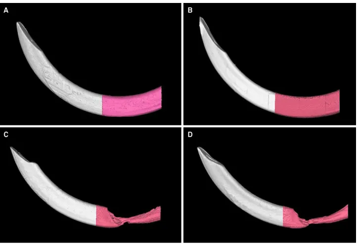
The three-dimensional reconstructions (Figure 2) clearly show the volume difference among the groups and the conformation of the incisors. The irradiated control and the irradiated resveratrol groups presented tooth defects, as well as diminished size.
discussion
Pat ie nt s u nde r r ad io t h e r apy t r e at m e nt are vulnerable to harmful side effects, such as disturbances that can impact the development of teeth. Tooth development begins in the embryonic
phase but is only complete after the irst decade
of life. During their developmental stages, the teeth are more susceptible to environmental disturbances, such as radiation, which can affect their normal development.17 In the present study,
the mandibular incisor of the rat was chosen, because of its continued growth and constant cell proliferation in the odontogenic region, replicating
the cell differentiation that occurs during tooth formation in humans.
The date chosen to sacriice the animals was based
on the continuous growth and eruption rate of the rodents’ incisors.5 A period of 30 days was chosen,
because it is adequate to evaluate the formation and mineralization of irradiated dental tissues. Moreover,
after 30 days, the undifferentiated irradiated cells of
the odontogenic region would then be positioned under the mandibular molars.
According to the present study results, radiation caused a significant defective formation of all mineralized materials composing the tooth structure. This alteration led to lower volumes of the evaluated mandibular incisor. Histological alterations in dental tissues caused by irradiation have been reported in prior literature.4,5,18 Nonetheless, the
literature on the effect of these defects in the entire dental structure is scant. Mature enamel defects are important because of their relation to compromised aesthetic, masticatory functions and heightened susceptibility to caries.4
Among the large range of radioprotective substances that have been studied, resveratrol has received much at tent ion because of its
antioxidant, anti-inlammatory, analgesic, antiviral,
cardioprotective and neuroprotective actions.19
However, i n t he present st udy, stat ist ically
signiicant differences could be observed between
the irradiated control and the irradiated resveratrol groups, demonstrating that resveratrol had no radioprotective effect on dental tissues. These
indings are not in agreement with the positive
effects previously reported in the literature, such as the radioprotection of bone marrow cells and even of normal uninvolved tissue in the head and neck region, as described by Xu et al.13
and Şimşek et al.20 on the protection of salivary
glands.14,19 These differences may be explained by
the different types of cells that were studied, as well as the administration of the medication performed intraperitoneally in these studies.
The selected dose (100 mg/kg) is in agreement with research that found positive radioprotective effects, such as the studies by Carsten et al.14 and
Şimşek et al.20 However, since no protection was
Table 1.Volumetric mean values (±SD) of the region of interest according to the evaluated groups (mm3).
Volume
Mean SD
Control 849.4A 121.1
Irradiated control 475.0B 137.6
Resveratrol 984.9A 98.6
Irradiated resveratrol 539.6B 155.7
observed in the present study, this dose may not be effective in dental tissues. In this sense, further investigation is needed to evaluate different concentrations and frequency of application of
this substance speciically for dental tissues, in an
endeavor to prove its potential as a radioprotector.
A positive inding of the present study was that the
administration of resveratrol itself showed no potential to modify dental structures, as indicated by the similar volumes of the resveratrol versus control groups (p > 0.05).
µCT technology was the method of choice of this study, since it provides high-resolution images and is a reliable and innovative technique that allows the assessment of volume and density of hard dental tissues, even during development. Moreover, µCT allows this assessment to be made without destroying the samples , and minimizes artifacts caused by sample preparation.15 The positive outcomes of the
research corroborate those by Dong et al.,15 who state
that µCT technology is a feasible technique for dental tissue evaluation, because it allows a quantitative evaluation of the structure as a whole. Furthermore, this study sought to shed light on the importance of research using innovative technology and imaging modalities incorporated into animal studies, by contributing more information and performing three-dimensional views of structures and tissues without destroying the samples.
Conclusion
The results of the present study demonstrate the deleterious effect of x-ray radiation on tooth formation, resulting in the reduced volume of dental structures. It was also observed that the dose of resveratrol used in this study had no effective radioprotective impact on dental tissues. Future studies are needed to evaluate different concentrations of this substance in an endeavor to prove its potential as a radioprotector. Figure 2. Three-dimensional reconstructions showing the differences in incisor volume among the groups: A- Control group, B- Resveratrol group, C- Irradiated control group, and D- Irradiated resveratrol group.
A B
1. McClure SA, Movahed R, Salama A, Ord RA. Maxillofacial metastases: a retrospective review of one institution’s
15-year experience. J Oral Maxillofac Surg. 2013;71(1):178-88.
doi:10.1016/j.joms.2012.04.009
2. Albuquerque RA, Morais VLL, Sobral APV. Sobral APV. [Protocol of odontological attendance of oncological pediatric patients: a literature review]. Rev Odontol UNESP.
2007;36(3):275-80. Portuguese.
3. Lopes NN, Petrilli AS, Caran EM, França CM, Chilvarquer I, Lederman H. Dental abnormalities in children submitted to
antineoplastic therapy. J Dent Child (Chic). 2006;73(3):140-5.
4. Ramos-Perez FMM, Espírito Santo AR, Perez DEC, Novaes PD, Bóscolo FN, Line SRP, et al. Ionizing radiation effects on the secretory-stage ameloblasts and enamel organic extracellular matrix. Radiat Environ Biophys.
2014;53(3):589-98. doi:10.1007/s00411-014-0539-4.
5. Araujo AMM, Gomes CC, Almeida SM, Klamt CB, Novaes PD. Effect of radiotherapy on the eruption rate and morphology of the odontogenic region of rat incisors. Arch Oral Biol. 2014;59(11):1242-1248. doi:10.1016/j.archoralbio.2014.07.004. 6. Kuntić VS, Stanković MB, Vujić ZB, Brborić JS,
Uskoković-Marković SM. Radioprotectors - the evergreen topic. Chem Biodivers. 2013;10(10):1791-803. doi:10.1002/cbdv.201300054.
7. Mishra S, Malhotra P, Gupta AK, Singh PK, Mishra AK, Javed S, et al. Novel method for screening of radioprotective agents providing protection to DNA ligase against gamma radiation induced damage. Int J Radiat Biol. 2014;90(2):187-92.
doi:10.3109/09553002.2014.868613.
8. Arora R, Gupta D, Chawla R, Sagar R, Sharma A, Kumar R, et al. Radioprotection by plant products: present status and future prospects. Phytother Res. 2005;19(1):1-22. doi:10.1002/ptr.1605 9. Jeandet P, Douillet-Breuil AC, Bessis R, Debord S, Sbaghi
M, Adrian M. Phytoalexins from the Vitaceae: biosynthesis, phytoalexin gene expression in transgenic plants, antifungal activity, and metabolism. J Agric Food Chem.
2002;50(10):2731-41. doi:10.1021/jf011429s
10. Ignatowicz E, Baer-Dubowska W. Resveratrol, a natural chemopreventive agent against degenerative diseases. Pol
J Pharmacol. 2001;53(6):557-69.
11. Frémont L. Biological effects of resveratrol. Life Sci.
2000;66(8):663-73. doi:10.1016/S0024-3205(99)00410-5
12. Baur JA, Sinclair DA. Therapeutic potential of resveratrol: the
in vivo evidence. Nat Rev Drug Discov. 2006 Jun;5(6):493-506. doi:10.1038/nrd2060
13. Xu L, Yang X, Cai J, Ma J, Cheng H, Zhao K, et al. Resveratrol
attenuates radiation-induced salivary gland dysfunction in
mice. Laryngoscope. 2013;123(11):E23-9. doi:10.1002/lary.24276
14. Carsten RE, Bachand AM, Bailey SM, Ullrich RL. Resveratrol reduces radiation-induced chromosome aberration frequencies in mouse bone marrow cells. Radiat Res.
2008;169(6):633-8. doi:10.1667/RR1190.1
15. Dong G, Dong Q, Liu Y, Lou B, Feng J, Wang K, et al. High-resolution micro-CT scanning as an innovative tool for evaluating dental hard tissue development. J Appl Clin
Med Phys. 2014;15(4):335-44. doi:10.1120/jacmp.v15i4.4956
16. Charan J, Kantharia ND. How to calculate sample size in
animal studies? J Pharmacol Pharmacother. 2013;4(4):303-6. doi:10.4103/0976-500X.119726
17. Nanci A. Ten Cate’s Oral Histology: Development, Structure and Function. 7th ed. St. Louis: Mosby; 2007.
18. English JA, Schlack CA, Ellinger F. Oral manifestations of ionizing radiation. II. Effect of 200 KV. x-ray on rat incisor teeth when administered locally to the head in
the 1,500 R. dose range. J Dent Res. 1954 Jun;33(3):377-8. doi:10.1177/00220345540330031101
19. Dobrzyńska MM. Resveratrol as promising natural
radioprotector. A review. Rocz Panstw Zakl Hig. 2013;64(4):255-62.
20. Şimşek G, Gürocak S, Karadaǧ N, Karabulut AB, Demirtaş
E, Karataş E, et al. Protective effects of resveratrol
on salivar y gland damage induced by total body
irradiation in rats. Laryngoscope. 2012;122(12):2743-8. doi:10.1002/lary.23609