abstract
Radiological and histopathological evaluation of
experimentally-induced periapical lesion in rats
Renata Cordeiro TEIXEIRA1, Cassia Maria Fischer RUBIRA1, Gerson Francisco ASSIS2, José Roberto Pereira LAURIS3,
Tania Mary CESTARI4, Izabel Regina Fischer RUBIRA-BULLEN5
1- DDS, MSc, PhD, Department of Stomatology, Bauru School of Dentistry, University of São Paulo, Bauru, SP, Brazil.
2- PhD, Associate Professor, Department of Biological Sciences, Bauru School of Dentistry, University of São Paulo, Bauru, SP, Brazil.
3- PhD, Associate Professor, Department of of Pediatric Dentistry, Orthodontics and Community Health, Bauru School of Dentistry, University of São Paulo, Bauru, SP, Brazil.
4- PhD, Department of Biological Sciences, Bauru School of Dentistry, University of São Paulo, Bauru, SP, Brazil.
5- DDS, MSc, PhD, Associate Professor, Department of Stomatology, Bauru School of Dentistry, University of São Paulo, Bauru, SP, Brazil.
Corresponding address: Renata Cordeiro Teixeira - Faculdade de Odontologia de Bauru - FOB/USP - Departamento de Estomatologia - Al. Dr. Octávio Pinheiro Brizolla, 9-75 - 17012-901 - Bauru - SP - Brazil - e-mail: [email protected]
Received: September 21, 2009 - Modiication: April 29, 2010 - Accepted: May 25, 2010
O
bjective: This study evaluated experimentally-induced periapical bone loss sites using digital radiographic and histopathologic parameters. Material and Methods:Twenty-seven Wistar rats were submitted to coronal opening of their mandibular right irst molars.
They were radiographed at 2, 15 and 30 days after the operative procedure by two digital radiographic storage phosphor plates (Digora®). The images were analyzed by creating
a region of interest at the periapical region of each tooth (ImageJ) and registering the
corresponding pixel values. After the sacriice, the specimens were submitted to microscopic analysis in order to conirm the pulpal and periapical status of the tooth. Results: There was signiicant statistically difference between the control and test sides in all the experimental
periods regarding the pixel values (two-way ANOVA; p<0.05). Conclusions: The microscopic analysis proved that a periapical disease development occurred during the experimental periods with an evolution from pulpal necrosis to periapical bone resorption.
Key words: Radiology. Periapical diseases. Histology. Rats.
IntroductIon
Digital intraoral radiographic systems have several advantages compared with conventional film-based radiography. These advantages, in Endodontics, include the potential of a lower radiation exposure to the patient and faster image display25. Moreover, it was introduced
the opportunity of evaluating the progression of periapical bone loss by digital tools as the pixel values3,15.
Radiographs do not always relect the extent of the destructive process in the periapical tissues and generally under-represent the size of a lesion12.
Studies have suggested that pathologic involvement of the cortical plate, or at least junctional trabeculae, is a prerequisite for radiographic detection of periapical pathoses16. Nevertheless, periapical
lesions confined to cancellous bone could be
detected radiographically using pixel values using one of the tools of the digital image, the histogram2.
Only a few studies employing animal models3,7,8 or
human biopsies10,21,23 have compared the diagnostic
and quantifying accuracy of radiographs to the gold standard represented by the histology. The aim of this study was to induce periapical disease in rats to assess the pixel values in different experimental periods to evaluate the bone loss progression.
MatErIaL and MEtHods
1: 2 days, Group 2: 15 days, Group 3: 30 days6. The
animals were submitted to general intramuscular anesthesia. In each animal, a cavity was made on the occlusal face of the mandibular irst molar on the right side with a spherical burr size ¼, at high speed without cooling28. The left side was set as control.
radiographic analysis
The material for study was obtained after euthanizing the animal with an anesthetic overdose. The right and left hemi-mandibles of each rat were separated and radiographed with two phosphor plates (PSPs/ Digora-Soredex-Tuusula, Finland). The exposition was performed according to the parameters: 70 kVp, 7 mA, focus-ilm distance of 30 cm, exposure time of 0.09 s. A standard aluminum stepwedge was set in each image, and an acrylic plate measuring 0.3x0.4x0.2 cm was positioned over the hemi-mandibles to simulate soft tissue. The stepwedge was used as a standard image technique, which is essential for control of day-to-day variations in the sensitivity of the detector29.
The radiographic evaluation was performed using the public domain software Image J (version 1.33 µ, National Institute of Health, Washington, DC) A region of interest (ROI/252 pixels) was opened in the periapical region of the every tooth, closest to the mesial root, without touching it. Only the periapical bone was enclosed in the ROI. Using the histogram tool, the pixel values of the ROIs were established. The normalized pixel values of the images (NPI) were then obtained by using the following equation29: NPI=PI/CR. Where PI is
the mean pixel intensity of the ROI, and CR is the pixel intensity of the ROI of the standard aluminum stepwedge. The pixel value of zero corresponded to black and the pixel value of 255 to white.
Histomorphometrical analysis
Hemi-mandibles were ixed in formalin 10%, decalciied in EDTA and processed histologically. Five semi-serial sections of each hemi-mandible with 5 µm thickness were cut and stained with hematoxylin and eosin. All histological sections were identiied with a random numerical sequence
and periapical tissues were histomorphometrically analyzed for both test and control sites. In the test site it was noted whether the tooth had pulpitis, necrosis or presented any sign of inlammation, and whether or not there was presence of periapical bone resorption. The percentages of bone tissue in the apical region of the root, soft tissue (connective tissue and inlammatory iniltrate, when present), bone marrow and inferior alveolar nerve were obtained in a digital image analysis system, composed of a Zeiss Axioskop II microscope, CCD-IRIS RGB – Sony camera (Sony DXC-151A RGB Video Camera-Sony Corporation, Tokyo, Japan) and Kontron KS300® software (Kontron Electronic
GMBM, Munich, Germany). For this purpose, an image was captured at 10x objective from each histologic slice, which contained the apical portion of the mesial root perpendicular to the alveolar nerve, positioned centrally. The studied area was established in numbers of pixels. Each image captured had 307,200 pixels and evaluated total area was 1536,000 pixels. The area (in pixels) was measured to quantify the area occupied by bone tissue in the apical region of the root, soft tissue (connective tissue and inlammatory iniltrate, when present), bone marrow and inferior alveolar nerve.
rEsuLts
radiographic analysis
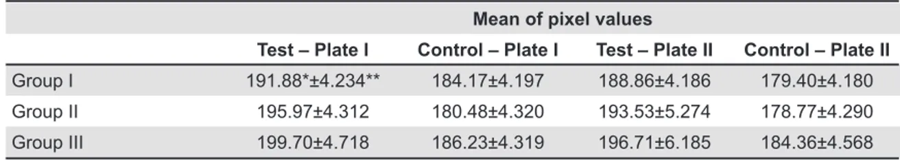
The radiographic analysis showed that the pixel values in the three experimental periods increased on the test side in comparison with the control side, indicating bone loss in a similar manner both PSPs plates used. The Student’s T test showed a statistically signiicant difference between control and test sites for the three groups tested (p<0.05) (Table 1). The inluence of the time on periapical disease was studied using one-way ANOVA and Tukey’s tests. ANOVA showed no statistically signiicant variation for any of the time intervals in Plate 1 (p>0.05), as well as for the control side of Plate 2 (p>0.05). This variation was only shown to be signiicant in Plate 2 (p<0.05) between the test groups 2 and 3 (Figures 1A, D, G, J).
Mean of pixel values
Test – Plate I Control – Plate I Test – Plate II Control – Plate II
Group I 191.88*±4.234** 184.17±4.197 188.86±4.186 179.40±4.180
Group II 195.97±4.312 180.48±4.320 193.53±5.274 178.77±4.290
Group III 199.70±4.718 186.23±4.319 196.71±6.185 184.36±4.568
*Mean pixel value from 9 animal samples.
**Standard deviation for mean pixel from 9 animal samples.
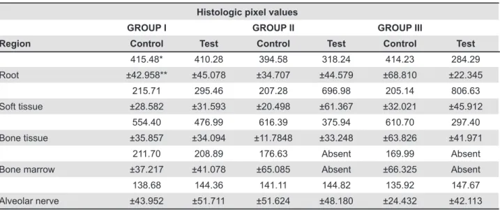
Histologic pixel values
GROUP I GROUP II GROUP III
Region Control Test Control Test Control Test
415.48* 410.28 394.58 318.24 414.23 284.29
Root ±42.958** ±45.078 ±34.707 ±44.579 ±68.810 ±22.345
215.71 295.46 207.28 696.98 205.14 806.63
Soft tissue ±28.582 ±31.593 ±20.498 ±61.367 ±32.021 ±45.912
554.40 476.99 616.39 375.94 610.70 297.40
Bone tissue ±35.857 ±34.094 ±11.7848 ±33.248 ±63.826 ±41.971
211.70 208.89 176.63 Absent 169.99 Absent
Bone marrow ±37.217 ±41.078 ±65.085 Absent ±66.325 Absent
138.68 144.36 141.11 144.82 135.92 147.67
Alveolar nerve ±43.952 ±51.711 ±51.624 ±48.180 ±24.432 ±42.113
*Mean pixel value from 9 animal samples.
**Standard deviation for mean pixel from 9 animal samples.
Table 2- Mean and standard deviation of the total number of pixels of each structure present in the periapical region of the
mesial root of the mandibular irst molar in the control and test groups in the experimental periods
Figure 1- Control group: A) radiographic image of hemi-mandible; B-C) photomicrograph of the left irst molar showing
healthy pulp (black arrow) and periapical tissues with cementum (ce), ibers of periodontal ligament (li) and alveolar bone (ab). Group 1: D) radiographic image of hemi-mandible similar at the control group; E-F) photomicrograph of the right irst molar exhibiting inlammatory cells (black arrow) in the periapical region. Group 2: G) radiographic image of hemi-mandible showing radiolucent area in the periapical region; H-I) photomicrograph of the right irst molar showing extensive bone (b)
resorption by osteoclasts (red arrow) and connective tissue (ct). Group 3: J) radiographic image of hemi-mandible exhibiting
extensive radiolucent area in the periapical region; K-L) photomicrography of the right irst molar showing abscess (red
Histopathological analysis
The histological analysis of the left irst molars of the rats, which served as control, showed no alterations in the pulp or periapical tissues, and evidenced that 100% of these teeth presented healthy pulp and periapical tissues (Figures 1B, C). The teeth submitted to coronal opening in group 1, showed in 100% of the cases, necrosis in the coronal region and pulp inlammation with the presence of inlammatory cells and increased vascularization in the medial and apical thirds. It was possible to observe inflammatory cells and small bone and cementum resorption in the periapical region (Figures 1E, F). Group 2 showed necrosis in the coronal and middle radicular pulp and extensive inlammatory process in the apical third. In the periapical region exhibited increase of bone resorption and replaced by conjunctive tissue rich in inlammatory cells (Figures 1H, I). The group 3 showed total pulp necrosis, extensive area of abscess and bone, cementum and dentine resorption and few bone trabeculae surrounded by conjunctive tissue in the apical portion (Figures 1K, L).
The area measured histomorphometrically was given in number of pixels. The total numbers of pixels obtained for each of the measured regions in the control and tests sides, in the various experimental periods are presented in Table 2. Two-way ANOVA and Tukey’s tests showed statistically signiicant difference between the test and control sides and among the experimental periods as well.
dIscussIon
In the present study, the periapical inlammation was induced by coronal opening of the mandibular right irst molar and conirmed that only coronal opening and pulp contact with the oral cavity were suficient for inducing periapical disease. Various other studies have described this technique for induction of pulpal and periapical inlammatory reactions in rat teeth19,28. The inlammatory process
was observed in the pulp region in the experimental period of 2 days. This is in agreement with the indings of a previous study that showed that pulp inlammation is already observed 6 h after the operative procedure19. In the present study, pulp
necrosis was observed in the experimental periods of 15 and 30 days. This is also in agreement with a previous study that showed the presence of complete pulp necrosis between the 8th and 20th
days27.
Hamachi, et al.16 (1995) afirmed that bone
resorption is present from the 3rd day after the
operative procedure. Bone resorption in the present study, although very subtle, could already be observed in the Group 1. Resorptive bone activity
was actively present on the 15th day (Group 2). Some
authors afirmed that this activity begins to diminish from then on, with a decrease in the number of osteoclasts1. Although the lesion appeared to be
stabilized, with diminished bone resorption, new bone formation did not occur due to the presence of aggressive factors30. In the 30-day experimental
period, a large area of resorption could be observed, but a smaller number of osteoclasts were present. The amount of periapical bone was minimal, and it was replaced by inlammatory tissue.
Statistical analysis showed a signiicant difference between test and control sides in the 3 experimental periods, except for the root region, in which this difference was not signiicant between the test and control sides, in the 2-day period, and between the experimental periods of 15 and 30 days. This was probably because resorption in periapical disease is greater in the bone region than in the root region, which was not very evident.
The present study conirmed radiographically the presence and intensity of bone resorption observed by the histological study. Jett, et al.18 (2004)and
Shrout, et al.26 (2003) removed medullar bone
from cadaver mandibles, radiographed the areas to measure the pixel value respectively. These studies showed that it is possible to detect bone loss in the mandibular region by analyzing the pixel value, even if it is not yet visible radiographically. Conversely, studies that compared images obtained by conventional radiographs (without pixel value analysis) and bone resorption seen microscopically, showed that this relationship is not established with precision4,14,
De Rossi, et al.9 (2007) concluded that although
image digitization could not improve the detection of the early stages of periapical lesions, it provides valuable quantitative assessment of extensive periapical lesions.
In order to diminish the noise level in the images captured directly, the phosphorus plates were covered by a protective envelope, as they are sensitive to light24. The images were always
downloaded immediately after the radiograph was taken, as the time between taking the radiograph and downloading the plates also inluences the pixel values5,13.
In the direct photostimulable phosphorus plate systems, such as Digora®, there is a correlation
between the radiation dose and the quantity of capture plate luminescence11,20,22. The capture
plates present less noise and a better quality image than the other direct systems, in addition to being more sensitive to small variations of the exposure source in comparison with conventional ilms, and should be used when small differences in contrast are important17,20. The noise and coeficient of
noise and small variation in the images in this study. In spite of resistance by many professionals, the high cost of the technology and the ethical implications because of the risk of manipulating the images, the use of digital imaging has increased and is constantly being perfected. Computerization tends to make this one the method of choice for daily radiographs.
concLusIons
The methodology applied was efficient for causing pulpitis and pulpal necrosis in the studied rats. Teeth with pulpitis microscopically presented periapical bone resorption, but the teeth with necrosis presented greater resorption. The control teeth presented healthy pulp, without signs of periapical bone resorption. The means of the pixel values in the areas of periapical disease induced in rats indicated greater bone resorption than the means of these values on the control side for the three experimental periods (p<0.05). Even small periapical bone resorption was already suficient for determining changes in the pixel values of that area in the direct digital method, when compared to their respective controls.
rEfErEncEs
1- Anan H, Akamine A, Maeda K. An enzyme histochemical study of the behavior of rat bone cells during experimental apical periodontitis. J Endod. 1993;19:83-6.
2- Attaelmanan A, Borg E, Gröndahl HG. Digitisation and display of intra-oral ilms. Dentomaxillofac Radiol. 2000;29:97-102.
3- Balto K, Müller R, Carrington DC, Dobeck J, Stashenko P.
Quantiication of periapical bone destruction in mice by
micro-computed tomography. J Dent Res. 2000;79:35-40.
4- Barkhordar RA, Hussain MZ, Hayashi C. Detection of interleukin-1 beta in human periapical lesions. Oral Surg Oral Med Oral Pathol. 1992;73:334-6.
5- Borg E, Attaelmanan A, Gröndahl HG. Image plate systems
differ in physical performance. Oral Surg Oral Med Oral Pathol Oral Radiol Endod. 2000;89:118-24.
6- Camps J, Pommel L, Bukiet F. Evaluation of periapical lesion healing by correction of gray values. J Endod. 2004;30:762-6.
7- Cotti E, Vargiu P, Dettori C, Mallarini G. Computerized
tomography in the management and follow-up of extensive periapical lesion. Endod Dent Traumatol. 1999;15:186-9. 8- Delano EO, Tyndall D, Ludlow JB, Trope M, Lost C. Quantitative radiographic follow-up of apical surgery: a radiometric and histologic correlation. J Endod. 1998;24:420-6.
9- De Rossi A, De Rossi M, Rocha LB, Silva LA, Rossi MA. Morphometric analysis of experimentally-induced periapical
lesions: radiographic vs histopathological indings. Dentomaxillofac
Radiol. 2007;36:211-7.
10- Farman AG, Avant SL, Scarfe WC, Farman TT, Green DB. In vivo
comparison of Visualix-2 and Ektaspeed Plus in the assessment of periradicular lesion dimensions. Oral Surg Oral Med Oral Pathol Oral Radiol Endod. 1998;85:203-9.
11- Farrier SL, Drage NA, Newcombe RG, Hayes SJ, Dummer PM.
A comparative study of image quality and radiation exposure for dental radiographs produced using a charge-coupled device and a phosphor plate system. Int Endod J. 2009;42:900-7.
12- Ferreira RI, Haiter-Neto F, Tabchoury CP, Paiva GA, Bóscolo
FN. Assessment of enamel demineralization using conventional, digital, and digitized radiography. Braz Oral Res. 2006;20:114-9. 13- Freitas P, Yaedú RY, Rubira-Bullen IR, Escarpinati M, Vieira MC, Schiabel H, et al. Reproducibility of pixel values for two photostimulable phosphor plates in consecutive standardized scannings. Braz Oral Res. 2006;20:207-13.
14- Genvert H, Miller H, Burn CG. Experimental production of
apical lesions of teeth in monkeys, and their relation to systemic disease. Yale J Biol Med. 1941;13:649-62.
15- Grecca FS, Leonardo MR, Silva LA, Tanomaru Filho M,
Borges MA. Radiographic evaluation of periradicular repair after endodontic treatment of dog's teeth with induced periradicular periodontitis. J Endod. 2001;27:610-2.
16- Hamachi T, Anan H, Akamine A, Fujise O, Maeda K. Detection of interleukin-1 beta mRNA in rat periapical lesions. J Endod. 1995;21:118-21.
17- Hildebolt CF, Fletcher G, Yokoyama-Crothers N, Conover GL,
Vannier MW. A comparison of the response of storage phosphor
and ilm radiography to small variations in X-ray exposure.
Dentomaxillofac Radiol. 1997;26:147-51.
18- Jett S, Shrout MK, Mailhot JM, Potter BJ, Borke JL. An evaluation of the origin of trabecular bone patterns using visual and digital image analysis. Oral Surg Oral Med Oral Pathol Oral Radiol Endod. 2004;98:598-604.
19- Kakehashi S, Stanley HR, Fitzgerald RJ. The effects of surgical exposures of dental pulps in germ-free and conventional laboratory rats. Oral Surg Oral Med Oral Pathol. 1965;20:340-9.
20- Kashima I. Computed radiography with photostimulable phosphor in oral and maxillofacial radiology. Oral Surg Oral Med Oral Pathol Oral Radiol Endod. 1995;80:577-98.
21- Laux M, Abbott PV, Pajarola G, Nair PN. Apical inlammatory
root resorption: a correlative radiographic and histological assessment. Int Endod J. 2000;33:483-93.
22- Lim KF, Loh EEM, Hong HY. Quantitative assessment of a new intra-oral digital imaging system. J Dent Res (Sp. Iss.). 1995;74:463.
23- Marmary Y, Koter T, Heling I. The effect of periapical rarefying osteitis on cortical and cancellous bone. A study comparing conventional radiographs with computed tomography. Dentomaxillofac Radiol. 1999;28:267-71.
24- Rubira-Bullen IRF, Escarpinati MC, Schiabel H, Vieira MAC, Rubira CMF, Lauris JRP. Evaluating noise in digitized radiographic images by means of histogram. J Appl Oral Sci. 2006;14:410-4.
25- Schmitd LB, Lima TC, Chinellato LEM, Bramante CM, Garcia RB, Moraes IG, et al. Comparison of radiographic measurements
obtained with conventional and indirect digital imaging during endodontic treatment. J Appl Oral Sci. 2008;16:167-70
26- Shrout MK, Jett S, Mailhot JM, Potter BJ, Borke JL, Hildebolt CF. Digital image analysis of cadaver mandibular trabecular bone patterns. J Periodontol. 2003;74:1342-7.
27- Stahl SS. Response of the periodontium, pulp, and salivary glands to gingival and tooth injury in young adult male rats. II. Pulp and periapical tissues. Oral Surg Oral Med Oral Pathol. 1960;13:734-42.
28- Tagger M, Massler M. Periapical tissue reactions after pulp exposure in rat molars. Oral Surg Oral Med Oral Pathol. 1975;39:304-17.
29- Tosoni GM, Lurie AG, Cowan AE, Burleson JA. Pixel intensity