Radiol Bras. 2016 Set/Out;49(5):281–287 281
Applicability of three-dimensional imaging techniques in fetal
medicine
*
Aplicabilidade da tecnologia tridimensional na medicina fetal
Werner Jr H, Santos JL, Belmonte S, Ribeiro G, Daltro P, Gasparetto EL, Marchiori E. Applicability of three-dimensional imaging techniques in fetal medicine. Radiol Bras. 2016 Set/Out;49(5):281–287.
Abstract
R e s u m o
Objective: To generate physical models of fetuses from images obtained with three-dimensional ultrasound (3D-US), magnetic reso-nance imaging (MRI), and, occasionally, computed tomography (CT), in order to guide additive manufacturing technology.
Materials and Methods: We used 3D-US images of 31 pregnant women, including 5 who were carrying twins. If abnormalities were detected by 3D-US, both MRI and in some cases CT scans were then immediately performed. The images were then exported to a workstation in DICOM format. A single observer performed slice-by-slice manual segmentation using a digital high resolution screen. Virtual 3D models were obtained from software that converts medical images into numerical models. Those models were then generated in physical form through the use of additive manufacturing techniques.
Results: Physical models based upon 3D-US, MRI, and CT images were successfully generated. The postnatal appearance of either the aborted fetus or the neonate closely resembled the physical models, particularly in cases of malformations.
Conclusion: The combined use of 3D-US, MRI, and CT could help improve our understanding of fetal anatomy. These three screening modalities can be used for educational purposes and as tools to enable parents to visualize their unborn baby. The images can be segmented and then applied, separately or jointly, in order to construct virtual and physical 3D models.
Keywords: Fetus; Fetal medicine; Three-dimensional technique; Ultrasound; Magnetic resonance imaging; Computed tomography.
Objetivo: Gerar modelos físicos de fetos utilizando imagens obtidas por ultrassonografia tridimensional (US3D), ressonância magnética (RM) e, em alguns casos, tomografia computadorizada (TC), para orientar a técnica de adição de camadas.
Materiais e Métodos: Foram usadas imagens obtidas de 31 gestantes, incluindo 5 casos de gestação gemelar. Os exames foram realizados usando US3D, RM e em alguns casos TC, e os arquivos foram exportados para uma estação de trabalho em formato DICOM. Um único observador realizou o processo de segmentação manual usando tela de alta resolução. Um software que converte imagens médicas em modelos numéricos foi utilizado para construir modelos virtuais 3D, que foram fisicamente materializados.
Resultados: Os modelos virtuais e físicos baseados na US3D, RM e TC realizados separadamente ou em conjunto foram concluídos com sucesso. A aparência pós-natal do feto abortado ou do recém-nascido se assemelhou muito com os modelos físicos, particular-mente nos casos de malformações.
Conclusão: O uso da US3D, RM e TC pode ajudar para melhor compreensão das características físicas do feto. Essas técnicas podem ser usadas com fins didáticos para auxiliar na abordagem multidisciplinar e na melhor compreensão dos pais. As imagens podem ser segmentadas e aplicadas separadamente ou combinadas para construir modelos virtuais 3D e físicos.
Unitermos: Feto; Medicina fetal; Tecnologia tridimensional; Ultrassonografia; Ressonância magnética; Tomografia computadorizada.
* Study conducted at the Alta Excelência Diagnóstica, the Clínica de Diagnóstico Por Imagem (CDPI) and the Instituto Fernandes Figueira (IFF), Rio de Janeiro, RJ, Brazil.
1. PhD, MD, Radiologist at the Alta Excelência Diagnóstica and at the Clínica de Diagnóstico Por Imagem (CDPI), Rio de Janeiro, RJ, Brazil.
2. PhD, Technologist at the Instituto Nacional de Tecnologia, Rio de Janeiro, RJ, Designer at the Center for Three-Dimensional Experimentation of the Pontifícia Universidade Católica do Rio de Janeiro (PUC-Rio), Rio de Janeiro, RJ, Brazil.
3. Biologist at the Center for Three-Dimensional Experimentation of the Pontifícia Universidade Católica do Rio de Janeiro (PUC-Rio), Rio de Janeiro, RJ, Brazil.
4. Designer at the Center for Three-Dimensional Experimentation of the Pontifícia Universidade Católica do Rio de Janeiro (PUC-Rio), Rio de Janeiro, RJ, Brazil.
5. PhD, MD, Radiologist at the Clínica de Diagnóstico Por Imagem (CDPI), Rio de Janeiro, RJ, Brazil.
6. PhD, Full Professor of Radiology at the Universidade Federal do Rio de Janeiro (UFRJ), Rio de Janeiro, RJ, Brazil.
Mailing address: Dr. Heron Werner Júnior. Alta Excelência Diagnóstica –
Radiolo-INTRODUCTION
A growing number of technological advancements in obtaining and viewing images through noninvasive techniques have brought major breakthroughs in medicine, especially in the diagnosis of fetal anomalies(1,2). In general, two types
of examinations are used in order to obtain images of the uterine cavity during pregnancy(1–3): ultrasound and
mag-netic resonance imaging (MRI). Computed tomography (CT) also provides detailed images of the fetus, especially of its skeleton, from the 30th week of pregnancy, although Heron Werner Júnior1, Jorge Lopes dos Santos2, Simone Belmonte3, Gerson Ribeiro4, Pedro Daltro1,
Emerson Leandro Gasparetto5, Edson Marchiori6
gia. Avenida Voluntários da Pátria, 423, Botafogo. Rio de Janeiro, RJ, Brasil, 22270-000. E-mail: [email protected].
its utility is restricted because it involves the use of ionizing radiation(4).
Three-dimensional (3D) virtual modeling has gained great momentum in recent years, due to the high perfor-mance of software applied in the fields of engineering, ar-chitecture, and design. It has been taking an increasingly user-friendly form, facilitating the visualization of 3D images(5–7).
The objective of this study was to develop virtual 3D models of fetuses during pregnancy from images obtained by ultrasound, MRI, and CT, alone or in combination.
MATERIALS AND METHODS
This study evaluated 31 pregnant women between Janu-ary 2008 and December 2014. The study was approved by the Research Ethics Committee of the Instituto Fernandes Figueira (IFF/Fiocruz). All the patients involved underwent 3D ultrasound (3D-US), alone or in combination with MRI, with no more than one day between the examinations in the latter case (Table 1). In all cases of suspected fetal malfor-mation based on a previous ultrasound, the combination of MRI and 3D-US was used. All 3D reconstructions for proto-typing were performed at National Institute of Technology and at the Center for Three-Dimensional Experimentation of the Pontifícia Universidade Católica do Rio de Janeiro, both of which are also located in the city of Rio de Janeiro, Brazil. The following inclusion criteria were applied: single-ton or multiple gestation with gestational age established by an ultrasound performed up to the 16th week of pregnancy or based on the date of the last menstrual period in women with a history of regular cycles; and fetuses with suspicion of abnormality or malformation, as identified by ultrasound. All of the pregnant women evaluated were at least 18 years of age and were examined between the 22nd and 37th weeks of gestation, some being scheduled for a second ex-amination, as necessary. Ultrasound and MRI examinations were performed and monitored by two professionals: a spe-cialist in gynecology, obstetrics, and fetal medicine; and a specialist in radiology.
The equipment used in the examinations were the Voluson 730 and Voluson E8 ultrasound systems (GE Medical Sys-tems/Kretztechnik GmbH, Zipf, Austria), with a 4–8 MHz transvaginal/transabdominal transducer. The MRI scans were obtained on one of two types of 1.5 T scanners (Magnetom Avanto and Aera; Siemens Healthcare, Erlangen, Germany). Patients were placed in the supine or left lateral decubitus
position, whichever made them more comfortable, and were introduced into the scanner feet first, in order to reduce the feeling of claustrophobia. A surface coil was positioned over the abdomen of the pregnant woman, and the following pro-tocol was applied: T2-weighted HASTE sequences—repeti-tion time/echo time (TR/TE), 140/140 ms; field of view (FOV), 300–200 mm; gap, 0; matrix, 256 × 256 mm; 4-mm slices; acquisition time, 18 seconds; and 40 slices in the axial, coronal, and sagittal planes of the fetus)—and 3D volumet-ric (TrueFISP) sequences—TR/TE, 3.02/1.34 ms; FOV, 340 mm; matrix, 256 × 90–256 mm; slices of 1.0–1.6 mm; ac-quisition time, 26 seconds; and 96–196 slices, preferably in the sagittal plane of the fetus. Each examination was com-pleted in 40 minutes or less(1–3).
In five cases of fetal skeletal malformations, we used files from CT scans obtained after the 30th week of pregnancy (Table 1). Those scans were obtained with a 64-channel multi-slice tomograph (Brilliance; Philips, Solingen, Germany), with the following parameters: 40 mAs, 120 kV, 64 slices/ rotation, a pitch of 0.75, and slices of 0.75 mm. That corre-sponds to a mean radiation dose of 3.12 mGy, dose-length product of 160.3 mGy.cm, and effective dose of 2.40 mSv(4,5). For the construction of the physical model from 3D-US, MRI, and CT data, the first step was to create the 3D virtual model of the fetus. All images generated by 3D-US, MRI, and CT were exported to a workstation in the DICOM format. The segmentation was then achieved by a technician with experience in 3D modeling, under the supervision of the physician in charge. The fetuses were reconstructed from thin slices that, collectively, generated a 3D surface, soft tissue information being obtained by 3D-US, MRI, or both, CT providing information only related to the skeletal struc-ture. Segmentation by 3D-US was performed in all cases (Tables 1, 2, and 3). For the segmentation of medical im-ages, we used the software Mimics, version 12 (Materialize, Leuven, Belgium), generating the final virtual model in the “wavefront object” and “standard triangular language” for-mats, the latter intended for 3D printing.
The process of reconstruction of fetuses in physical models from ultrasound, MRI, and CT images generated a patent (serial number PI08090521).
RESULTS
The physical models generated were considered satis-factory in all cases (Figures 1, 2, 3, and 4). The average
Table 1—Summary of the five cases in which CT was used.
Case
1 2 3 4 5
Gestational age (weeks)
34 34 34 34 35
Method
3D-US / CT 3D-US / CT 3D-US / MRI / CT 3D-US / MRI / CT 3D-US / MRI / CT Diagnosis
Hypoplasia of the left femur
Hypoplasia of the femur and left tibia, together with left fibular agenesis Achondrogenesis
Thoraco-omphalopagus
Amputation of the legs, ectrodactyly of the right hand, and syndactyly of the left hand
Technique
SLA SLA SLA, ZCorp
ZCorp ZCorp
Table 2—Summary of four cases of multiple pregnancy with the use of 3D-US and MRI. Case 6 7 8 9
Gestational age (weeks)
31 28 27 27
Method
3D-US / MRI 3D-US / MRI 3D-US / MRI 3D-US / MRI Diagnosis
Twin pregnancy (one fetus with agenesis of the corpus callosum) Twin pregnancy (one fetus with ventricular dilatation)
Diencephalic syndrome Triplet pregnancy Technique SLA, ZCorp SLA, ZCorp ZCorp ZCorp
SLA, stereolithography, liquid-based system; ZCorp, powder-based system.
Table 3—Summary of 21 cases of singleton gestation involving 3D-US associated with MRI.
Case(s) 10 11 12, 13 14 15, 16 17 18 19 20 21 22, 23 24 25 26 27 28 29 30 31
Gestational age (weeks)
26 29 28, 32 31 26, 25 34 26 30 26 22 27, 28 37 32 26 29 28 30 32 32 Method
3D-US / MRI 3D-US / MRI 3D-US / MRI 3D-US / MRI 3D-US / MRI 3D-US / MRI 3D-US / MRI 3D-US / MRI 3D-US / MRI 3D-US / MRI 3D-US / MRI 3D-US / MRI 3D-US / MRI 3D-US / MRI 3D-US / MRI 3D-US / MRI 3D-US / MRI 3D-US / MRI 3D-US / MRI Diagnosis
Chiari II
Agenesis of the corpus callosum Cleft lip
Diaphragmatic hernia Alobar holoprosencephaly
Hydrocephalus Trisomy 21 Sacrococcygeal teratoma type III Beckwith–Wiedemann syndrome Encephalocele Lymphangioma Cervical teratoma Apert syndrome Thanatophoric dysplasia Translocation 7;15 Retrognathism
Left radial agenesis and omphalocele Esophageal atresia
Epignathus
Tecnnique
SLA, ZCorp SLA, ZCorp, FDM
ZCorp SLA, ZCorp SLA, ZCorp SLA, ZCorp ZCorp SLA, ZCorp ZCorp ZCorp ZCorp ZCorp, Objet Connex
ZCorp ZCorp ZCorp ZCorp ZCorp ZCorp, Objet Connex
ZCorp
SLA, stereolithography, liquid-based system; ZCorp, powder-based system; FDM, fusion deposition modeling.
Table 4—Estimated time and cost of manufacturing in all 31 of the cases evalu-ated.
Case(s)
1, 2, 8, 9, 10, 11, 16, 23, 24, 25, 29, 30, 31
12, 19, 20, 21, 22 14, 26
13, 28 6, 15, 17, 18 3
4 5, 7, 27
Estimated time (hours) 22–26 2–4 5–7 1–2 4–5 11 28 7–8 Estimated cost (US$) 1300–2500 80–120 200–400 30–80 150–250 800 1900 280–500
printing time and cost for each process are summarized in Table 4. CT provided high-resolution images of the fetal skel-eton. MRI images showed high contrast between the organs and external surface. The physical models obtained by 3D-US provided excellent data for the impressions of the face, ears, hands and feet.
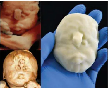
A combination of methods for the construction of physical models was successfully devised. In the case of a 25-week fetus with alobar holoprosencephaly and proboscis (case 16, Table 3), which had been assessed by 3D-US and MRI on
the same day, the body was modeled based on the MRI file, whereas the face and the extremities were modeled based on the 3D-US file. In that case, the 3D-US was instrumental in the evaluation of the extremities and the face (Figure 4).
DISCUSSION
Additive manufacturing technology allows the conver-sion of a virtual 3D model to a physical model, with precise dimensions, in a process that is fast and easy. The process transfers a 3D data file, obtained by superimposing individu-ally segmented layers, to an additive manufacturing device, or 3D printer, which constructs physical models by super-imposing thin layers of raw materials(6–9).
Werner et al.(10) introduced the use of physical models
in fetal disease research, an area in which studies involving digital (3D) modeling are scarce(11–14). The results suggest
a new possibility in the interaction between the parents and the fetus during prenatal monitoring, physically recreating
the interior of the uterus during pregnancy, demonstrating the actual size of the fetus, as well as its anatomy.
One of the main concerns of this study was to obtain high-quality images that can be manipulated with 3D soft-ware, without a loss of accuracy. Fetal movements during Figure 2.Case 18. Fetus with trisomy 21. Whole-body virtual and physical model obtained by MRI.
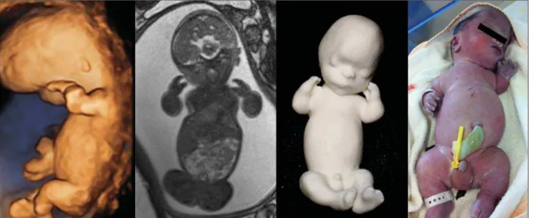
Figure 3.Case 29. Fetus with left radial agenesis and ompha-locele. Whole-body virtual and physical model obtained by MRI.
Figure 4D. Polydactyly of the foot identified by 3D-US, by MRI, and in the physical model.
Figure 4B. Whole-body 3D reconstruction from the MRI and physical model.
Figure 4.Case 16. A: Fetus at the 25th week of gestation, showing alobar holo-prosencephaly and proboscis. Face obtained by 3D-US/3D MRI, and physical model obtained from the ultrasound data.
Figure 4C. Fetal profile obtained by 3D-US and physical model. Notice the pro-boscis (arrow).
the acquisition of images constituted one of the main diffi-culties, especially in the MRI evaluation. This problem is minimized in ultrasound, because the image is acquired in
real time and can be frozen during movement. However, in some cases, the lower resolution of contrast ultrasound cre-ated difficulties due to the limits of the gray scale. The qual-ity of the process is directly associated with the accuracy of the mathematical data that will be used in order to generate the physical model. The images are acquired in slices, which are superimposed for the construction of the model.
The physical models have an impact on the planning of medical interventions(15,16). The can also be used in fetal
medicine for educational purposes(11,14,17,18). The act of combining images obtained by different methods (ultrasound and MRI) can result in better understanding, on the part of the parents and of a multidisciplinary medical team, in evalu-ating certain types of diseases(11–14).
Previous studies have employed ultrasound and 3D models. Blaas et al.(19) calculated the volumes of embryos
and fetuses in the first trimester of pregnancy, transforming the embryo/fetus area into a virtual model. In another study, conducted by Nelson et al.(20), 3D-US data were converted
the advantages of 3D visualization over traditional images. In 3D visualization, the area of interest can be clearly evalu-ated and manipulevalu-ated by the observer, who can thus appre-ciate the physical characteristics of the object and their spa-tial relationships. Therefore, the additive manufacturing device, or 3D printer, came to function as a 3D display de-vice, representing a powerful tool to facilitate the visualiza-tion of various anatomical structures. The models generated represented an important means of communicating, in a tan-gible way, with pregnant women, providing them with in-formation that is more easily understood.
Based on those experiments, the study of 3D fetal mod-eling started using CT files related to fetuses with a gesta-tional age over 30 weeks in order to build physical models of the fetal skeleton(7,11). The result was a series of
connect-ing structures of bones in a virtual 3D environment(6). To
maintain the integrity of the entire virtual skeleton, with preservation of its shape and spatial coordinates, modeling was performed with software (Autodesk Maya; Alias/ Wavefront, Santa Barbara, CA, USA) that allowed a physi-cal model to be produced without losing the accurate posi-tioning of its different parts. The next challenge was the delimitation of the entire outer surface of the fetal body based on the slices obtained by CT. This interactive visual process detected the limits of fetal body parts using a digital stylus, which interacts directly with the computer screen. The re-sulting layers of the entire fetal surface were superimposed, generating a 3D volumetric model.
Based on the results obtained with the CT files, studies using files obtained from fetal MRI scans were initiated. Although manual segmentation was used, the biggest prob-lem with the MRI-based technique was the thickness of the slices and the smaller number of slices. The main difference between the data obtained by CT and those obtained by MRI was the quality of the contrast between the organs. The gray-scale contrast between the organs is greater in MRI. This greater sharpness allowed easy visual separation of the rel-evant areas on a graphics processing screen with a variable-pressure stylus. On CT scans, only the skeleton was easily identifiable. However, despite the better contrast obtained with MRI, there was at first a limitation on the number of slices obtained (approximately 30–40), making the accuracy of the final result questionable. However, with the use of the TrueFISP sequence, which offered a larger number of slices (> 90), the fetal outline obtained became clearer. The TrueFISP technique allowed the slice thickness to be reduced from 4 mm to approximately 1 mm. In the case of MRI, it was easier to obtain images of better quality at later gestational ages, when there is less interference from motion artifacts. The biggest challenge was in the construction of mod-els using ultrasound(7). The ultrasound modality allows a
faster scan of the fetus, the image being automatically trans-formed into a virtual 3D image on the screen(21–23). Up to
the 18th week of gestation, ultrasound allows complete view-ing of the fetal body. Thereafter, the fetal body parts are
visualized as separate blocks to be joined. The tomographic ultrasound imaging function (ultrasound images in CT form) of the 4D View GE software was used in order to render the 3D-US images, obtaining results similar to those obtained by MRI. The images were exported to the Mimics software for 3D image reconstruction, maintaining the accuracy and reliability. Thus, the MRI protocol was adopted for the pro-cessing of ultrasound images.
The mastery of the ultrasound reconstruction technique opened up the possibility of combining the MRI and ultra-sound files when they were acquired on the same day(11). In
that way, all 3D files obtained by ultrasound, MRI, and, in some cases, CT could be combined. As exemplified in case 13 of the present study, it became possible to combine 3D-US images of the face, hands, or feet with fetal body images obtained by MRI, and the necessary biometric proportions can be maintained by means of various measures for both techniques.
As for the cost of production of the physical models, the four manufacturing techniques adopted in this case study differed in relation to the construction and the materials used, which are the main items to be considered in the calculation of costs(11). The most widely used technique involved
print-ers using ZCorp (plaster-based composite) powder. The physical models resulting from that process are the least expensive, especially when compared with those resulting from processes such as stereolithography and selective laser sintering, which use a laser beam for hardening a photosen-sitive resin layer or sintering polyamide powder(6,7).
CONCLUSION
The segmentation and reconstruction techniques devel-oped for fetal modeling can be applied to the construction of virtual and physical models obtained from ultrasound, MRI, and CT images, individually or in combination.
On the basis of the results of this study, we believe that the physical models will, in the near future, facilitate the tactile and interactive study of complex abnormalities in various disciplines. These techniques may also be useful for prospective parents, to recreate a 3D model with the physi-cal characteristics of the fetus, allowing a more direct emo-tional connection with the unborn child.
REFERENCES
1. Antunes EG, Werner H, Daltro PA, et al. Evaluation of fetal cervi-cal lymphangioma by magnetic resonance imaging and correlation with sonographic findings. Radiol Bras. 2009;42:299–302. 2. Daltro P, Werner H, Gasparetto TD, et al. Congenital chest
mal-formations: a multimodality approach with emphasis on fetal MR imaging. Radiographics. 2010;30:385–95.
3. Hellinger JC, Epelman M. Fetal MRI in the third dimension. Ap-plied Radiology. 2010;39:8–22.
4. Cassart M, Massez A, Cos T, et al. Contribution of three-dimen-sional computed tomography in the assessment of fetal skeletal dysplasia. Ultrasound Obstet Gynecol. 2007;29:537–43. 5. Dos Santos JL, Werner H, Fontes R, et al. Additive manufactured
imaging and computed tomography scan data. In.: Hoque ME, editor. Rapid prototyping technology – principles and functional requirements. Rijeka, Croatia: InTech; 2011. p. 179–92. 6. Werner H, dos Santos JR. Tecnologias 3D. Rio de Janeiro, RJ:
Re-vinter; 2010.
7. Lopes J, Brancaglion Jr A, Azevedo SA, et al. Tecnologias 3D – desvendando o passado, modelando o futuro. Rio de Janeiro, RJ: Lexikon Editora Digital; 2013.
8. Willis A, Speicher J, Cooper DB. Rapid prototyping 3D objects from scanned measurement data. Image and Vision Computing. 2007;25:1174–84.
9. Ferreira C, Santos J, Silva J. Exemplos de aplicações da prototipa-gem rápida. In: Volpato N, Ahrens C, Ferreira C, et al., editors. Prototipagem rápida – tecnologias e aplicações. São Paulo, SP: Editora Blucher; 2007. p. 195–224.
10. Werner H, dos Santos JR, Fontes R, et al. The use of rapid proto-typing didactic models in the study of fetal malformations. Ultra-sound Obstet Gynecol. 2008;32:955–6.
11. Werner H, dos Santos JR, Fontes R, et al. Additive manufacturing models of fetuses built from three-dimensional ultrasound, mag-netic resonance imaging and computed tomography scan data. Ul-trasound Obstet Gynecol. 2010;36:355–61.
12. Werner H, Lopes J, Tonni G, et al. Physical model from 3D ultra-sound and magnetic resonance imaging scan data reconstruction of lumbosacral myelomeningocele in a fetus with Chiari II malforma-tion. Childs Nerv Syst. 2015;31:511–3.
13. Werner H, Rolo LC, Araujo Júnior E, et al. Manufacturing models of fetal malformations built from 3-dimensional ultrasound, mag-netic resonance imaging, and computed tomography scan data. Ultrasound Q. 2014;30:69–75.
14. Werner H, Dos Santos JL, Araujo Júnior E. Physical models of the foetus created using magnetic resonance imaging, computed
to-mography, and ultrasound data: history, description, and potential uses. Rev Bras Ginecol Obstet. 2015;37:149–51.
15. Armillotta A, Bonhoeffer P, Dubini G, et al. Use of rapid prototyping models in the planning of percutaneous pulmonary valved stent im-plantation. Proc Inst Mech Eng H. 2007;221:407–16.
16. Robiony M, Salvo I, Costa F, et al. Virtual reality surgical planning for maxillofacial distraction osteogenesis: the role of reverse engi-neering rapid prototyping and cooperative work. J Oral Maxillofac Surg. 2007;65:1198–208.
17. Werner H, Dos Santos JRL, Fontes R, et al. Virtual bronchoscopy in the fetus. Ultrasound Obstet Gynecol. 2011;37:113–5. 18. Werner H, Lopes dos Santos JR, Fontes R, et al. Virtual
broncho-scopy for evaluating cervical tumors of the fetus. Ultrasound Obstet Gynecol. 2013;41:90–4.
19. Blaas HG, Taipale P, Torp H, et al. Three-dimensional ultrasound volume calculations of human embryos and young fetuses: a study of the volumetry of compound structures and its reproducibility. Ultrasound Obstet Gynecol. 2006;27:640–6.
20. Nelson TR, Bailey MJ. Solid object visualization of 3D ultrasound data. J Med Imaging. 2000;3982:26–34.
21. Araujo Júnior E, Simioni C, Nardozza LMM, et al. Prenatal diag-nosis of Beckwith-Wiedemann syndrome by two- and three-dimen-sional ultrasonography. Radiol Bras. 2013;46:379–81.
22. Araujo Júnior E, Santana EFM, Nardozza LMM, et al. Assessment of embryo/fetus during pregnancy by three-dimensional ultrasonog-raphy using the HD live software: iconographic essay. Radiol Bras. 2015;48:52–5.