C
a s eR
e p o Rt2 0 0 Arq Bras Oftalmol. 2016;79(3):200-1 http://dx.doi.org/10.5935/0004-2749.20160058
INTRODUCTION
Polypoidal choroidal vasculopathy (PCV) is a disorder that is characterized by dilatation of the choroidal vessels. This disorder was initially reported as idiopathic PCV in 1990(1). Angioid streaks (AS)
are breaks in the Bruch’s membrane that result in irregular radial or concentric lines around the optic disc; these are mostly associated with pseudoxanthoma elasticum (PXE)(2). In literature, PCV and AS are
rarely described in the same patient(3-5), and visual impairment usually
occurs because of complications, such as choroidal neovasculariza-tion (CNV) during the natural course of both diseases(2,5). Visual acuity
can remain unaffected if hemorrhage, subretinal or intraretinal exu-dation, or pigment epithelial detachments do not develop. Herein we present a case of silent PCV in a patient with AS.
CASE REPORT
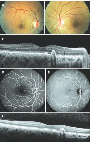
A 26-year-old woman was admitted to our clinic for routine oph thalmic examination. Her past ocular and medical histories were unremarkable. At presentation, visual acuities were 20/20 for both eyes, and anterior segment findings for both eyes were normal. Fundoscopy disclosed AS radiating from the optic disc and bilateral changes in the retinal pigment epithelium (RPE) that were evident on the nasal side of the fovea of the right eye (Figure 1 A, B). Optical co-herence tomography (OCT) showed a steep, dome-shaped RPE ele-vation with moderate hyperreflectivity beneath the RPE (Figure 1 C). Due to the suspicion of PCV, we performed fluorescein angiography (FA) and indocyanine green angiography (ICGA). FA demonstrated irregular hyperfluorescence around the optic disc associated with AS in both eyes and increasing hyperfluorescent foci in the nasal side of the fovea of the right eye (Figure 1 D). ICGA showed a focal area of
hyperfluorescence surrounded by a hypofluorescent halo in the right macula (Figure 1 E). Neovascularization was excluded by the imaging modalities, and no associated systemic conditions related to AS were found on consultation.
With these findings, we diagnosed the patient with AS in both eyes and PCV in the right eye. Because she had no symptoms and no intraretinal or subretinal fluid was seen on imaging (Figure 1 F), our initial plan was to monitor the patient. The PCV remained silent with no leakage, and no decrease in vision was seen during the 2 years of follow-up.
DISCUSSION
PCV is a localized enlargement of the choroidal vasculature that forms polyps and originates from the inner choroid(1). Hyalinization
of vessels as well as plasma and/or fibrin exudation are the his to-pa thological features of PCV(6). Serous or hemorrhagic pigment
epi thelial detachments, subretinal hemorrhages, and exudation are the common signs of PCV, and these secondary complications cause visual disturbances(6). If the patient has no symptoms and there are
no ophthalmoscopic signs, imaging modalities, especially ICGA, can assist physicians in diagnosing the lesion.
PCV has been shown to be associated with pathologies such as tilted disc, high myopia, retinitis pigmentosa, central serous chorio-retinopathy, and AS(7-9).
Few cases with coexistence of AS and PCV have been reported in literature(3-5). PCV has been detected at initial examination in some
of the cases in literature, and some have developed PCV as observed during the follow-up of CNV due to AS(3-5). The first reported case was
initially treated for CNV as a complication of AS in both eyes, and the
Silent polypoidal choroidal vasculopathy in a patient with angioid streaks
Vasculopatia polipoidal de coróide quiescente em um paciente com estrias angióides
Zafer CebeCi1, Serife bayraktar1, Merih Oray1, Nur kir1
Submitted for publication: August 3, 2015 Accepted for publication: September 23, 2015
1 Department of Ophthalmology, Istanbul Faculty of Medicine, Istanbul University, Istanbul, Turkey.
Funding: No specific financial support was available for this study.
Disclosure of potential conflicts of interest: None of the authors have any potential conflict of interest to disclose.
Corresponding author: Zafer Cebeci. Istanbul Tip Fakultesi, Goz Hastaliklari A.D. Capa, Istanbul 34390 - Turkey - E-mail: [email protected]
ABSTRACT
We present a case of silent polypoidal choroidal vasculopathy (PCV) in a patient with angioid streaks. PCV was detected during a routine ophthalmic examination and confirmed by fluorescein angiography, indocyanine green angiography, and optical coherence tomography. After 2 years of follow-up, the PCV remained silent without any complications. We report this rare coexistence and review literature on this topic.
Keywords: Polyps; Choroid; Choroidal neovascularization; Fluorescein angiogra-phy; Indocyanine green; Retinal pigment epithelium; Tomography, optical cohe-rence; Vascular endothelial growth factor A; Angioid streaks
RESUMO
Nós apresentamos um relato de vasculopatia polipoidal de coroide (PCV) em pacien-te com estrias angióides. Vasculopatia polipoidal de coroide depacien-tectada em exame oftalmológico de rotina e confirmado por angiofluoresceinografia, angiografia com indocianina verde e tomografia de coerência óptica. Após 2 anos de seguimento a vasculopatia polipoidal de coroide permaneceu quiescente, sem qualquer complicação. Nós relatamos esta coexistência rara e apresentamos revisão da literatura.
Ce b e C i Z, e ta l.
2 0 1
Arq Bras Oftalmol. 2016;79(3):200-1
the lesions were far away from the fovea, they did not require any treatment, which suggests that vascular anomalies causing PCV can be initially detected in patients with AS. Further, some patients may have a predisposition for developing polypoidal lesions.
A 59-year-old male patient with a history of PXE and CNV due to AS developed PCV 1 year after the diagnosis of neovascularization; the patient had already undergone nine intravitreal anti-VEGF injec-tions for CNV(5).
AS is usually related to a systemic condition, with PXE being the most common. Smooth muscle cells play a role in the systemic pa-thological changes of PXE(10). Abnormalities in the smooth muscle
cells of the choroidal vascular structure lead to dilatations that form PCV, and these two entities can show similarities in their pathogene-sis(3). Moreover, alterations of Bruch’s membrane, which simplify the
development of CNV in patients with AS, may also facilitate compli-cations due to polyps. It is also important to keep in mind that there were no associated systemic diseases in the present case, but the patient is still young and there is a possibility that she may develop systemic findings in future. The reported PCV cases in patients with AS have been diagnosed at older ages(3-5). In addition to the pathology
of Bruch’s membrane in AS, which usually progresses over many years, aging may contribute to the impairment of Bruch’s membrane; the-refore, complications may easily occur.
PCV lesions can stay silent and do not affect visual acuity if there are no signs of leakage or hemorrhage from the polypoidal lesions(6).
Our patient did not report visual loss, and her PCV was diagnosed incidentally. Ophthalmoscopic examination and imaging modalities confirmed the diagnosis of PCV, and she did not develop any com-plications during the 2 years of follow-up.
In conclusion, patients with AS should be followed-up not only for the development of CNV but should also be followed-up routinely for polypoidal lesions. Vision-threatening complications can occur during the natural course of PCV, and imaging techniques such as ICGA and OCT are the most important tools for the diagnosis of this rare association.
REFERENCES
1. Yannuzzi LA, Sorenson J, Spaide RF, Lipson B. Idiopathic polypoidal choroidal vascu-lopathy (IPCV). Retina. 1990;10(1):1-8.
2. Gliem M, Finger RP, Fimmers R, Brinkmann CK, Holz FG, Charbel Issa P. Treatment of choroidal neovascularization due to angioid streaks: a comprehensive review. Retina. 2013;33(37):1300-14.
3. Baillif-Gostoli S, Quaranta-El Maftouhi M, Mauget-Faÿsse M. Polypoidal choroidal vas-culopathy in a patient with angioid streaks secondary to pseudoxanthoma elasticum. Graefes Arch Clin Exp Ophthalmol. 2010;248(12):1845-8.
4. Nakagawa S, Yamashiro K, Tsujikawa A, Otani A, Tamura H, Ooto S, et al. The time course changes of choroidal neovascularization in angioid streaks. Retina. 2013;33(4): 825-33.
5. Khan S, Engelbert M, Imamura Y, Freund KB. Polypoidal choroidal vasculopathy: si mul-taneous indocyanine green angiography and eye-tracked spectral domain optical coherence tomography findings. Retina. 2012(6);32:1057-68.
6. Nowak-Sliwinska P, van den Bergh H, Sickenberg M, Koh AH. Photodynamic therapy for polypoidal choroidal vasculopathy. Prog Retin Eye Res. 2013;37:182-99. 7. Nakanishi H, Tsujikawa A, Gotoh N, Hayashi H, Iwama D, Tamura H, et al. Macular
com-plications on the border of an inferior staphyloma associated with tilted disc syndrome. Retina. 2008;28(10):1493-501.
8. Ishida T, Moriyama M, Morohoshi K, Furuse Y, Fukuda T, Ohno-Matsui K, et al. Polypoi-dal choroiPolypoi-dal vasculopathy in a case with retinitis pigmentosa. Int Ophthalmol. 2013; 33(3):305-8.
9. Toyama T, Ohtomo K, Noda Y, Ueta T. Polypoidal choroidal vasculopathy and history of central serous chorioretinopathy. Eye (Lond). 2014;28(8):992-7.
10. Gheduzzi D, Sammarco R, Quaglino D, Bercovitch L, Terry S, Taylor W, et al. Extracuta-neous ultrastructural alterations in pseudoxanthoma elasticum. Ultrastruct Pathol. 2003;27(6):375-84.
Figure 1. A) Color fundus photography of the right eye demonstrated radial and cir cumferential angioid streaks (AS) around the optic disc as well as pigment epithelial changes at the nasal side of the fovea. B) Color fundus photography of the left eye showed AS around the optic disc. C) Optical cohorence tomography (OCT) scan through the retinal pigment epithelium (RPE) changes in the right macula at the initial visit demonstrated sharply elevated RPE with moderate hyperrelectivity under it. D) Fluorescein angiography showed dye leakage nasal to the macula in the right fundus. E) Indocyanine green angiography showed a foci of hyperluorescence surrounded by a hypoluorescent halo in the right macula. F) OCT scan of the same area after 2 years of followup showed a polypoidal lesion.
A
C
D
F
E B
patient developed PCV temporally to the macula and in the nasal side of the fovea of the right eye during the follow-up period(3).
In a series of 44 cases with AS, PCV was found in six eyes of five patients(4). PCV was identified in two eyes at initial examination, and