https://doi.org/10.1590/0004-282X20170171 ARTICLE
Syndromic craniosynostosis:
neuropsycholinguistic abilities and imaging
analysis of the central nervous system
Craniossinostoses sindrômicas: habilidades neuropsicolinguísticas e análise por imagem
do sistema nervoso central
Luciana Paula Maximino1,2, Luis Gustavo Ducati4, Dagma Venturini Marques Abramides1, Camila de Castro
Corrêa3, Patrícia Fernandes Garcia1, Adriano Yacubian Fernandes1,4
Within the ield of craniofacial anomalies, there is a het
-erogeneous group of disorders represented by craniosynosto
-ses, which occur due to premature fusion of one or more cra
-nial sutures and may cause esthetic and functional damage1.
With a prevalence of one in each 2,500 live births, the cranio
-synostosis may occur both as isolated disorders or as part of
syndromes. Most syndromic craniosynostosis have autoso
-mal dominant inheritance2, which highlights the importance
of genetic counseling for these patients.
he most frequent syndromic craniosynostosis include Crouzon, Apert, Saethre-Chotzen, Pfeifer and Muenke syndromes; the irst three accounting for nearly two thirds of syndromic cases2.
1Universidade de São Paulo, Faculdade de Odontologia de Bauru, Departamento de Fonoaudiologia e Audiologia, Bauru SP, Brasil; 2Universidade de São Paulo, Hospital para Reabilitação para Anomalias Craniofaciais, Bauru SP, Brasil;
3Universidade Estadual Paulista, Faculdade de Medicina de Botucatu, Departamento de Oftalmotogia, Otorrinilaringologia e Cirurgia de Cabeça e Pescoço,
Botucatu SP, Brasil;
4Universidade Estadual Paulista, Faculdade de Medicina de Botucatu, Departamento de Neurologia, Psicologia e Psiquiatria, Botucatu SP, Brasil.
Correspondence: Luciana Paula Maximino; Al. Octávio Pinheiro Brisola, 9 / 75; 17012-901 Botucatu SP, Brasil; E-mail: [email protected]
Conflict of interest: There is no conflict of interest to declare.
Support: FAPESP2000/080803 and CNPQ 307043/2008-8.
Received 05 August 2016; Received in final form 09 July 2017; Accepted 13 September 2017. ABSTRACT
Objective: To characterize patients with syndromic craniosynostosis with respect to their neuropsycholinguistic abilities and to present these findings together with the brain abnormalities. Methods: Eighteen patients with a diagnosis of syndromic craniosynostosis were studied. Eight patients had Apert syndrome and 10 had Crouzon syndrome. They were submitted to phonological evaluation, neuropsychological evaluation and magnetic resonance imaging of the brain. The phonological evaluation was done by behavioral observation of the language, the Peabody test, Token test and a school achievement test. The neuropsychological evaluation included the WISC III and WAIS tests.
Results: Abnormalities in language abilities were observed and the school achievement test showed abnormalities in 66.67% of the patients. A normal intelligence quotient was observed in 39.3% of the patients, and congenital abnormalities of the central nervous system were observed in 46.4% of the patients. Conclusion: Abnormalities of language abilities were observed in the majority of patients with syndromic craniosynostosis, and low cognitive performance was also observed.
Keywords: acrocephalosyndactylia; craniofacial dysostosis; central nervous system; neuropsychology; language.
RESUMO
Objetivo: Caracterizar as habilidades neuropsicolinguísticas de indivíduos com craniossinostoses sindrômicas e apresentar esses achados com as anomalias do sistema nervoso central. Métodos: Participaram do estudo 18 sujeitos com diagnóstico clínico de craniossinostose sindrômica, 44,4% com a síndrome de Apert e 55,6% síndrome de Crouzon. Todos os sujeitos foram submetidos a avaliação fonoaudiológica, psicológica e exames de ressonância magnética do encéfalo. A avaliação fonoaudiológica foi contemplada pela Observação Comportamental da Linguagem, Teste Peabody (TVIP), Teste Token e Teste de Desempenho Escolar (TDE); enquanto a psicológica utilizou a WISC-III e a WAIS.
Resultados: Observou-se alteração nas habilidades de linguagem em todos os protocolos utilizados, sendo o TDE o que apresentou maior porcentagem de alteração (66,67%).A avaliação cognitiva evidenciou quociente de inteligência dentro da média em 39,3% dos sujeitos, enquanto que 46,4% apresentaram malformações congênitas do sistema nervoso central. Conclusão: Constatou-se alterações nas habilidades de linguagem na maioria dos sujeitos com craniossinostoses sindrômicas, bem como o baixo desempenho cognitivo.
Apert syndrome is a congenital disorder characterized primarily by craniosynostosis, midface hypoplasia, and syn
-dactyly of the hands and feet with a tendency to fusion of bony structures. Most cases are sporadic, but autosomal dominant inheritance has been reported3
. Crouzon syn
-drome is an autosomal dominant disorder characterized by craniosynostosis causing secondary alterations of the facial bones and facial structure. Common features include hyper
-telorism, exophthalmos and external strabismus, parrot-beaked nose, short upper lip, hypoplastic maxilla, and a rela
-tive mandibular prognathism4.
hey share other characteristics beyond craniosynosto
-sis, including cranial base anomalies, abnormal facies, limb anomalies and mutation of the ibroblast growth factor receptor 2 gene2. Additionally, there is frequent occurrence
of increased intracranial pressure, hydrocephaly, optical atrophy, breathing problems, speech and hearing disorders, obstructive sleep apnea and visual impairment5,6.
Surgical treatment may be required, for esthetic reasons and neurological complications6. In the treatment of these
disorders, craniofacial surgery for cranial decompression per
-formed in the irst year of life is fundamental to avoid intra
-cranial hypertension, which may have deleterious efects on the cognitive and linguistic development7,8.
Regardless of the type or etiology, among craniofacial anomalies, this group represents a signiicant array of pathol
-ogies that may impair diferent functions of the central ner
-vous system (CNS) during development of the children9.
hese impairments imply the need for multidisciplinary care, with a varied staf of specialists, including plastic surgeons, neurosurgeons, geneticists, dentists, neurologists, speech-language pathologists, ear, nose and throat doctors, orthope
-dists, social workers, and others10.
Within these complex disorders that afect the craniofa
-cial structures, it is possible to observe anatomical and func
-tional interferences that may cause language delays and/or disorders11
. he hypothesis of the present study was that, in addition to the cognitive alterations, the language altera
-tions that may also be associated with these condi-tions may include language or learning disorders. Language impair
-ment presents as deicits in comprehension and change in at least one aspect of language, such as phonology, syntax, semantics, and pragmatics12.
Learning disability is a broad term. It is a condition when a child’s achievement is substantially below what one might expect for that child. It does not include problems that are primarily the result of intellectual disabilities, emotional dis
-turbance, visual, hearing, or emotional disabilities. hese children, despite having an average or above average level of intelligence, have diiculty acquiring basic academic skills, such as the luent reading of words, correct spelling, written expression and mathematical operations13.
Assessment of the linguistic and cognitive integrity by speech-language and psychological evaluations, herein called
neuropsycholinguistic, are fundamental to rule out any lan
-guage and learning disorders in syndromic craniosynostosis11,14.
his study evaluated the neuropsycholinguistic abilities and morphology of the CNS in patients with syndromic craniosynos
-tosis. he aim of this study was to characterize this population with regard to their neuropsycholinguistic aspects and to pres
-ent these indings together with the brain abnormalities.
METHODS
Ethical aspects
his study was conducted from 2008 to 2011 at the Hospital for Rehabilitation of Craniofacial Anomalies of the University of São Paulo, Bauru, São Paulo (a tertiary refer
-ence center for craniofacial anomalies), after approval by the Institutional Review Board (n. 288/2006). All criteria of Regulation 196/96 were met. All patients or legal caretakers agreeing to participate in the study signed an informed con
-sent form. All the patients who were evaluated were regularly enrolled at this hospital and met the inclusion criteria.
Sample
he study was conducted on 18 patients with a clinical diagnosis of syndromic craniosynostosis, with a mean age of 18.75 years (standard deviation 64.38; minimum 6.33 years; maximum 31.25 years). here was predominance of a low socioeconomic level (83.3%), ranging from low to high15. he
percentage of patients with a diagnosis of syndromic cra
-niosynostosis was 44.4% Apert syndrome (AS) and 55.6% Crouzon syndrome (CS), as described in Table 1.
Inclusion criteria: Patients receiving regular treatment
at the hospital where the study was conducted; diagnosis of syndromic craniosynostosis; availability to perform all evalu
-ations planned in the study.
Exclusion criteria:Hearing impairment: sensorineural or
conductive hearing loss.
Procedures
All patients were submitted to speech-language and psy
-chological analyses and magnetic resonance imaging (MRI) of the brain. It should be highlighted that all analyses were performed according to the chronological age of patients.
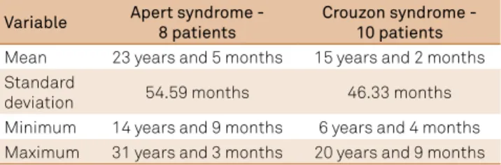
Table 1. Sample distribution among Apert and Crouzon syndromes, with information on the number of patients and mean age.
Variable Apert syndrome - 8 patients
Crouzon syndrome - 10 patients
Mean 23 years and 5 months 15 years and 2 months Standard
deviation 54.59 months 46.33 months
Auditory and speech analysis
Initially an audiological evaluation was performed, which was a prerequisite for continuation of the other evaluations. his was comprised clinical ear inspection, threshold tone audiometry16 and tympanometry17
. All patients were required to have results within the normal range.
he speech-language analysis was performed by behavioral observation of language (qualitative analysis)18, as well as uti
-lization of standardized protocols that allowed quantiication. Concerning the behavioral observation of language, the parameters of each speciic age range were considered, tak
-ing into account language reception and expression. Table 2 shows the details of observation of each language level19.
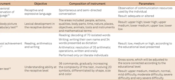
he speech-language analyses performed, the instru
-ments employed, as well as their objectives, composition and parameters for analysis18,20-22 are shown in Table 3.
Psychological analysis
he cognitive analysis was obtained using the Wechsler Intelligence Scale for Children – III23, a standardized test
that measures intellectual functioning in children aged six to 16 years, and the Wechsler Adult Intelligence Scale24, a test
designed to measure intelligence in adults and older teenag
-ers. he intelligence quotients (IQ) were obtained as verbal IQ, performance IQ and full IQ. he scales have a mean (aver
-age) standard score of 100. Scores from 90–110 are considered
average. Just outside of that range is the high average range (110–119), the low average range (80–89), and the borderline range is 70–79. Preschool children were assessed using the form L-M of the Stanford-Binet scale25.
A numerical value of 70 was considered to be the divid
-ing line between patients with satisfactory IQ (equal to or greater than 70) and those with unsatisfactory IQ (below 70), as suggested by the World Health Organization26. Although
the most updated deinition of intellectual disability consists of IQ measurement plus an adaptive scale, in this study we adopted the measure of the IQ only, to establish correlations.
Neuroimaging examination
he MRIs were obtained in a 0.5T scanner (Flexart, TOSHIBA, Japan) in sequences T1, T1 inversion recovery, T2 and Flair, in sagittal, coronal and axial planes, and later evaluated by a neurologist. It is important to explain that as the MRI scans were done at low resolutions (0.5T) it was not fea
-sible to evaluate small malformations such as focal dysplasias.
Analysis of results
he results were tabulated and scored according to the guide
-lines and standardization of tests employed for speech-language and cognitive analysis. he Student’s t-test, Tukey test, analy
-sis of variance, chi-square test and Spearman’s correlation were applied for comparison and correlation between variables.
Table 2. Description of behaviors analyzed for each language component in receptive and expressive language19.
Language components Expressive Receptive
Phonology Production of speech sounds Hearing, sound discrimination and processing
Syntax Utilization of grammatical structures of language Understanding of grammatical structures of language Semantics Utilization of vocabulary, meaning and concept Understanding of vocabulary, meaning and concept
Pragmatics Functional utilization of language as communication
means, coherent responses, maintenance of topic Understanding of language
Table 3. Instruments employed for evaluation of receptive and expressive aspects of language, presenting the instrument name, objective, composition and parameters.
Instrument Objective Composition of instrument Parameters
Behavioral observation of language18
Receptive and expressive language
Spontaneous and semi-directed conversation
Observation of communication resources used by the individual
Result: adequate or altered
Peabody picture vocabulary test20
Lexical development in the receptive domain
The areas included: people, actions, qualities, body parts, time, nature, places, objectives, animals, tools and instruments and mathematical terms
Result: upper high; lower high; upper medium; lower medium; upper low; lower low
School achievement test21
Reading, arithmetics and writing
Reading: decoding of 70 isolated words
Result: low, medium or high, according to the educational level presented
Writing: writing their own name and 34 words presented as dictation
Arithmetics: resolution of 35 arithmetic operations, written and orally
Performed only on literate individuals
Token test22 Understanding ability at
the receptive level
36 commands, gradually increasing the complexity of the test, involving 20 symbols, differentiated by shape, size and color
Gross score, which will be adjusted to the score corrected according to the educational level
RESULTS
In the present sample, ive patients (27.78%) did not pres
-ent with impairm-ent of spoken language abilities, of whom four had CS and one had AS. Figure 1 shows the occurrence of disorders for each ability assessed by the behavioral obser
-vation of language.
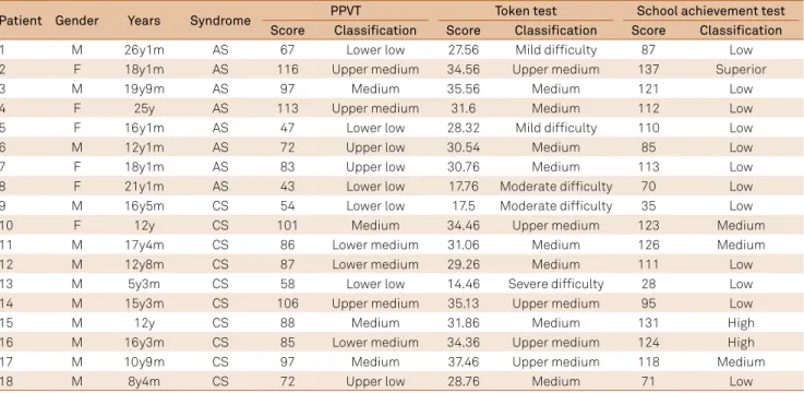
The results of quantitative standardized tests are presented in Table 4. The Peabody Picture Vocabulary Test (PPVT) showed more diffuse distribution of scores, the Token test was altered in 27.78% of patients and the school achievement test revealed low scores for most patients (66.67%).
Considering the indings of the clinical and standard
-ized speech-language evaluation, it was possible to infer the
speech-language diagnostic hypothesis, in which 44.5% of the sample presented with a learning disorder and 16.7% had a language disorder, while 33.4% of the sample did not pres
-ent with language alterations. One pati-ent had diiculties with written language (Patient 3).
Table 5 shows the intelligence quotient (IQ) results (ver
-bal, performance and full IQs), which revealed IQs within the average in 50% of patients analyzed, and four patients (22.2%) exhibited intellectual disability.
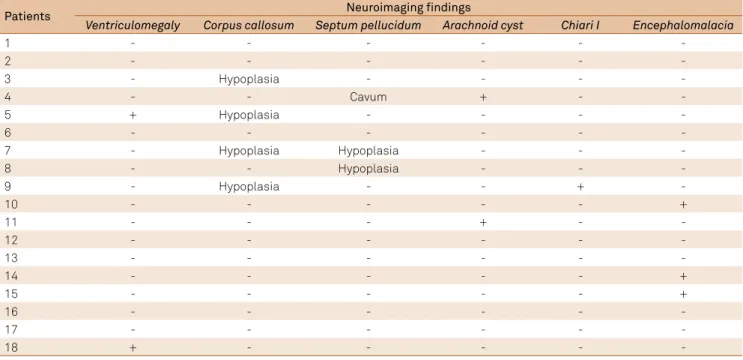
Concerning the CNS disorders, it should be mentioned that encephalomalacia is related to surgical complications, rather than a congenital disorder of the brain. hus, both congenital and acquired disorders were observed on the MRI, showing that 61.1% of individuals exhibited abnormalities of the CNS (Table 6).
In the statistical analysis of variables, the results indi
-cated a correlation between the IQ and speech-language diagnosis (p = 0.002).
he speech-language diagnostic hypothesis revealed an association between the results of the PPVT (p = 0.046), Token test (p = 0.004) and school achievement test (p = 0.001); as well as the diagnosis of language disorder (0.002) and learning disorder (0.021). hus, it may be inferred that the results obtained by complementary evaluation were sensi
-tive enough to deine the hypothesis, conirming the ind
-ings (Table 7).
Concerning the morphological alterations of the CNS, the results revealed an association between hypoplasia of the corpus callosum and the indings of the PPVT test (p = 0.037), i.e., the performance of the patients.
Figure. Performance in oral language abilities observed by the behavioral observation of language.
Adequate Altered
18
16
12
8
4
0 14
10
6
2
Phonology Syntax Semantics Pragmatics Reception
Table 4. Results of standardized tests: PPVT, Token test and school achievement test, according to the classification proposed and score achieved of each patient, specifying age and gender.
Patient Gender Years Syndrome PPVT Token test School achievement test
Score Classification Score Classification Score Classification
1 M 26y1m AS 67 Lower low 27.56 Mild difficulty 87 Low
2 F 18y1m AS 116 Upper medium 34.56 Upper medium 137 Superior
3 M 19y9m AS 97 Medium 35.56 Medium 121 Low
4 F 25y AS 113 Upper medium 31.6 Medium 112 Low
5 F 16y1m AS 47 Lower low 28.32 Mild difficulty 110 Low
6 M 12y1m AS 72 Upper low 30.54 Medium 85 Low
7 F 18y1m AS 83 Upper low 30.76 Medium 113 Low
8 F 21y1m AS 43 Lower low 17.76 Moderate difficulty 70 Low
9 M 16y5m CS 54 Lower low 17.5 Moderate difficulty 35 Low
10 F 12y CS 101 Medium 34.46 Upper medium 123 Medium
11 M 17y4m CS 86 Lower medium 31.06 Medium 126 Medium
12 M 12y8m CS 87 Lower medium 29.26 Medium 111 Low
13 M 5y3m CS 58 Lower low 14.46 Severe difficulty 28 Low
14 M 15y3m CS 106 Upper medium 35.13 Upper medium 95 Low
15 M 12y CS 88 Medium 31.86 Medium 131 High
16 M 16y3m CS 85 Lower medium 34.36 Upper medium 124 High
17 M 10y9m CS 97 Medium 37.46 Upper medium 118 Medium
18 M 8y4m CS 72 Upper low 28.76 Medium 71 Low
DISCUSSION
Syndromic craniosynostosis, especially AS and CS, are diagnosed by clinical evaluation by geneticists, tak
-ing into account the phenotypic aspects, ideally add-ing a genetic investigation by molecular biology analysis. Our cohort had casual equal distribution of genders, with a predominance of patients with CS (Table 1). No studies were found in the literature indicating the specific occur
-rence of CS.
Despite the existence of studies attempting to correlate factors that interfere with neuropsychological development in AS and CS, there are few studies in the literature speciic to CS, possibly because the intellectual disability in this group is much lower compared to those with AS. Patients with AS presented with mild and irregular intellectual disability, with varied alterations in some brain structures, besides the inlu
-ence of the socioeconomic level and educational level of the parents27. In the present study, the prevalence of intellectual
disability in patients with AS was 22.2%.
Table 5. Description of values of psychological evaluation of the verbal intelligence quotient (VIQ); performance intelligence quotient (PIQ) and full intelligence quotient (FIQ) of patients in the sample.
Patients Syndrome TEST VIQ PIQ FIQ Final classification
1 AS WAIS 71 74 71 Borderline
2 AS WISC-III 100 108 104 Average
3 AS WISC-III 65 71 65 Intellectual disability
4 AS WAIS 78 68 70 Borderline
5 AS WISC-III 75 73 72 Borderline
6 AS Terman-Merril - - 84 Average
7 AS WISC-III 74 86 78 Borderline
8 AS WISC-III 48 51 47 Intellectual disability
9 CS WISC-III 48 49 46 Intellectual disability
10 CS WISC-III 95 93 93 Average
11 CS WISC-III 80 72 74 Borderline
12 CS WISC-III 91 98 93 Average
13 CS WISC-III 57 61 56 Intellectual disability
14 CS WISC-III 92 95 93 Average
15 CS WISC-III 101 100 101 Average
16 CS WISC-III 101 97 98 Average
17 CS WISC-III 112 90 102 Average
18 CS WISC-III 72 96 82 Average
(-): not performed; AS: Apert syndrome; CS: Crouzon syndrome; WISC: Wechsler Intelligence Scale for Children; WAIS: Wechsler Adult Intelligence Scale.
Table 6. Findings of magnetic resonance imaging of the brain indicating the disorders observed in patients in the sample.
Patients Neuroimaging findings
Ventriculomegaly Corpus callosum Septum pellucidum Arachnoid cyst Chiari I Encephalomalacia
1 - - -
-2 - - -
-3 - Hypoplasia - - -
-4 - - Cavum + -
-5 + Hypoplasia - - -
-6 - - -
-7 - Hypoplasia Hypoplasia - -
-8 - - Hypoplasia - -
-9 - Hypoplasia - - +
-10 - - - +
11 - - - + -
-12 - - -
-13 - - -
-14 - - - +
15 - - - +
16 - - -
-17 - - -
-18 + - - - -
he literature reveals the need for more thorough neu
-ropsychological evaluation for patients with AS, consider
-ing the heterogeneity of cognitive alterations28, as was also
observed in this study (Table 3) in the diferent age ranges. his wide age range implies interferences from the efects of cranial deformities and also from the treatments received.
Congenital malformations of the CNS were observed in 61.1% of patients with syndromic craniosynostosis (Table 4). he literature describes alterations in the MRI of patients with AS of 55.6%29, compared to 42.8%30 and 40%31
in patients with CS.
his study did not ind signiicant correlation between the MRI indings and the IQ and language abilities (Table 5). It should be highlighted that all patients with AS27 and CS30
with normal brain structures exhibited IQs above 70, show
-ing a tendency.
he language abilities were altered in 72.3% of the sample (Figure). his marked diiculty is reported in the literature, indicating problems in both expressive and receptive lan
-guage32, as well as speciic alterations in the syntactic level of
expressive language33.
he standardized instruments that allow quantitative analysis in speech-language pathology do not address all age ranges; therefore, the added use of behavioral observation of language was necessary, so all data could be combined to guide the language diagnosis.
Speciically, 44.44% of patients exhibited an altered per
-formance in the PPVT (Table 2), while the literature indicates alterations in 100% of cases of syndromic craniosynostosis33.
Understanding was altered, as indicated by the Token test in 27.78% and 58% in the behavioral observation of lan
-guage. Corroborating this inding, Shipster et al.34
found that understanding was altered in 40% of children with AS.
he result of the school achievement test revealed low scores in 12 patients (66.67%), characterizing speciic cases of learning disorders (Table 2).
he speech-language analysis allowed characterization of the close interaction between developmental aspects and IQ, hence the utilization of the term neuropsycholinguistic development was pertinent. he patients analyzed showed a relationship between low IQ and language disorders, with smaller global impairment for patients with only learning disorders, as previously reported in the literature in studies on patients with AS35, CS14,31 and Saethre-Chotzen syndrome9.
he understanding of language and learning disorders observed in patients with syndromic craniosynostosis, relating to the several factors investigated, allows a better therapeutic approach and contributes to the understanding of neuropsy
-cholinguistic disorders, addressing the parallelism between biological aspects (neuronal connectivity and brain circuits as a whole) and environmental aspects (adequate stimulation by healthy afective and challenging cognitive interactions).
he limitations of the evaluation of speech-language abil
-ities across a wide age range, as in the present study, should be noted. In an attempt to overcome this limitation, the study included behavioral observations of language. Further studies are warranted to follow up and better understand the communication abilities of patients with syndromic cranio
-synostosis, as well as speciic studies for validation of new instruments for assessment.
In conclusion, alterations in language abilities were present in most patients with syndromic craniosynostoses, as well as morphological anomalies of the CNS. Low cogni
-tive performance was observed in a few patients. It should be highlighted that learning disorders were correlated with milder cognitive alterations.
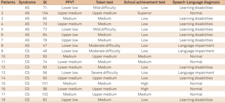
Table 7. Summarized findings of language tests with the respective speech-language diagnoses.
Patients Syndrome QI PPVT Token test School achievement test Speech-Language diagnosis
1 AS 71 Lower low Mild difficulty Low Learning disabilities
2 AS 104 Upper medium Upper medium Superior Normal
3 AS 65 Medium Medium Low Learning disabilities
4 AS 70 Upper medium Medium Low Learning disabilities
5 AS 72 Lower low Mild difficulty Low Learning disabilities
6 AS 84 Upper low Medium Low Learning disabilities
7 AS 78 Upper low Medium Low Learning disabilities
8 AS 47 Lower low Moderate difficulty Low Language impairment
9 CS 46 Lower low Moderate difficulty Low Language impairment
10 CS 93 Medium Upper medium Medium Normal
11 CS 74 Lower medium Medium Medium Normal
12 CS 93 Lower medium Medium Low Learning disabilities
13 CS 56 Lower low Severe difficulty Low Language impairment
14 CS 93 Upper medium Upper medium Low Learning disabilities
15 CS 101 Medium Medium High Normal
16 CS 98 Lower medium Upper medium High Normal
17 CS 102 Medium Upper medium Medium Normal
18 CS 82 Upper low Medium Low Learning disabilities
1. Senarath-Yapa K, Chung MT, McArdle A, Wong VW, Quarto N, Longaker MT et al. Craniosynostosis: molecular pathways and future pharmacologic therapy. Organogenesis. 2012;8(4):103-13. https://doi.org/10.4161/org.23307
2. Panigrahi I. Craniosynostosis genetics: the mystery unfolds. Indian J Hum Genet. 2011;17(2):48-53. https://doi.org/10.4103/0971-6866.86171
3. Mantilla-Capacho JM, Arnaud L, Díaz-Rodriguez M, Barros-Nuñez P. Apert syndrome with preaxial polydactyly showing the typical mutation Ser252Trp in the FGFR2 gene. Genet Couns. 2005;16(4):403-6.
4. Glaser RL, Jiang W, Boyadjiev SA, Tran AK, Zachary AA, Van Maldergem L et al. Paternal origin of FGFR2 mutations in sporadic cases of Crouzon syndrome and Pfeiffer syndrome. Am J Hum Genet. 2000;66(3):768-77. https://doi.org/10.1086/302831
5. Jong T, Rijken BF, Lequin MH, Veelen ML, Mathijssen IM. Brain and ventricular volume in patients with syndromic and complex craniosynostosis. Childs Nerv Syst. 2012;28(1):137-40. https://doi.org/10.1007/s00381-011-1614-7
6. Khanna PC, Thapa MM, Iyer RS, Prasad SS. Pictorial essay: the many faces of craniosynostosis. Indian J Radiol Imaging. 2011;21(1):49-56. https://doi.org/10.4103/0971-3026.76055
7. Marucci DD, Dunaway DJ, Jones BM, Hayward RD. Raised intracranial pressure in Apert syndrome. Plast Reconstr Surg. 2008;122(4):1162-8. https://doi.org/10.1097/PRS.0b013e31818458f0
8. Flapper WJ, Anderson PJ, Roberts RM, David DJ. Intellectual outcomes following protocol management in Crouzon, Pfeiffer, and Muenke syndromes. J Craniofac Surg. 2009;20(4):1252-5. https://doi.org/10.1097/SCS.0b013e3181acdf9a
9. Lamônica DA, Maximino LP, Feniman MR, Silva GK, Zanchetta S, Abramides DV et al. Saethre-Chotzen syndrome, Pro136His TWIST mutation, hearing loss, and external and middle ear structural anomalies: report on a Brazilian family. Cleft Palate Craniofac J. 2010;47(5):548-52. https://doi.org/10.1597/08-251.1
10. Agochukwu NB, Solomon BD, Muenke M. Impact of genetics on the diagnosis and clinical management of syndromic craniosynostoses. Childs Nerv Syst. 2012;28(9):1447-63. https://doi.org/10.1007/s00381-012-1756-2
11. Arduino-Meirelles AP, Lacerda CBF,
Gil-da-Silva-Lopes VL. Developmental aspects of oral language in craniosynostosis. Pro Fono. 2006;18(2):213-20. https://doi.org/10.1590/S0104-56872006000200011
12. American Speech-Language-Hearing Association. Spoken language disorders. 2016 (cited 2017 Apr 6). Available from: http://www.asha. org/Practice-Portal/Clinical-Topics/Spoken-Language-Disorders/
13. National Joint Committee for Learning Disabilities. Learning disabilities: an overview. 2008 (cited 2017 Apr 6). Avaliable from: http:// www.ldonline.org/article/Learning_Disabilities%3A_An_Overview
14. Godoy JF, Spinardi ACP, Ducati LG, Abramides DVM, Feniman MR, Yacubian-Fernandes A et al. Achados neuropsicolinguísticos na síndrome de Crouzon: relato de caso. Rev Soc Bras Fonoaudiol. 2010;15(4):594-7. https://doi.org/10.1590/S1516-80342010000400020
15. Graciano MIG, Lehfeld NAS, Neves Filho A. Critérios de avaliação para a classificação sócio-econômica: elementos de atualização. Serv Social Realidade. 1999;8(1):109-28.
16. Martínez Fernández A, Alañón Fernández MA, Ayala Martínez LF, Alvarez Alvarez AB, Miranda León MT, Sainz Quevedo M. [Comparative study between auditory steady-state responses, auditory brain-stem responses and liminar tonal audiometry]. Acta Otorrinolaringol Esp. 2007;58(7):290-5. Spanish. https://doi.org/10.1016/S2173-5735(07)70353-8
17. Jerger J. Clinical experience with impedance audiometry. Arch Otolaryngol. 1970;92(4):311-24. https://doi.org/10.1001/archotol.1970.04310040005002
18. Hage SRV, Pereira TC, Zorzi JL. [Behavioral Observation Protocol: reference values for a quantitative analysis]. Rev CEFAC. 2012;14(4):677-90. Portuguese. https://doi.org/10.1590/S1516-18462012005000068
19. Weiss AL, Tomblin JB, Robin DA. Language disorders. In: Tomblin JB, Morris HL, Spriestersbach DC. Diagnosis in speech-language pathology. 2nd ed. San Diego: Singular; 2000. paginação
20. Dunn LM, Padilla ER, Lugo DE, Dunn LM. Teste de vocabulário por imagens Peabody. Circle Pines: American Guidance Service, 1986.
21. Stein LM. Teste de desempenho escolar: manual para aplicação e interpretação. São Paulo: Casa do Psicólogo; 1994.
22. De Renzi E, Faglioni P. Normative data and screening power of a shortened version of the Token Test. Cortex. 1978;14(1):41-9. https://doi.org/10.1016/S0010-9452(78)80006-9
23. Wechsler D. WISC-III - Escala de Inteligência Wechsler para crianças: manual. 3aed. São Paulo: Casa do Psicólogo; 2002.
24. Wechsler D. WAIS-III Escala de Inteligência Wechsler para adultos: manual. 3a ed. São Paulo: Casa do Psicólogo; 2004.
25. Terman LM, Merril MA. Medida de la inteligencia. Madrid: Espasa Calpe; 1979.
26. World Health Organization - WHO. The ICD-10 classification of mental and behavioral disorders: clinical descriptors and diagnostic guidelines. Geneva: World Health Organization; 1992.
27. Yacubian-Fernandes A, Palhares A, Giglio A, Gabarra RC, Zanini S, Portela L et al. Apert syndrome: factors involved in the cognitive development. Arq Neuropsiquiatr. 2005;63(4):963-8. https://doi.org/10.1590/S0004-282X2005000600011
28. Da Costa AC, Savarirayan R, Wrennall JA, Walters I, Gardiner N, Tucker A et al. Neuropsychological diversity in Apert syndrome: a comparision of cognitive profiles. Ann Plast Surg. 2005;54(4):450-5. https://doi.org/10.1097/01.sap.0000149387.95212.df
29. Yacubian-Fernandes A, Palhares A, Giglio A, Gabarra RC, Zanini S, Portela L et al. Apert syndrome: analysis of associated brain malformations and conformational changes determined by surgical treatment. J Neuroradiol. 2004;31(2):116-22. https://doi.org/10.1016/S0150-9861(04)96978-7
30. Yacubian-Fernandes A, Ducati LG, Silva MV, Abramides DVM, Perosa GB, Palhares A et al. [Crouzon syndrome: factors related to the neuropsychological development and to the quality of life]. Arq Neuropsiquiatr. 2007;65(2B):467-71. Portuguese. https://doi.org/10.1590/S0004-282X2007000300020
31. Ducati LG. {Evaluation of factors related to neuropsychological development and language acquisition in patients with Crouzon syndrome]. [dissertação]. Bauru: Hospital de Reabilitação de Anomalias Craniofaciais; 2008. Portuguese.
32. Misquiatti ARN. Avaliação de linguagem em indivíduos com síndrome de Apert, Crouzon e Pfeiffer [dissertação]. São Paulo: Pontifícia Universidade Católica de São Paulo; 1996.
33. Elfenbein JL, Waziri M, Morris HL. Verbal communication skills of children with craniofacial anomalies. Cleft Palate J. 1981;18(1):59-64.
34. Shipster C, Hearst D, Dockrell JE, Kilby E, Hayward R. Speech and language skills and cognitive functioning in children with Apert syndrome: a pilot study. Int J Lang Commun Disord. 2002;37(3):325-43. https://doi.org/10.1080/13682820210138816
35. Garcia PF. [Language profile characterization of individuals with Apert syndrome]. [dissertation]. Bauru: Universidade de São Paulo; 2010.