*Correspondence: A. Hensel, University of Münster, Institute for Pharmaceu-tical Biology, Hittorfstr. 56, D-48149 Münster, Germany. e-mail: ahensel@ uni-muenster.de
A
vol. 45, n. 2, abr./jun., 2009
In vitro
investigations of
Cynara scolymus
L. extract on cell
physiology of HepG2 liver cells
Gesine Löhr, Alexandra Deters, Andreas Hensel
*Institute for Pharmaceutical Biology, University of Münster, Germany
The objective of this study was the investigation of a potential inluence of artichoke leaf extract (ALE) on the cell physiology and gene expression of phase I/II enzymes of human liver cells HepG2 and investigation on potential cell protective effects against ethanol-induced cell toxicity against HepG2 cells. Cell biological assays under in vitro conditions using HepG2 liver cells and investigation of mitochondrial activity (MTT test), proliferation assay (BrdU incorporation ELISA), LDH as toxicity marker, gene expression analysis by RT-PCR and enzyme activity of glutationtransferase. Artichocke extract, containing 27% caffeoylquinic acids and 7% lavonoids induced mitochondrial activity, proliferation and total protein content under in vitro
conditions in human liver cells HepG2. These effects could not be correlated to the well-known artichoke secondary compounds cynarin, caffeic acid, chlorogenic acid, luteolin and luteolin-7-O-glucoside. The lavones luteolin and luteolin-7-O-glucoside had inhibitory effects at 100 µg/mL level on HepG2 cells, with luteolin being a signiicant stronger inhibitor compared to the respective glucoside. Artichoke leaf extract had minor stimulating effect on gene expression of CYP1A2, while CYP3A4, GGT, GPX2, GSR and GST were slightly inhibited. GST inhibition under in vitro conditions was also shown by quantiication
of GST enzyme activity. Induction of gene expression of CYP1A2 was shown to be supraadditive after simultaneous application of ethanol plus artichoke extract. Artichoke leaf extract exhibited cell protective effects against ethanol-induced toxicity within cotreatment under in vitro conditions. Also H2O2 damage was signiicantly inhibited by simultaneous artichoke incubation. Pre- and posttreatments did not exert protective effects. DMSO-induced toxicity was signiicantly reduced by pre-, post- and cotreatment with artichoke extract and especially with luteolin-7-O-glucoside, indicating a direct interaction with the toxifying
agent and an induction of repair mechanisms.
Uniterms:Cynara scolymus L. Asteraceae. Artichoke. Liver cells. Cell proliferation. Gene expression.
O objetivo deste estudo foi a investigação de uma potente inluência do extrato das folhas da alcachofra (ALE) na isiologia celular e na expressão gênica de enzimas de fase I/II de células hepáticas humanas HepG2 e investigação no potencial efeito protetor celular em células HepG2 contra toxicidade celular induzida por etanol. Ensaios biológicos de células em condições in vitro usando células de fígado HepG2
e investigação da atividade mitocondrial (teste MTT), ensaio de proliferação, LDH como marcador de toxicidade, análise de expressão gênica por RT-PCR e atividade da enzima glutationa transferase. O extrato da alcachofra, contendo 27% de ácidos cafeoilquínico e 7% de lavonóides, induzem a atividade mitocondrial, proliferação e o teor de proteína total em condições in vitro em células hepáticas humanas
HepG2. Estes efeitos não podem ser correlacionados aos compostos secundários conhecidos da alcachofra, cinarina, ácido cafeico, ácido clorogênico, luteolina e luteolin-7-O-glicosídeo. As lavonas luteolina e
pela incubação simultânea do extrato de alcachofra. Pré e pós-tratamento não exerceram efeitos protetores. Toxicidade induzida por DMSO foi signiicativamente reduzida por pré, pós e co-tratamento com extrato de alcachofra e especialmente com luteína-7-O-glicosídeo, indicando uma interação direta com o agente
toxicante e a indução dos mecanismos de reparo.
Unitermos: Cynara scolymus L. Asteraceae. Alcachofra. Células hepáticas. Proliferação cellular.
Expressão gênica.
INTRODUCTION
Cynara scolymus L. is an herbal medicinal product widely used in functional foods and phytotherapeutics for digestive complaints, adjuvant treatment of moderate hyperlipidaemia and hepato-biliary disorders in traditional
European medicine. For review of established clinical use see (ESCOP Monograph, 2003). Traditionally, hepatostimu
-lating effects are described; for review see (Brand, 1992).
The herbal material is phytochemically
charac-terized by caffeoylquinic acids, e.g. chlorogenic acid and 1,5-dicaffeoylquinic acid (cynarin), lavonoids (e.g. luteolin-7-O-glucoside and 7-O-rutinoside) and bitter
tasting sesquiterpene lactones (e.g. cynaropicrin). Various
aliphatic acids and caffeic acid derivatives are described. Concerning the mode of action for treatment of
hypercholesterinaemia (Brand, 1990) the reduction of elevated serum cholesterol levels by artichoke is due to
an inhibition of incorporation of acetate into the
non-saponiiable lipid fraction and thus will reduce cholesterol biosynthesis (Gebhard, 1995; Gebhard, 1996). As assessed
by a recent Cochrane review [5] the clinical effects publi-shed unto now indicate a clear tendency towards lower
serum cholesterol during artichoke therapy. Other beneits as lipid-lowering eficacy are reported, the evidence ho -wever is not compelling.
Other traditional uses of artichoke leaf extract are digestive complaints and hepatobiliary disturbances (for review see EScop Monograph, 2003). These indications
are due to an increased secretion of bilary substances into
bile canali (Matuschowski et al. 1997; Gebhard, 2001),
leading to a choleretic effect (Matuschowski et al., 2005).
Clinical data on the use of artichoke on this topic are pro
-mising and are reviewed in the above mentioned ESCOP monograph. Other traditional uses of artichoke leaf extract are hepatoprotective (Adzet et al., 1987; Preziosi, 1962,
Wojcicki, 1978; Gebhard 1997) and liver regenerating effects (Maros et al., 1968).
Because literature does not answer the question on
the cellular mode of action concerning hepatoactivity the aim of the following study was the in vitro investigation
of artichoke extracts and isolated compounds on cell
physiology of human liver cells and to evaluate potential
hepatoprotective effects against different exogenous noxes on physiological and gene expression level.
MATERIALS AND METHODS
General
Standardized artichoke extract was obtained from Casellamed, Köln, Germany and was derived from a com
-mercialized extract use for Hepar®SL forte by extraction with aliphatic alcohols (Wittemer et al, 2005). This extract contained 27.5% caffeoylquinic acids with 10.3% speci
-ied as 3-caffeoylquinic acids. The sum of lavonoids in the extract was 7.1% with 5.8% of luteolin-7-O-glucosid. Cynarin, chlorogenic acid, caffeic acid, luteolin and
luteolin-7-O-glucosid were obtained by Casellamed AG
with purity > 95%. If not stated otherwise, biological as
-says were performed in 3 independent as-says in replicates.
Testing on human liver cell line
HepG2 (ATCCC HB-8065) cell line, clon H20, capable to express phase I and II metabolizing enzymes
under standard cell culture conditions, was obtained from
Prof. Mersch-Sundermann, University of Giessen, Germa
-ny, and was cultured as described previously (Dauer et al.,
2003; Hofmann et al., 2007). Cells were routinely grown
in low glucose (1 g/L) Dulbecco`s Modiied Eagle`s Me
-dium (DMEM) with L-glutamine and 25 mM Hepes (PAA, Cölbe, Germany) supplemented with 15 % (v/v) heat-inactivated fetal calf serum and gentamycin (30 µg/mL)
in a humidified atmosphere at 37+/-0.5°C, 5% CO2
(Incubator Binder AG, Germany). The medium was
changed every 3-5 days. Test-substances were dissolved at 1 mg/mL in HepG2-medium (if necessary under addition of DMSO as cosolvent, respective control solutions in the same way) and iltered through 0.2 µm cellulose-acetate (Iwaki Glass, Tokyo, Japan).
Cell assays
Incubation with test compounds occurred over 48 hours: cells were seeded into 96-well cell plates (Greiner,
24 h, the medium was removed and cells were exposed to 100 and 10 µg/mL of test compounds for 48 h. Cell viability was quantiied using the MTT-test according to Mosmann, 1983). Cell proliferation was quantified by BrdU-Incorporation ELISA [18]. Extracytosolic LDH (Porstmann et al., 1985) was quantiied by cytotoxicity
assay (Roche Diagnostics, Mannheim, Germany). For gene expression analysis, cells were incubated together with test compounds for 24 hours. Total RNA was iso
-lated with Perfect RNA Eukaryotic Mini Kit (Eppendorf, Hamburg) according to the instructions of the manufac
-turer. The amount of total RNA was estimated by optical density at 260 nm. Equal amounts of RNA were used for reverse transcriptase polymerase chain reaction (RT-PCR) done with TaqMan® Reverse Transcription Reagents® (Applied Biosystems, Darmstadt). Real-Time-PCR was carried out using the TaqMan® Universal PCR Master Mix and speciic TaqMan® gene expression assays (from Applied Biosystems, Darmstadt, Germany):
For GST no distinct isoforms were screened but the protein coding gene type GST SCLo6A1 from the HGNC 23613 primary source with a validated RefSeq status from human gene identiication number Hs.388874 from chromosome No. 5 Matuschowsky et al., 1997). Glyceraldehyd-3-phosphate dehydrogenase was used as
the internal reference. Data were quantiied by the 7300 Real-time PCR System RQ Study software (Applied Bio
-systems, Darmstadt). Gene expression was monitored in 3 independent assays.
Protein biochemistry
Protein isolation: Adherent cells were washed two times with ice cold water, followed by treatment with lyses
buffer (NaPP buffer, 50 mM, pH 6.8, 5% SDS, 40 mM DTT, 5mM EDTA, 5 mM EGTA, 15% glycerine, 0.1%
Triton®X-100). Cells were harvested, transferred into a
tube and lysed by sonication for 3 x 10 sec. Supernatant was used after centrifugation (12.000xg) for protein de
-termination according to (Lowry et al., 1951). GST was
determined using the GST determination of Novagen (San Diego, U.S.A) (Suzuki et al., 2003).
RESULTS
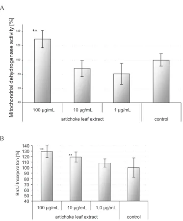
An artichocke leaf extract (ALE) containing 27% caffeoylquinic acids and 7% flavonoids (Wittemer et al, 2005) signii cantly increased the mitochondrial de-signiicantly increased the mitochondrial de -hydrogenase activities of human liver HepG2 after 48 h incubation time at a 100 µg/mL level, while lower doses
were ineffective (Fig. 1A). This was confirmed by the results of proliferation test: ALE signiicantly increased
proliferation rate at doses of 100 µg/mL to about 130% proliferation, and at 10 µg/mL to 120%; lower doses were
ineffective (Fig. 1B). Additionally a signiicant increase in protein content in treated HepG2 cells was measured: ALE
at 100 µg/mL signiicantly induced the protein content of
the cells to about 130% (data not shown), relecting the
increase in cell number and energy turn over.
genes Assay ID
GPX Hs00702173_s1 CYP1A2 Hs00167927_m1 CYP3A4 Hs00604506_m1
GGT1 Hs00359124_g1
GSR Hs00167317_m1 GST Hs00542846_m1
GAPDH Hs99999905_m1
FIGURE 1 - Inluence of artichoke leaf extract (ALE) at 1, 10
and 100 µg/mL level on mitochondrial dehydrogenase activity
(A) and mitotic cell proliferation (B) of HepG2 cells after 48
hours incubation. Mitochondrial activity determined by MTT
test, proliferation determination by BrdU incorporation ELISA. The bars represent standard errors SD from 3 independent
experiments with n = 10 replicates each with *p < 0.05,
No necrotic and apoptotic cytotoxicity of ALE was
observed within the test range of 1 to 100 µg/mL as measured
by LDH determination and inspection on apoptotic bodies. To investigate the inluence of isolated artichoke compounds on HepG2 cells reference compounds luteolin, luteolin-7-O-glucosid, caffeic acid, chlorogenic acid and cynarin were tested at 100, 10 and 1 µg/mL level. Within MTT assay cynarin, chlorogenic acid and caffeic acid did
not exhibit any signiicant effect. Luteolin and luteolin-7 -O-glucosid at 100 µg/mL (3.5 x 10⁻⁷ resp. 2.3 x 10⁻⁷ mol/L)
signiicantly decreased mitochondrial activity (Fig. 2A),
related to the calculation of absolute amounts of 100 µg/mL of both compounds. Also when the measured data were
normalized to equimolar concentrations (3.5 x 10⁻⁷ mol/L
Luteolin with 84 % inhibition, 3.5 x 10⁻⁷ mol/L,
luteolin-7-O-glucoside with 54 % inhibition) signiicantly higher inhibition of mitochondrial activity was obvious for lute-olin compared to the respective glycoside. This was also
relected within the respective proliferation assays (Fig. 2B). The inhibitory effect of the flavone-glucosid was signiicantly lower than that of the free aglycon luteolin.
The mRNA expression of different phase I and II en-he mRNA expression of different phase I and II en
-zymes of HepG2 cells was determined by quantitative
RT-PCR after a 24 h incubation time of cells with and without ALE in serum-free media. Gene expression was monitored in 3 independent assays for glutathione-S-transferase GST, glutathionperoxidase GPX, glutathionreductase GSR, γ -glutamyltransferase GGT1 and the cytochrome P450
iso-forms CYP1A2 and CYP3A4. These CYPs were selected because CYP3A4 is the main hepatic cytochrom (about 30%) beside CYP1A2 (about 10 to 12%). Measured test values were related to the expression of the endogenous control glycerol-3-phosphate dehydrogenase (GAPDH). mRNA expression for CYP1A2 was slightly increased in the presence of artichoke extract ALE (100 µg/mL) to 1.76
+/- 2.4. Because of the high variability this effect is as
-sessed not to be signiicant. Signiicant inhibition of gene expression by ALE was seen for CYP3A4 (0.23 +/- 0.39), for GGT1 (0.59+/- 0.21), for GPX2 (0.50 +/- 0.30), for GSR (0.72 +/- 0.23) and GST (0.22 +/-0.28).
The inhibition of glutathione-S-transferase GST gene expression was conirmed by determination of the respective enzyme activity. Therefore HepG2 cells were incubated with ALE (100, 10 µg/mL) for 48 h. Total pro
-tein was isolated from cell lysate and GST activity was de
-termined after conjugation of 1-chloro-2,4-dinitrobenzene CDNB to endogenous GST. GST activity was calculated relative to the total cell protein content against a GST calibration curve in ALE-treated cells and untreated con
-trol. As shown in Fig. 3 ALE inhibited GST activity in a
dose-dependent manner.
Summarizing at this point, artichoke extract is as -sessed to have stimulating activity on the proliferation and the energy turn-over of liver cells, accompanied by effects
on metabolising enzymes.
In order to investigate if artichoke extract has also anti-toxic effects against exogenous chemicals, ALE was tested as cytoprotective activity against the toxifying effects of ethanol (marker for unspeciic enzyme toxicity), hydrogenperoxide (oxidative toxicity) and dimethylsulfo -FIGURE 2 - Inluence of luteolin and luteolin-7-O-glucoside
at 1, 10, 100 µg/mL on mitochondrial dehydrogenase activity
(A) and mitotic cell proliferation (B) of HepG2 cells after 48
hours of incubation. Mitochondrial activity was determined by MTT test, proliferation was determined by BrdU incorporation
ELISA. The bars represent SD from independent assay with
n = 10 replicates with *p < 0.05, **p < 0.01 compared to the
untreated control group.
FIGURE 3 -Concentration-dependent inhibition of
glutathione-S-transferase (GST) in HepG2 cells after 48 h treatment with ALE relative to the total protein concentration. The bars
xid (necrotic toxicity). In a preliminary study the respec
-tive concentrations and incubation times were veriied to induce a cell toxicity, equivalent to about 50% reduction of MTT activity. Ethanol was used in the following studies at a 25 to 50 mM level, H2O2 at 1 to 10 mM, and DMSO at 3.5
and 5% concentration over a 6 to 24 hours incubation time. As shown in Table I, cotreatment of HepG2 cells with ALE (100 µg/mL) and ethanol (25 and 50 mM) for
48 hours showed a signiicant protective effect. Also at
10 µg level the respective data showed (not signiicant) tendencies towards cytoprotective effects.
Also cotreatment of HepG2 cells with ALE (100 µg/mL) and H2O2 (1, 5, 10 mM) for 24 hours sho
-wed a signiicant protective effect against the low-dose hydrogen peroxide induced toxicity: while only minor antioxidative effects of ALE were seen against strong cell
damage induced by 10 mM H2O2, clear effects of ALE were observed against 5 mM H2O2.
Coincubation of HepG2 cells with DMSO (3.5 and 5%) and ALE for 48 hours (10, 100 µg/mL) indicated
reduced DMSO-induced toxicity at the 100 µg/mL level,
while lower doses did not protect from DMSO-mediated cell toxicity.
Similar test systems, but with pre- and postincuba -tion of cells with ethanol, H2O2 or DMSO prior or after
an ALE-treatment, did not reduce the toxic effects of the chemicals towards HepG2 cells.
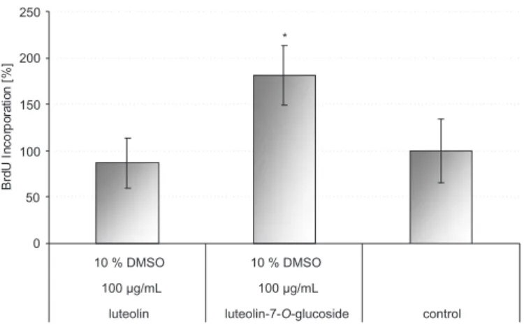
In addition to these experiments with artichoke extract, a potential cytoprotective effect of the lavonole reference compounds luteolin and luteolin-7-O-glucoside
was investigated against DMSO as exogenous stress fac
-tor. For that purpose one cell group, serving as a control, was treated with DMSO 10%. Proliferation behaviour of this group - with a signiicantly reduced vitality in contrast to an untreated control - was calculated as 100%. Coin
-cubation of two cell groups with luteolin + DMSO 10% resp. luteolin-7-O-glucoside + DMSO 10% indicated that
the lavone-glycoside signiicantly protects cells against DMSO-related toxicity (Fig. 4).
For investigation of the cytoprotective effects of ALE against ethanol-induced cell damage the mRNA ex-he mRNA ex
-pression of different phase I and II liver enzymes was de
-termined after 3 different treatment cycles of HepG2 cells over a 24 h incubation time (pre-, post- and cotreatment
FIGURE 4 - Proliferation (BrdU-ELISA) of HepG2 cells within
48 h cotreatment with DMSO 10% and 100 µg/mL luteolin (0.35
µM/mL) resp. luteolin-7-O-glucoside (0.22 µM/mL).*p < 0.05
compared to the DMSO 10% control group.
TABLE I -Inluence of different pre-, post- and cotreatment regimes of ethanol, hydrogen peroxide and DMSO in combination
with artichocke extract (ALE) on mitochondrial activity of HepG2 cells. Mitochondrial activity was determined by MTT test and
measured values were related to the respective control groups (100%). **p < 0.01. ⇒: followed by treatment with compounds
indicated
Type of treatment of HepG2 cells Mitochondrial Activity (%)
Ethanol 50 mM; 48 h ⇒ ALE 100 µg; 48 h 313 +/- 38% **
Ethanol 25 mM; 48 h ⇒ ALE 100 µg; 48 h 178 +/- 77% **
Ethanol 50 mM; 48 h ⇒ ALE 10 µg; 48 h 283 +/- 111%
Ethanol 25mM; 48 h ⇒ ALE 10 µg; 48 h 168 +/- 60%
H2O2 10 mM; 24 h ⇒ ALE 100 µg; 24 h 116 +/- 2%
H2O2 5 mM; 24 h ⇒ ALE 100 µg; 24 h 263 +/- 92% **
H2O2 1 mM; 24 h ⇒ ALE 100 µg; 24 h 107 +/- 12%
DMSO 5%; 48 h ⇒ ALE 100 µg; 48 h 100 +/- 12 %
DMSO 5%; 48 h ⇒ ALE 10 µg; 48 h 97 +/- 7 %
DMSO 3.5%; 48 h ⇒ ALE 100 µg; 48 h 137 +/- 6 % **
of cells with ethanol 50 mM and ALE 100 µg/mL). Gene
expression was monitored for GST, GPX, GSR, GGT1, CYP1A2 and CYP3A4 and all values were related to the expression of the endogenous control GAPDH. As shown in Table II ethanol 50 mM increased mRNA levels of CYP1A2 signiicantly. Pre- and posttreatment experiments with ALE revealed no big differences between the differ -ent test groups. In contrast to that, a thirtyfold increase in
CYP1A2 was observed when coincubation was performed with ethanol plus ALE for a 24 h incubation period, indi
-cating a strong inluence of the artichoke-ethanol mixture
on metabolic activity of the liver cells.
Significant influences of ethanol and/or ALE on expression of GST, GPX, GSR, GGT1 were not observed.
CONCLUSIONS
Extracts of artichoke are widely used for its choleret-xtracts of artichoke are widely used for its choleret -ic and antihypercholesterinaem-ic effects. Within literature potential hepatoprotective effects are described, mainly us-ing in vivo rat models (Gebhard, 1997). In vitro studies of
hepatoprotective effects of artichoke extracts on cultured rat hepatocytes are shown against t-butylhydroperoxide-induced peroxidation of membrane lipids (Gebhard, 1997).
Additionally cyanarin and caffeic acid were shown to
reduce the carbontetrachloride-induced leakage of liver enzymes glutamic oxaloacetic transaminase and glutamic pyruvic transaminase (Adzet et al., 1987).
These data correlate with our investigations,
indica-ting artichoke extract to have a direct effect on liver cells. Cell physiology of HepG2 cells is clearly up-regulated,
with increased mitochondrial activity, leading to a higher energy status, inducing an increased protein synthesis and ending in a higher mitogenic activity. On the other side
it is absolutely unclear which compounds from artichoke
are responsible for these effects. In our investigations neither cynarin, chlorogenic acid or caffeic acid had any
inluence on the energy status of the cells. From that point
of view artichoke extract speciications for preparations marketed with the indication “hepatoprotection” should not be standardized on these compounds. On the other side the lavones were shown to have strong inhibitory effects on liver cells with luteolin being a quite strong inductor of apoptosis by inhibition of topoisomerase II (Cantero et al., 2006). It seems interesting that luteolin-7-O-glucoside
has signiicantly less toxic effects on HepG2 cells rather
than the free aglycon luteolin, probably by a reduced
absorption into the cells. For manufacture of commercial artichoke leaf extracts specifications therefore should
focus on the amount of luteolin-glucoside beside that of the free aglycon.
Because the protein content of the cells is increased
during incubation with artichoke extract it could be ar
-gued that also the metabolizing enzymes are upregulated unspeciically. This is clearly not the case when looking at the gene expression data of phase I and II enzymes expressed in HepG2 cells. Only one cytochrome mRNA, namely CYP1A2 is upregulated to a minor degree which is assessed to have no clinical relevance. All other enzymes monitored were slightly inhibited. This was also conirmed by biochemical determination of enzyme activity of GST which was signiicantly down-regulated by artichoke ex
-tract. If this enzyme inhibition has any clinical relevance
this has to be investigated by in vivo studies in future.
Beside the direct effect of artichoke extract on HepG2 cells, pronounced cell protection by ALE was obvious. This was not only due to the known antioxi
-dative effects of artichoke which were obvious in our studies against hydrogen peroxide and in previous studies (Gebhard, 1997) against t-BHP. Cell protection of ALE
can also be discussed on ethanol-induced cell damage over metabolic pathways as well as against necrotic membrane
disturbance, induced by compounds as DMSO. In general direct interactions between toxifying agents and ALE can be claimed as shown by the respective cotreatment expe -riments. On the other side also indirect effects are induced TABLE II - Inluence of artichoke extract (ALE, 100 µg/mL) in pre-, post- and cotreatment experiments with ethanol (50 mM) on
relative gene expression on cytochromes CYP1A2 and CYP3A4 by quantitative real time PCR (TaqMan® assay), normalized to
the house keeping gene GAPDH and untreated controls. ⇒: followed by treatment with compounds indicated
Treatment of HepG2 cells CYP1A2 CYP3A4
Ethanol 50 mM; 1 h ⇒ ALE 100 µg; 23 h 3.3 1.0
Control experiment: Ethanol 50 mM; 1 h ⇒ Medium only; 23 h 3.0 2.5
ALE 100 µg; 23 h ⇒ Ethanol 50 mM; 1 h 7.1 1.7
Control experiment: Medium only; 23 h ⇒ Ethanol 50 mM; 1 h 6.2 1.0
Ethanol 50 mM; 24 h + ALE 100 µg; 24 h 29.8 3.9
by artichoke extracts. The posttreatment experiments with ALE after damage with DMSO clearly conirmed the in -duction of repair mechanisms. A prophylactic application
of artichoke extract does not seem to have an effect. On the
other side it has to be pointed out that the positive effects
of ALE against EtOH-, H2O2- and DMSO-damaged liver
cells is not due to a detoxiicant effect but more due to an stimulation of metabolic capacity of the cells. Sum
-marizing artichoke leaf extract may have the potential to
be used as hepatoprotective agent. Prospective research will identify those compounds, which are responsible for described effects.
ACKNOWLEDGEMENTS
The HepG2 cell line was a generous gift from Prof. Dr. Mersch-Sundermann is acknowledged. Financial su
-pport by Lichtwer AG / Casellamed AG is acknowledged.
REFERENCES
ADZET, T.; CAMARASA, J.; LAGUNA, J. Hepatoprotective
activity of polyphenolic compounds from Cynara scolymus
against CCl4 toxicity in isolated rat hepatocytes. J. Nat.
Prod., v.50, p.612-617, 1987.
BRAND, N.; Cynara. In: HÄNSEL, R.; SÉLLER, K.,
RIMPLER, H., SCHNEIDER, G., (Eds.). Hagers Handbuch
der Pharmazeutischen Praxis, Drogen. 5. ed. Berlin,
Heidelberg, New York: Springer Publishers, 1992. v.4, p.1117-1131.
CANTERO, G.; CAMPANELLA, C.; MATEOS, S.; CORTES, F. Topoisomerase II inhibition and high yield of endoreduplication induced by the lavonoids luteolin and
quercetin. Mutagenesis, v.21, p.321-325, 2006.
DAUER, A.; HENSEL, A.; LHOSTE, F.; KNASMUELLER, S.; MERSCH-SUNDERMANN, V. Genotoxic and
antigenotoxic effects of catechin and tannins from
the bark of Hamamelis virginiana L. in metabolically competent, human hepatoma cells (HepG2) using single
cell electrophoresis. Phytochem., v.63, p.199-207, 2003.
ESCOP Monograph – The Scientiic Foundation for Herbal
Medicinal Products. Cynarae folium - Artichoke leaf. 2.ed.
New York: Thieme Publishers, 2003.
GEBHARD, R. Anticholestatic activity of flavonoids from
artichoke (Cynara scolymus L.) and their metabolites. Med.
Sci. Mon., v.7, supl.1, p.316-320, 2001.
GEBHARD, R. Antioxidative and protective properties of
extracts from leaves of the artichoke (Cynara scolymus L.)
against hydroperoxide-induced oxidative stress in cultured
rat hepatocytes. Tox. Apl. Pharmacol., v.144, p.279-286,
1997.
GEBHARD, R. Artischockenextrakt – in vitro Nachweis einer
Hemmwirkung auf die Cholesterolbiosynthese. Medizin
Welt, v.46, p.348-350, 1995.
GEBHARD, R. Neue Erkenntnisse zur Wirkung von
Artischockenextrakte. Z. Allgemeinmed., v.72, p.20-23,
1996.
HOFMANN, T.; DETERS, A.; MÜLLER, G.; STARK,
T.; WITTSCHIER, N.; HENSEL, A. Occurence of
N-Phenylpropenoyl-L-amino acids in different herbal drugs
and inluence on human keratinocytes, human liver cells
and against adhesion of H. pylori to human stomach. Planta
Med., v.73, p.142-150, 2007.
LOWRY, O.H.; NIRA, J.; ROSEBROUGH, N.J.; FARR, A.L.; RANDALL, R.J. Protein measurement with the Folin
phenol reagent. J. Biol. Chem. v.193, p.265-275, 1951.
MAROS, T.; SERES-STRUMR, L.; RAIZ, G.; RETEJI, C.;
KOVACS, V.V.; HINTS, M. Wirkung der Cynara scolymus
-Extrakte auf die Regeneration der Rattenleber Drug Res.,
v.18, p.884-886, 1968.
MARTIN, A.; CLYNES, M. Comparison of 5 microplate
colorimetric assays for in vitro cytotoxicity testing and cell
proliferation assays. Cytotechnology, v.11, p.9-58, 1993.
MATUSCHOWSKI, P.; GUMBINGER, H.G.; NAHRSTEDT,
A.; WINTERHOFF, H. Testing of Cynara scolymus L. in
the isolated perfunded rat liver. Planta Med., v. 63, p.
55-56, 1997.,
MATUSCHOWSKI, P.; NAHRSTEDT, A.; WINTERHOFF, H . P h a r m a k o l o g i s c h e U n t e r s u c h u n g e n e i n e s
Frischpflanzenpresssaftes aus Cynara scolymus auf
choleretische Wirkung. Z. Phytother., v.26, p.216-221, 2005.
MOSMANN, M. Rapid colorimetric assay for cellular growth and survival: applications to Proliferation and cytotoxicity
PITTLER, M.H.; THOMPSON, C.J.; ERNST, E. Artichoke leaf extract for treating hypercholesterolaemia (Review).
The Cochrane Database of Syst. Rev., volume?, Issue 3,
pages?, 2002.
PORSTMANN, T.; TERNYK, T.; AVRAMEAS, S. Quantiication of 5-bromo-2’-deoxyuridine into DNA: an enzyme immunoassay for the assessment of the lymphoid
cell proliferative response. J. Immunol. Methods, v.82,
p.169-179, 1985.
PREZIOSI, P. Dal Cynara scolymus allacito 1,4-dicaffeilchinico.
Il Fármaco (ed. Sc.), v. 17, p. 701-745, 1962.
STOEV, S.D.; STEFANOV, M.; DENEB, S.; RADIC, B.;, DOMINAN, A.M.; PERAICA, M. Experimental mycotoxicosis in Chickens induced by ochratoxin A and penicillic acid and intervention with natural plant extracts.
Vet. Res. Común., v.28, p.724-46, 2004.
SUZUKI, T.; ONOGAWA, T.; ASANDO, N.; MIZUTAMARI,
H.; MAKKAICHI, T.; TANEMOTO, M.; et al.Identiication
and characterization of novel rat and human gonad-speciic
organic anion transporters. Mol. Endocrinol., v.17,
p.1203-1215, 2003.
WITTEMER, S.M.; PLOCH, M.; WINDECK, T.; MÜLLER,
S.C.; DREWELOW, B.; DERENDORF, H.; e t
al.Bioavailability and pharmacokinetic properties of
caffeoylquinic acids and lavonoids after oral administration
of artichocke leaf extracts in human. Phytomedicine, v.12,
p.25-38, 2005.
WOJCICKI, J. Effect of 1,5-dicaffeylquinic acid (cynarine) on
colesterol levels in serum and liver of acute ethanol-induced
rats. Drug Alc. Dep., v.3, p.143-145, 1978.
Received for publication on 22th december 2008