Arq Neuropsiquiatr 2010;68(6):888-892
Management of supratentorial
epidural hematoma in children
Report on 49 patients
Wellingson Silva Paiva, Almir Ferreira de Andrade, Luis Mathias Júnior, Vinicius Monteiro de Paula Guirado,
Robson Luis Amorim, Nadia Nader Magrini, Manoel Jacobsen Teixeira
ABSTRACT
Traumatic head injury is a common cause of mortality and acquired neurological impairment in children. However, pediatric epidural hematomas (EDHs) are not common and few series have studied the evolution of these patients. In this study, we present the results from a sample of patients with EDH with long-term follow-up. Method: Between January 2006 and December 2008, 49 patients with traumatic EDH were treated at our unit. Clinical course, radiological findings and outcomes were evaluated. Neurological status was assessed using the Glasgow Coma Scale (GCS). The patients’ ages ranged from one day to 16 years. The mean follow-up was six months. Results: On admission, most of the patients presented mild trauma and 57% had a GCS of 13-15. The most common symptom was irritability. The most frequent mechanisms of injury were: falling from a height in 29 cases and motor vehicle accidents in 16 cases. Three of these patients presented GCS 3, but only one died. We found a late neurological deficit in nine patients. Conclusion: These lesions may occur following mild head trauma and in alert children with nonfocal neurological examinations. However, in children presenting irritability with subgaleal hematomas and a history of loss of consciousness, skull computed tomography must be performed.
Key words: epidural hematoma, head trauma, children.
Tratamento de hematoma epidural supratentorial em crianças: avaliação em 49 pacientes RESUMO
Trauma craniocerebral é uma causa frequente de mortalidade e comprometimento neurológico adquirido em crianças. No entanto, hematomas epidurais (HED) são raros em pacientes pediátricos, com poucas series estudando a evolução destes pacientes. Neste estudo, os autores apresentam os resultados de uma casuística de pacientes com HED acompanhados em longo prazo. Método: Entre janeiro de 2006 e dezembro de 2008, 49 pacientes com HED foram tratados em nossa unidade. Curso clínico, achados radiológicos, e resultados foram avaliados. O estado neurológico foi avaliado com o Glasgow Coma Scale (GCS). A idade variou de 1 dia a 16 anos. A média de acompanhamento foi de 6 meses. Resultados: Na admissão, a maioria dos pacientes apresentava trauma leve e 57% estavam com GCS de 13-15. O sintoma mais comum foi irritabilidade. Os mecanismos de trauma mais frequentes foram queda de altura em 29 casos e acidentes de trânsito em 16 casos. Três destes pacientes apresentavam GCS 3, mas somente um morreu. Verificou-se déficit neurológico tardio em nove pacientes. Conclusão: Esta lesão pode ocorrer após traumas leves e em crianças alerta com exames neurológicos não focais. No entanto, em crianças com irritabilidade com hematoma subgaleal e história de perda de consciência, tomografia do crânio deve ser realizada.
Palavras-chave: criança, hematoma epidural, trauma craniocerebral.
Correspondence
Wellingson Silva Paiva Rua Teodoro Sampaio 498 / 66 05406-000 São Paulo SP - Brazil E-mail: [email protected]
Received 24 January 2010 Received in final form 31 May 2010 Accepted 7 June 2010
Traumatic head injury is a common cause of mortal-ity and acquired neurological impairment in children1.
However, pediatric acute epidural hematomas (EDHs) are not common and, when they occur, they often present atypically2,3. he reported incidence of EDH after head
trauma in hospitalized children ranges from 1% to 6%3,4.
However some authors have reported an increased inci-dence of unassociated EDH over recent years5,6. Its
diag-nosis can be quite challenging because its clinical presen-tation is usually subtle and nonspeciic. In this study, we present the results from a sample of children with EDH with long-term follow-up.
METHOD
All children (less than 16 years of age) diagnosed with traumatic epidural hematoma on computed tomography (CT) scans who were admitted to our hospital over the study period were included. Between January 2006 and December 2008, 49 patients with traumatic EDH were treated. Clinical course, radiological indings and out-comes were evaluated. Neurological status was assessed using the Glasgow Coma Scale (GCS). All the patients were treated in accordance with a standard advanced trauma life support protocol if they presented directly to the emergency department of our institution. he indi-cation for ventilation was a neurological GCS score <8, or respiratory or cardiovascular impairment. he trauma workup was started in accordance with the clinical status and the mechanism of the trauma, and it usually includ-ed head and body CT scans and abdominal ultrasound. he patients’ ages ranged from one day to 16 years. he mean follow-up was six months. Patients with sponta-neous EDH, EDH of unknown etiology or infratentorial EDH were excluded from our current study.
Patient management was either surgical or conserva-tive based on the children’s clinical condition, GCS score, evidence of midline shift on the initial head CT scan and size of the EDH. Conservative management consisted of close observation in either a neonatal or a pediatric inten-sive care environment, with heart rate, respiratory rate and oxygen saturation monitoring in addition to frequent neu-rological clinical examinations and serial head CT scans.
RESULTS
Over this period, 49 patients with epidural hemato-mas were admitted to our hospital. On admission, most of the patients (57%) had a GCS of 13-15, and 33% of the patients had severe traumatic brain injury (TBI) (Table 1). Among the patients with mild head trauma, the most common presenting symptom was irritability, which oc-curred in 19/28 (68%) of them (Table 2). hirty patients (61%) patients had a skull fracture. here were 26 males and 23 females. With regard to the lateralization of the
Table 1. Patient distribution according to Glasgow Coma Scale (GCS) score.
TBI classiication Number of patients
GCS 13-15 28
GCS 9-12 5
GCS 3-8 16
TBI: traumatic brain injury.
Table 2. Patient distribution according to symptoms at hospital admission.
Symptom Number of patients
Irritability 28
Subgaleal hematoma 23
Lethargy 5
Coma 16
Pupil abnormality 4
Hemiparesis 1
Table 3. Patient distribution according to epidemiological and radiological features.
Glasgow Coma Scale (GCS) Number of patients
DEATH (GCS 1) 1
GCS 2 0
GCS 3 2
GCS 4 7
GCS 5 39
Table 4. Patient distribution according to outcome and follow-up.
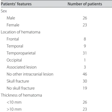
Patients’ features Number of patients
Sex
Male 26
Female 23
Location of hematoma
Frontal 8
Temporal 9
Temporoparietal 31
Occipital 1
Associated lesion 3
No other intracranial lesion 46
Skull fracture 30
No skull fracture 19
Thickness of hematoma
<10 mm 26
hematoma in our study, 29/49 (59.1%) were located on the right side, and the remaining 40.9% were located on the left side, while the temporal-parietal area was the most common anatomical location for the hematoma (Table 3).
Skull fractures were seen on CT in 30 patients (61%). he most common sites were the parietal bone (18) and the temporal bone (8). he size of the EDH was found to be more than 2 cm in 19/49 patients, between 1 and 2 cm in 4/49 and less than 1 cm along its largest diame-ter in 26/49.
Two children with epidural hematomas sufered acute neurological deterioration, which occurred within 8 hours of the head trauma.
Over the patients’ evolution, we found that one tient died. In terms of the Glasgow Coma Scale, 39 pa-tients presented complete recovery, while seven papa-tients had GCS 4 and two patients had severe neurological se-quelae. he group of patients who underwent craniotomy had the worst prognosis: these patients presented large hematomas and lower scores on the GCS. Eleven patients in the surgical group had severe TBI, while only ive pa-tients undergoing medical management had severe TBI (Table 4).
he mechanisms of injury in our series were: falling from a height in 29 cases (59%), motor vehicle accidents in 16 cases (33%), obstetric maneuver during delivery in one case (2%), and domestic accident/aggression in three cases (6%) (Figure).
Associated lesions (subdural or contusion hematoma) were found in seven patients, and ive of these were in the surgical group, presenting worse evolution during follow-up. Only ive patients presented associated systemic inju-ries: two cases of upper limb fractures, one case of low-er limb fracture, one case of pulmonary contusion and one case of closed abdominal trauma that did not need surgery.
DISCUSSION
It is well known that acute epidural hematomas in children are a rare and potentially life-threatening com-plication resulting from head injuries. hese intracrani-al lesions in children represent 2 to 3% of intracrani-all head inju-ry complications1,6.
Consistently with other series, falls were the most common cause of trauma in our series. Pasaoglu et al.7
found that falls were the most common underlying mech-anism in 63% of their pediatric cases, and Ersahin et al. 8
identiied falls as the most common mechanism of inju-ry in 62% of their pediatric cases. It is noteworthy is that almost 60% of the EDHs in our study were related to ac-cidents at home or within a familial environment. Only roughly one third of the accidents were traic-related.
Among the patients admitted with mild head trau-ma, 68% presented symptoms of irritability alone and al-most 60% were alert with non-focal neurological exam-inations. Previous studies on patients with head trauma also demonstrated that serious intracranial injury may oc-cur following minor head trauma among alert patients. his group of patients represents a great challenge for pe-diatric emergency teams. Miller et al.9 reported that 183
patients with intracranial hemorrhage after “minor” head injury arrived in an alert condition with a GCS score of 15; 57% had no clinical signs. Likewise, Cook et al.10
re-ported that among 100 patients with traumatic epidural hematoma and GCS of 14 or 15, 40% had nausea or vom-iting but no focal neurological signs. Although it is desir-able to have cranial CT scans for all patients with TBI, it can be recognized that this is not viable everywhere. he criteria for indicating CT scans involve not only medical and legal issues but also cost11.herefore, some authors
have advised that CT scans should only be performed on subsets of patients who are considered to be at higher risk of developing intracranial lesions12,13. Others have
main-tained that failure to systematically perform CT scans for TBI implies a certain rate of misdiagnosis that has to be weighed against the reduction in cost14. hus, protocols
for the use of CT scans to assess cases of mild head inju-ry (MHI) vainju-ry among institutions and countries, depend-ing on the local cost-efectiveness analysis. In our institu-tion, as reported on previous occasions, there is a liber-al policy. Hence, craniliber-al CT scans are advised for liber-almost all patients with MHI whose trauma is perceived as non-trivial by the attending emergency physician or neuro-surgeon, using criteria that include GCS, trauma mecha-nism, age and lucidity11,15.
he criteria for how to select cases for either conser-vative or surgical treatment remain controversial. Chen et al.16 suggested that a hematoma volume larger than 30 ml
with a thickness of more than 15 mm and a midline shift of more than 5 mm constitutes a strong indication for 0
5 10 15 20 25 30 35
Delivery Maneuver Domestic
Aggression Traffic
Accident Falls
1 3
16 29
surgical treatment. Similarly, Bejjani et al.17 found that the
most important radiographic parameters dictating surgi-cal evacuation were a maximum hematoma diameter of more than 18 mm and a midline shift of more than 4 mm. Our policy for treating EDH is somewhat diferent. In our series, 23 patients (47%) underwent a neurosurgical op-eration and 26 underwent conservative treatment. he criteria adopted for conservative management were he-matoma thickness less than 10 mm and midline shift less than 5 mm. In two patients in whom we observed neu-rological deterioration and increasing hematoma thick-ness, we performed emergency craniotomy with hema-toma evacuation, and these patients presented GCS 5 in the follow-up. We believe that conservative management can only be performed in an intensive care unit environ-ment in hospitals with neurosurgical attendance. A new skull CT scan needs to be performed 12-24 hours after the irst examination.
Children sustaining cranial trauma associated with EDH constitute a heterogeneous group with a variety of clinical outcomes18. he outcomes among the children
in our series were encouraging. Despite potentially life-threatening intracranial hemorrhages, there was only one death: a patient who was admitted with a GCS score of 3 points and nonreactive pupils. here was also relative-ly low morbidity: no children were in a vegetative state. he approximate mortality rate was 5%, although mortal-ity in the literature consulted ranged from 0% to 12%19,20
and was even higher in the pre-CT era18. From our series,
we believe that if lesions are treated early, and aggressive-ly in cases with worse consciousness, improved outcomes can be achieved.
In a similar study conducted in our unit two decades ago, surgical treatment was performed more often, pos-sibly because it was more diicult to repeat CT scans at that time. hus, craniotomy was performed on 74% of patients, while in our current study, craniotomy was re-quired in 46% of the cases. In the same study covering the period from 1987 to 1991, most of the patients managed without surgery underwent CT more than 12 hours af-ter the trauma (60%), whereas in our series, only two pa-tients underwent CT more than 12 hours after the trau-ma, and the two cases were from another institution. he time taken to admit cases to hospital has also decreased, thus indicating greater organization within the rescue sys-tem 20 years on. An X-ray of the head was performed on 43 patients before the CT scan in the earlier series, while in the present series, only three cases underwent X-ray imaging before tomography, and two of these were from another institution. his also indicates that access to to-mography has become simpliied, along with a tendency not to use skull X-rays in head trauma cases.
Comparing the results from these two studies in our
unit (1987-1991 versus 2006-2008)6, we found that falls
continued to be a common cause of trauma, but that there had been signiicant growth in the proportion of cases due to automobile accidents (from 1.7% to 33%). his can perhaps be explained by the large increase in the vehicle leet in our city that has taken place over this in-terval. Regarding the outcomes, there was no signiicant diference in the mortality rate (3.4% versus 2.5%) over the decades that separated the samples. However, a smaller proportion of aggressive treatment (craniotomy) was re-quired to achieve equivalent functional outcomes.
One of our patients was a full-term newborn with a skull fracture and large epidural hematoma secondary to forceps delivery. EDH in newborn infants is a very rare condition. In a study on a series of newborns with epi-dural hematomas, Heyman et al.21 stated that when the
hematoma was more than 1 cm thick and more than 4 cm long in the anteroposterior orientation or associat-ed with a depressassociat-ed cranial fracture, surgery should be performed, given that it is well known that most brain damage is caused by mechanical pressure. In this patient of ours, the dimensions of the hematoma and its asso-ciation with a depressed skull fracture indicated surgi-cal treatment.
EDH can be managed with excellent outcomes, even in children. his lesion may occur after relatively minor head trauma and in alert children with nonfocal neuro-logical examinations. However, in children presenting ir-ritability with subgaleal hematoma and a history of loss of consciousness, skull CT must be performed. he neu-rological condition and the size of the EDH seen on head CT scans are the most commonly used criteria for cor-rect management.
REFERENCES
1. Parslow RC, Morris KP, Tasker RC, Forsyth RJ, Hawley CA. On behalf of the UK, Peadiatric Traumatic, Brain Injury, Study Steering, Group and the Pae-diatric, Intensive Care, Society Study Group. Epidemiology of traumatic brain injury in children receiving intensive care in the UK. Arch Dis Child 2005;90:1182-1187.
2. Duhaime AC, Alario AJ, Lewander WJ, et al. Head injury in very young children: mechanisms, injury types, and ophthalmologic indings in 100 hospitalized patients younger than 2 years of age. Pediatrics 1992;90: 179-185.
3. Reece RM, Sege R. Childhood head injuries. Accidental of inlicted? Arch Pediatr Adolesc Med 2000;154:11-15.
4. Maggi G, Aliberti F, Petrone G, et al. Extradural hematomas in children. J Neurosurg Sci 1998;42:95-99.
5. Stieg PE, Kase CS. Intracranial hemorrhage: diagnosis and emergency man-agement. Neurol Clin 1998;16:373-390.
6. dos Santos AL, Plese JP, Ciquini JO, et al. Extradural hematomas in children. Pediatr Neurosurg 1994;21:50-54.
7. Pasaoglu A, Orhon C, Koc K, Selcuklu A, Akdemir H, Uzunoglu H. Traumatic extradural haematomas in pediatric age group. Acta Neurochir (Wien) 1990;106:136-139
8. Ersahin Y, Mutluer S, Guzelbag E. Extradural hematoma: analysis of 146 cases. Childs Nerv Syst 1993;9:96-99.
10. Cook RJ, Borsch WC, Fearnside MR, et al. Outcome prediction in extradural haematomas. Acta Nourochir 1988;95:98-94.
11. de Andrade AF, de Almeida AN, Bor-Seng-Shu E, Lourenço L, Mandel M, Marino R Jr. The value of cranial computed tomography in high-risk, mildly head-injured patients. Surg Neurol 2006;65 (Suppl 1):S10-S13.
12. Haydel MJ, Preston CA, Mills TJ, Luber S, Blaudeau E, DeBlieux PM. Indica-tions for computed tomography in patients with minor head injury. N Engl J Med 2000;343:100-105.
13. Stiell IG, Wells GA, Vandemheen K, et al. The Canadian CT Head Rule for pa-tients with minor head injury. Lancet 2001;357:1391-1396.
14. Ibanez J, Arikan F, Pedraza S, et al. Reliability of clinical guidelines in the de-tection of patients at risk following mild head injury: results of a prospective study. Neurosurgery 2004;100:825-834.
15. de Andrade AF, Marino R, Ciquini O, Figueiredo EG, Machado AG. Guide-lines for neurosurgical trauma in Brazil. World J Surg 2001;25:1186-1201. 16. Chen TY, Wong CW, Chang CN, et al. The expectant treatment of
as-ymptomatic supratentorial epidural hematomas. Neurosurgery 1993;32: 176-179.
17. Bejjani GK, Donahue DJ, Rusin J, et al. Radiological and clinical criteria for the management of epidural hematomas in children. Pediatr Neurosurg 1996;25:302-308.
18. Gerlach R, Dittrich S, Schneider W, Ackermann H, Seifert V, Kieslich M. Trau-matic epidural hematomas in children and adolescents: outcome analysis in 39 consecutive unselected cases. Pediatr Emerg Care 2009;25:164-169. 19. Browne GJ, Lam LT. Isolated extradural hematoma in children presenting
to an emergency department in Australia. Pediatr Emerg Care 2002;18: 86-90.
20. Bullock MR, Chesnut R, Ghajar J, et al. Surgical management of acute epi-dural hematomas. Neurosurgery 2006;58 (Suppl):S7-S15.