O R I G I N A L P A P E R
Analysis of the activation mechanism of
Pseudomonas stutzeri
cytochrome
c
peroxidase through an electron transfer chain
P. M. Paes de Sousa•D. Rodrigues•C. G. Timo´teo • M. L. Simo˜es Gonc¸alves•G. W. Pettigrew•I. Moura• J. J. G. Moura• M. M. Correia dos Santos
Received: 22 December 2010 / Accepted: 12 April 2011 / Published online: 6 May 2011
ÓSBIC 2011
Abstract The activation mechanism of Pseudomonas stutzeri cytochrome c peroxidase (CCP) was probed through the mediated electrochemical catalysis by its physiological electron donor,P. stutzericytochromec-551. A comparative study was carried out, by performing assays with the enzyme in the resting oxidized state as well as in the mixed-valence activated form, using cyclic voltam-metry and a pyrolytic graphite membrane electrode. In the presence of both the enzyme and hydrogen peroxide, the peak-like signal of cytochrome c-551 is converted into a sigmoidal wave form characteristic of an ErC0i catalytic
mechanism. An intermolecular electron transfer rate con-stant of (4±1) 9105M-1s-1 was estimated for both forms of the enzyme, as well as a similar Michaelis– Menten constant. These results show that neither the
intermolecular electron transfer nor the catalytic activity is kinetically controlled by the activation mechanism of CCP in the case of the P. stutzeri enzyme. Direct enzyme catalysis using protein film voltammetry was unsuccessful for the analysis of the activation mechanism, since
P. stutzeriCCP undergoes an undesirable interaction with the pyrolytic graphite surface. This interaction, previously reported for the Paracoccus pantotrophus CCP, induces the formation of a non-native conformation state of the electron-transferring haem, which has a redox potential 200 mV lower than that of the native state and maintains peroxidatic activity.
Keywords Cytochromecperoxidase
Cytochromec-551 Pseudomonas stutzeri
Mediated catalysis Activation mechanism
Introduction
The incomplete reduction of molecular oxygen to water results in the formation of hydrogen peroxide, a species that can induce cell damage or death owing to its ability to form free radicals. In biological processes, catalases dis-mutate the hydrogen peroxide molecule or peroxidases reduce it to water [1].
Bacterial cytochrome cperoxidases (CCPs) are dimeric dihaem proteins that catalyse the reduction of hydrogen peroxide to water using small monohaem cytochromes as electron donors [1]. Well-characterized members of this group are the CCPs from Pseudomonas aeruginosa[2–5],
Paracoccus pantotrophus[6–8],Pseudomonas nautica[9],
Nitrosomonas europaea [10], Methylococcus capsulatus
[11],Rhodobacter capsulatus[12] andPseudomonas stutzeri
[13]. In most of these enzymes, in the resting oxidized state P. M. Paes de Sousa (&)C. G. Timo´teoI. Moura
J. J. G. Moura
ReQuimte, Centro de Quı´mica Fina e Biotecnologia, Departamento de Quı´mica,
Faculdade de Cieˆncias e Tecnologia, Universidade Nova de Lisboa, 2829-516 Caparica, Portugal e-mail: [email protected]
D. RodriguesM. L. Simo˜es Gonc¸alves M. M. Correia dos Santos (&)
Centro de Quı´mica Estrutural, Instituto Superior Te´cnico, Av. Rovisco Pais, 1049-001 Lisbon, Portugal e-mail: [email protected]
G. W. Pettigrew
Royal (Dick) School of Veterinary Studies, University of Edinburgh,
Summerhall,
(Fig.1, state A) one of the haems is in a low-spin/high-spin thermal equilibrium, has a high redox potential, reflecting a histidine–methionine coordination, and is believed to function as the electron-transferring haem (E haem). The second haem (P haem), with a low redox potential and a low-spin bishistidine coordination in the fully oxidized state, is thought to be the catalytic centre where hydrogen peroxide is reduced to water. Reduction of the E haem induces a series of complex spin state and coordination changes, producing a mixed-valence enzyme in which the P haem has become high-spin pentacoordinated, providing access to the substrate (Fig.1, state B) [14]. Calcium ions are required for the conversion of the mixed-valence enzyme into the activated state, but members of the bac-terial CCP family differ in their affinity for this cation. The enzymes fromP. nautica[9] andP. pantotrophus[15–17] behave very similarly, with the activation mechanism hin-dered in the as-isolated conditions, whereas the enzymes fromR. capsulatus[12] andP. stutzeri[13] do not require addition of further Ca2? to the state in which they are isolated for full activity. With the enzyme in the active form, the catalytic cycle is completed in the presence of hydrogen peroxide (Fig.1, states E and F).
In fact,P. stutzeri CCP seems to be purified in a form with tightly bound Ca2?, in which the calcium binding site responsible for dimer formation and enzyme activation, i.e. change of the low-potential haem to a high-spin pentaco-ordinated state, is fully occupied. Spectroscopic studies suggested that this form is readily active upon reduction by sodium ascorbate [13]. The affinity for calcium ions in the mixed-valence state is so high that Ca2?returns to it from
the EGTA which was added to empty the site in the oxi-dized state of the enzyme (Fig.1, states C and D). Steady-state kinetic studies of P. stutzeri CCP using either its physiological partner (P. stutzeri cytochromec-551) or horse heart cytochromecindicated that preincubation with Ca2?has no effect on the activity of the enzyme [13].
The use of voltammetric techniques is well proved in unravelling important aspects of the chemistry of metal-loproteins and metalloenzymes [18]. An especially attrac-tive approach is protein film voltammetry (PFV), a strategy where the protein is adsorbed at (ideally) monolayer cov-erage on a suitable electrode [19]. In PFV, besides the extremely small sample amounts that can be used, diffusion limitations are avoided, direct electron transfer is achieved and important information about the intrinsic thermody-namic and kinetic properties of the protein can be obtained. In the particular case of direct electron transfer of redox enzymes, the replacement of the physiological redox partner by the electrode and the direct correlation between current and catalytic activity are powerful tools for the study of the catalytic mechanism and its redox-linked chemistry. However, the use of PFV may be hindered if adsorption at the electrode surface is not successfully achieved, affecting the enzyme’s activity [20]. In the case of bacterial CCPs, we recently reported the formation of an altered form of the E haem whenP. pantotrophusCCP is directly adsorbed on a pyrolytic graphite (PG) electrode [21], a preferential surface for PFV studies [18]. The altered form has a redox potential approximately 300 mV lower than that of the native state and the enzyme main-tains peroxidatic activity.
Fig. 1 Model for the mechanism of activation and turnover ofPseudomonas stutzericytochromecperoxidase (CCP).EGTAethylene glycol
An alternative approach is mediated enzyme electro-chemistry, where, similarly to steady-state kinetics, the assays are performed with enzyme, cosubstrate (mediator) and substrate all present in solution. The mediator is the only species that interacts with the electrode and the electrochemical signal is directly related to the rate of its consumption during the catalytic reaction. In many cases, the mediators are stable molecules of low molecular weight that can easily exchange electrons with the elec-trode, and act as substituents of the redox metalloprotein natural partner [22]. When the physiological electron mediator is known, the natural electron transfer relay can be mimicked.
The use of membrane electrodes, a configuration where the proteins are entrapped between a dialysis membrane and the electrode surface, has been shown to be a very interesting strategy for this kind of study [23–25]. As happens in PFV, only small amounts of protein are nec-essary (3–4ll) and, owing to instantaneous dialysis, vari-ous experimental parameters can be rapidly investigated [26, 27]. Moreover, the theoretical features have been analysed in detail and quantitative information about the redox processes can be easily obtained [28].
We have recently used cyclic voltammetry and a membrane electrode to analyse the mediated catalysis of
P. pantotrophusCCP by one of its physiological donors,
P. pantotrophus pseudoazurin [29]. We now report the mediated electrochemistry of P. stutzeri CCP, using its physiological partner,P. stutzericytochromec-551, as an electron donor. To probe the activation mechanism of the enzyme, both the oxidized and the mixed-valence forms were analysed. Direct electrochemistry of the enzyme was also investigated.
Materials and methods
Reagents
Poly(L-lysine) hydrobromide was obtained from Sigma
(MW15,000–30,000). All other chemicals were pro
anal-ysis grade and solutions were prepared with Milli-Q water. Protein purification
P. stutzeri CCP and P. stutzeri cytochrome c-551 were isolated and purified as described before [13]. In this particular purification, a fraction of the enzyme was obtained in the mixed-valence, already active state, without addition of any chemical reductant at any point during the procedure. The concentration of the proteins was determined spectrophotometrically using the extinc-tion coefficients at 408 nm (e=252 mM-1cm-1) and
551 nm (e=30.89 mM-1cm-1) for the oxidized enzyme and the reduced cytochrome, respectively [13].
Procedure
The cyclic voltammograms were collected with an EG&G PAR potentiostat/galvanostat (model 273A) controlled via the 270 software. The scan rate varied between 5 and 200 mV s-1. Catalytic assays with the enzyme directly adsorbed on the electrode surface were performed with an electrode rotation rate of 3,000 rpm.
A conventional three-electrode configuration cell was used, with a carbon rod auxiliary electrode and an Ag/AgCl reference electrode (205 mV vs. the standard hydrogen electrode, SHE). Throughout this article, all potential val-ues are referred to the SHE and are affected by an error of 5–10 mV. The working electrode was a basal plane PG electrode with a 0.2-cm nominal radius. The area of the electrode was determined from its response in a known concentration of the ferrocyanide/ferricyanide couple [30] and was found to be close to the nominal value.
Electrode preparation
Prior to each experiment, the PG electrode was polished by hand on a polishing cloth (Buehler 40-7212) with an aqueous alumina slurry (0.3 lm, Buehler 40-6363-006), sonicated for 1 min and rinsed very well with Milli-Q water.
For the direct electrochemistry of CCP, a 4-ll drop of working solution (containing the protein) was deposited on the electrode surface and left to dry at room temperature for 30 min.
The membrane configuration, prepared with a nega-tively charged Spectra/Por MWCO 3500 membrane, was used for the analysis of cytochromec-551 and the mediated catalysis of CCP. Briefly, a small volume (4 ll) of the protein(s) solution was deposited on a square piece (about twice the diameter of the electrode’s body) of the dialysis membrane, then the electrode was pressed against the membrane and a rubber ring was fitted around the electrode body so that the entrapped solution formed a uniform thin layer.
Electrolyte and working solution
In all experiments the supporting electrolyte was 10 mMN -(2-hydroxyethyl)piperazine-N0-ethanesulfonic acid (HEPES) buffer, pH 7.5±0.1. In most cases 150 mM NaCl was used, which was replaced by 0.1 mg ml-1poly(L-lysine)
and 1lMP. stutzeriCCP (mediated catalysis). The effect of substrate concentration was analysed for different hydrogen peroxide concentrations between 10 and 150lM. In the experiments with a saturating substrate concentration, a concentration of 500lM (direct electrochemistry) or 125lM (mediated catalysis) was present in the electrolyte solution.
All solutions were deaerated for 30 min with high-purity nitrogen, and all measurements were performed at least in duplicate in a temperature-controlled room (T=20± 1°C).
Results and discussion
Direct electrochemistry ofP. stutzeriCCP
The direct electrochemistry of P. stutzeri CCP in non-turnover conditions was analysed at a PG electrode in 10 mM HEPES buffer, pH 7.5, without the addition of calcium ions. Although the enzyme has twoc-type haems, cyclic voltammograms ofP. stutzeriCCP adsorbed on the electrode surface revealed just one cathodic peak with an anodic counterpart, in the potential range ?800 to -600 mV and for scan rates,v, between 5 and 200 mV s-1 (Fig.2). The signal visible at potentials above ?0.1 V is also present in the blanks and is therefore meaningless for the study presented. The analysis of the voltammograms showed that the electrochemical response arises from a diffusionless one-electron redox process where both the oxidized and the reduced forms are adsorbed. A formal
potential E00
= -125 mV was estimated at pH 7.5. Differential pulse voltammograms revealed two peaks with peak potentials Ep= -107 mV versus SHE and
Ep= -244 mV versus SHE. Only the more positive one
persisted upon successive scans, corresponding to the signal observed in cyclic voltammetry.
In the presence of hydrogen peroxide only one cata-lytic wave develops in the same potential range, with
Ecat = -136 mV versus SHE and a current that increases
with increasing substrate concentration. Experiments performed in the absence of enzyme demonstrated that the direct reduction of hydrogen peroxide does not occur within that potential window. It is clear from Fig.2 that the shape of the cyclic voltammograms is not sigmoidal. Although the forward current tends to level off to a limiting value, in the reverse scan the current decreases considerably. Moreover, in successive scans the catalytic signal decreases until the initial non-turnover voltam-mogram is restored.
Similar behaviour was observed for the P. pantotro-phus CCP at the same type of electrode surface [21]. Namely, just one non-turnover signal persists on suc-cessive scans at a potential that does not match the preliminary data from a potentiometric titration, where redox transitions at about ?60 and -300 mV at pH 7.5 were estimated for the E and P haems of P. stutzeri
CCP, respectively. Also, only a cathodic catalytic signal is detected in the presence of hydrogen peroxide, at a potential close to that of the signal without substrate, behaviour distinct from that observed for the N. euro-paea CCP, which displays catalytic activity at a high redox potential (more than 500 mV vs. SHE) [31].
At pH 7.5 the enzyme has an overall negative charge since its pI is 5.4 [32]. From examination of the elec-trostatic surface potential of P. stutzeri CCP, it is also clear that a negatively charged region surrounds the per-oxidatic centre, whereas the surface of the protein around the E haem is much more hydrophobic (C. Timo´teo, unpublished results). Therefore, the interaction of the enzyme with the negatively charged PG surface most likely occurs through hydrophobic interactions and/or hydrogen bonding between the electrode surface and neighbouring residues of the E haem. This is also sup-ported by the fact that the electrochemical signal dete-riorates in the presence of the positively charged coadsorbate poly(L-lysine), in spite of the acidic
iso-electric point of the enzyme and the negative charge of the electrode at pH 7 [33].
Taking into account the electrochemical behaviour described and its resemblance to that verified for the
Paracoccusenzyme [21], one can only attribute the signal observed forP. stutzeriCCP to a non-native conformation state of the E haem, induced by interaction with the PG
Fig. 2 Cyclic voltammograms (v=50, 100 and 200 mV s-1) of
110lM P. stutzeri CCP adsorbed at a pyrolytic graphite (PG) electrode, in 10 mMN-(2-hydroxyethyl)piperazine-N0-ethanesulfonic
surface. This altered form of the E haem has a redox potential about 200 mV lower than that of the native state and also displays peroxidatic activity, as observed for
P. pantotrophusCPP.
Direct enzyme voltammetry is not feasible but infor-mation regarding the activation mechanism can be obtained through the interaction with the natural partner, cyto-chrome c-551 from P. stutzeri. The electrochemical behaviour of the cytochrome was first analysed and then the mediated electrocatalysis of P. stutzeri CCP was investigated.
Direct electrochemistry ofP. stutzericytochromec-551 The direct electrochemistry of P. stutzeri cytochrome
c-551 was obtained for the first time at a PG membrane electrode. Addition of NaCl to the electrolyte solution or of poly(L-lysine) to the working solution was necessary to
obtain a well-defined electrochemical response. Direct electrochemistry of cytochromec-551 fromP. aeruginosa
at a PG electrode has been reported [24], but in that case no modification of the electrode was necessary. Although both proteins have acidic isoelectric points [13, 24], from examination of the electrostatic surface potential, it is clear that the region around the haem is more positive in the
P. aeruginosacytochrome (Protein Daka Bank entry 451C) than in the P. stutzeri protein (Protein Data Bank entry 1COR). Therefore, the presence of positively charged species is an important condition for the reduction of
P. stutzeri cytochrome c-551 at an electrode that bears a net negative charge at pH 7.5 [33], whereas in the case of cytochrome c-551 from P. aeruginosa no promoter was required. When the NaCl concentration was varied in the electrolyte (0–650 mM) the current increased by 60% up to 150 mM, staying constant for higher salt concentrations (data not shown). Similar results were obtained with poly(L-lysine). In all subsequent experiments 150 mM
NaCl was present in the electrolyte solution.
A typical cyclic voltammogram of P. stutzeri cyto-chrome c-551 is shown in Fig.3 (full line). The voltam-mograms are not affected by the presence of the enzyme in the entrapped solution. In all circumstances, a reversible one-electron transfer reaction occurs at least for
v\50 mV s-1withipc/ipa close to 1 andEpa andEpc being independent of the scan rate. A membrane configuration was used, but thin layer conditions were only verified for
v\10 mV s-1. Above this value, ip varies linearly with
v1/2 with a null intercept, and the diffusion coefficient
D=(1.0±0.1)910-6cm2s-1 was determined using the Randles–Sˇevcˇik equation [28], in perfect agreement with known values for other cytochromes of similar molecular mass [34]. As to the peak-to-peak separation,
DEp=Epa -Epc, it is close to the theoretical value for a
one-electron Nernstian reaction controlled by diffusion. Departure from reversibility is clearly observed for the highest scan rates through the increase of this parameter. However, for all scan rates the average (Epa?Epc)/2 is
constant within the experimental error, and a formal reduction potential E00 =219 mV could be estimated at pH 7.5. This value is in accordance with the formal potential reported in [35] for this cytochrome, showing that the native state of the protein is preserved in the solution entrapped in the membrane electrode.
Catalytic activity ofP. stutzeriCCP withP. stutzeri
cytochromec-551 as an electron donor
The catalytic activity ofP. stutzeriCCP towardsP. stutzeri
cytochrome c-551 was investigated in the same experi-mental conditions using the oxidized and the mixed-valence (half-reduced) forms of the enzyme.
Voltammograms from solutions containing only
P. stutzeriCCP either in the presence or in the absence of hydrogen peroxide were indistinguishable from the back-ground current (data not shown) in the potential range used (?0.5 to-0.1 V). However, when both 100lMP. stutzeri
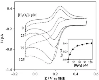
cytochromec-551 and 1lMP. stutzeriCCP are present, it is clear from Fig. 3that, for assays with the oxidized form, the peak current of cytochrome c-551 increases with suc-cessive additions of hydrogen peroxide, until the peak-shaped signal is converted into a sigmoidal wave form. This change in shape is the result of the electrocatalytic reaction with the enzyme, i.e. in the entrapped solution
Fig. 3 Cyclic voltammograms (v=20 mV s-1) of 100lMP.
bothP. stutzeriCCP and its physiologic donor were able to orient themselves in a way that the formation of the tran-sient complex occurred and the intermolecular electron transfer took place.
For hydrogen peroxide concentrations higher than 125lM the current decreases. Similar behaviour was observed in the mediated catalysis ofP. pantotrophusCCP byP. pantotrophuspseudoazurin or horse heart cytochrome
c [25, 29], most probably due to the inactivation of the enzyme in the presence of high substrate concentrations.
The catalytic current of the sigmoidal wave, developed when a saturating hydrogen peroxide concentration (125lM) is added to the electrolyte, is independent of the scan rate up to 50 mV s-1. The half-wave potential of this wave,E1/2=218 mV=E0
0
[28], confirms that the actual transfer process is the catalysed reduction of cytochrome
c-551. As previously described [36], this behaviour is consistent with a reaction mechanism involving an initial heterogeneous electron transfer reaction at the electrode (Fig.4, step 1), followed by homogeneous chemical reac-tions: the oxidized form of cytochrome c-551 is regener-ated by CCP (Fig.4, step 2), which, in turn, is recycled by hydrogen peroxide (Fig.4, step 3), i.e. a ErC0i catalytic
reaction scheme [28].
For each hydrogen peroxide concentration, the catalytic current icat was calculated as the difference between the
experimental current in the presence and absence of sub-strate (both measured at the same potential) and plotted as a function of hydrogen peroxide concentration. The results, shown in the inset in Fig.3, were non-linearly fitted to the Michaelis–Menten equation for concentrations up to 125lM:
icat¼
icatmaxCH2O2
CH2O2þKM
; ð1Þ
whereicatmaxis the catalytic current observed at the
max-imum rate andKM is the Michaelis–Menten constant. As
can be seen in Fig.3, the fit shows that the experimental data are in good agreement with Eq.1and lead to aKMof
(25±2) lM. It must be pointed out that for low concen-trations the catalytic currents are affected by the substrate
diffusion since a stationary electrode was used, leading to an overestimation of the Michaelis–Menten constant [37]. In fact, a KM of 2lM was estimated from steady-state
kinetics, although horse heart cytochromecwas used as the electron donor [13]. However, when the differences between the two types of assays are taken into account, the results can be considered to be in reasonable agreement.
Since diffusion control was verified,icatmaxis given by
the equation
icatmax ¼nFAD1=2Cc551k
01=2; ð
2Þ
where Cc551 is the concentration of the cytochromec-551
(mediator) andk0is the pseudo-first-order rate constant for the intermolecular electron transfer, i.e.k0=kCCCP, where
CCCP is the concentration of the enzyme. As long as the
electron transfer betweenP. stutzericytochromec-551 and
P. stutzeri CCP is the rate-limiting step in the overall process, the second-order rate constantkcan be determined by Eq.2. Competition between the intermolecular and the enzyme–substrate electron transfer reactions can be evaluated through the kinetic parameter r given by Limoges et al. [38]:
r¼
kCc552
kcatCH2O2
CH2O2þKM
: ð3Þ
An estimation ofrcan be obtained withKM=2lM and
kcat=88 s -1
reported forP. stutzeri CCP at pH 7.5 with horse heart cytochrome c as the electron donor [13], and taking as an approximation forkthe value reported for the electron transfer between P. pantotrophus pseudoazurin andP. pantotrophus CCP, 19105M-1s-1. For 125lM hydrogen peroxide r &0.1, indicating that in principle Eq.2 can be used. A rate constant
k=k0/CCCP=(4±1)9105M -1
s-1 was determined with Eq. 2 for the intermolecular electron transfer reaction between fully oxidized P. stutzeri CCP and its physiological donor P. stutzeri cytochrome c-551.
Alternatively,kcan be estimated from the slope of a plot of (icat/ip) versus (1/v)1/2, which should obey a linear
relationship with a null intercept if the intermolecular electron transfer betweenP. stutzericytochromec-551 and the enzyme limits the overall reaction rate. Cyclic vol-tammograms were recorded for scan rates in the range 5–100 mV s-1 in the absence of substrate, and at 20 mV s-1in the presence of saturating hydrogen peroxide (125 lM). In the absence of substrate, the peak current is given by the equation
ip ¼2:69105n3=2AD1=2Ccytv1=2; ð4Þ
whereas the catalytic current is given by Eq.2. From the slope (0.20±0.01) of the (icat/ip) versus (1/v)1/2 plot, a
pseudo-first-order rate constantk0=(0.31±0.03) s-1was
Fig. 4 Mediation scheme for CCP: the electrode reduces cytochrome
determined, which corresponds to an intermolecular elec-tron transfer rate constantk=(3.1±0.3)9105M-1s-1 for the mediated catalysis of oxidizedP. stutzeri CCP by
P. stutzericytochromec-551.
The good agreement between the k values using dif-ferent approaches is an indication that the conditions required to analyse the data according to the model depicted in Fig.4were met.
A similar set of experiments was performed with the mixed-valence enzyme. As mentioned (see ‘‘Materials and methods’’), a fraction of the enzyme was purified in the mixed-valence state, but to guarantee that the high-potential haem ofP. stutzeri CCP was reduced, experiments in the presence of sodium ascorbate were also performed [16]. As observed for the oxidized form, when bothP. stutzeri cyto-chromec-551 and mixed-valenceP. stutzeriCCP are present in solution, the peak-shaped voltammograms transform, in the presence of increasing amounts of hydrogen peroxide, into a sigmoidal wave characteristic of a catalytic ErC0i
mechanism. Fitting the variation of the catalytic current with hydrogen peroxide concentration to the Michaelis–Menten equation (Eq.1), we estimated values ofKM=(32±4)lM
and k=(5.7±0.5)9105M-1s-1 for the Michaelis– Menten and intermolecular electron transfer rate constants, respectively.
Figure5 shows the fit of the experimental i–E curve obtained for the mixed-valence enzyme in the presence of 125lM hydrogen peroxide to the theoretical wave for a catalytic ErC0i mechanism [28,38]:
i¼ nFACc552 Dk
0C
CCP
ð Þ1=2
1þexp nF
RTðEE1=2Þ
: ð5Þ
Fitting of the forward branch of the wave to Eq.5leads to the half-wave potential E1/2=208 mV (=E0
0
) and k0 = 0.45 s-1, which corresponds tok=4.59105M-1s-1.
The similarity of the results obtained forKMandk, using
either the fully oxidized or the mixed-valence CCP, shows that the oxidation state of the high-potential haem of the enzyme has no effect on the mediated catalysis by
P. stutzeri cytochrome c-551. These results support the proposal that the enzyme is readily active upon reduction, since the (tightly) Ca2?binding site is already filled in the resting oxidized state.
Conclusions
The activation mechanism of P. stutzeri CCP was inves-tigated using the physiological partner P. stutzeri cyto-chrome c-551 as an electron relay for mediated catalysis. A PG membrane electrode was used and this configuration was demonstrated once more to be a useful option for the electrochemical analysis of biological systems. The catal-ysis was analysed by cyclic voltammetry for both the oxidized enzyme and the mixed-valence enzyme. All experiments were done in the absence of added calcium. Similar intermolecular rate constants were estimated for the two forms ofP. stutzeriCCP.
Previous spectroscopic studies had suggested that
P. stutzeriCCP needs calcium ions to be active but, unlike other CCPs, is isolated in a dimeric form with one tightly bound Ca?2 and is active as soon as the mixed-valence state is attained. Further characterization ofP. stutzeriCCP and cytochrome c-551 to elucidate the nature of the intermolecular electron transfer and the catalytic mecha-nism has now been reported. The electrochemical results presented for the mediated catalysis of oxidized and mixed-valenceP. stutzeri CCP confirm that the activation mech-anism of the enzyme is fast, since no differences were detected between the results obtained for each form.
Analysis of the activation mechanism through direct electron transfer ofP. stutzeriCCP was not possible, since an altered form of the enzyme is induced by interaction with the PG surface, as observed for the P. pantotrophus
CCP.
Acknowledgments This work was performed within the research
project PPCDT/QUI/55743/2004. D.R. thanks Fundac¸a˜o para a Cieˆncia e Tecnologia for financial support (POCI/QUI/55743/2004). P.M.P.S. and C.G.T. would like to thank the Cieˆncia 2007 (FCT-MCTES) programme.
References
1. Halliwell B, Gutteridge JMC (eds) (1989) Free radicals in biol-ogy and medicine. Oxford Science Publications, Oxford
Fig. 5 Catalytic curve obtained for 1lM mixed-valence enzyme and
2. Ellfolk N, Soininen R (1970) Acta Chem Scand 24:2126–2136 3. Foote N, Thompson AC, Barber D, Greenwood C (1983)
Bio-chem J 209:701–707
4. Foote N, Turner R, Brittain T, Greenwood C (1992) Biochem J 283:839–843
5. Ellfolk N (1983) Biochim Biophys Acta 743:23–30
6. Goodhew CF, Wilson IBH, Hunter DJB, Pettigrew GW (1990) Biochem J 271:707–712
7. Pettigrew GW (1991) Biochim Biophys Acta 1058:25–27 8. Hu W, Van Driessche G, Devreese B, Goodhew CF, McGinnity
DF, Saunders N, Fu¨lo¨p V, Pettigrew GW, Van Beeumen J (1997) Biochemistry 36:7958–7966
9. Alves T, Besson S, Duarte L, Pettigrew GW, Girio FMF, De-vreese B, Vanderberghe I, Van Beeumen J, Fauque G, Moura I (1999) Biochim Biophys Acta 1434:248–259
10. Arciero DM, Hooper AB (1994) J Biol Chem 269:11878–11886 11. Zahn JA, Arciero DM, Hooper AB, Coats JR, DiSpirito AA
(1997) Arch Microbiol 168:363–372
12. De Smet L, Pettigrew GW, Van Beeumen JJ (2001) Eur J Bio-chem 268:6559–6568
13. Timo´teo CG, Tavares P, Goodhew CF, Duarte LC, Jumel K, Gı´rio FMF, Harding S, Pettigrew GW, Moura I (2003) J Biol Inorg Chem 8:29–37
14. Pettigrew GW, Echalier A, Pauleta SR (2006) J Inorg Biochem 100:551–567
15. Gilmour R, Goodhew CF, Pettigrew GW, Prazeres S, Moura I, Moura JJG (1993) Biochem J 294:745–752
16. Gilmour R, Goodhew CF, Pettigrew GW, Prazeres S, Moura I, Moura JJG (1994) Biochem J 300:907–914
17. Gilmour R, Prazeres S, McGinnity DF, Goodhew CF, Moura JJG, Moura I, Pettigrew GW (1995) Eur J Biochem 234:878–886 18. Le´ger C, Bertrand P (2008) Chem Rev 108:2379–2438 19. Butt JN, Armstrong FA (2008) In: Hammerich O, Ulstrup J (eds)
Bioinorganic electrochemistry. Springer, Dordrecht, pp 91–128 20. Chen KI, McEwan AG, Bernhardt PV (2009) J Biol Inorg Chem
14:409–419
21. Paes de Sousa PM, Pauleta SR, Simo˜es Gonc¸alves ML, Pettigrew GW, Moura I, Moura JJG, Correia dos Santos MM (2011) J Biol Inorg Chem 16:209–215
22. Save´ant JM (2006) Elements of molecular and biomolecular electrochemistry. Willey, Hoboken
23. Ferapontova EE, Ruzgas T, Gorton L (2003) Anal Chem 75:4841–4850
24. Lojou E, Cutruzzola` F, Tegoni M, Bianco P (2003) Electrochim Acta 48:1055–1064
25. Paes de Sousa PM, Pauleta SR, Rodrigues D, Simo˜es Gonc¸alves ML, Pettigrew GW, Moura I, Moura JJG, Correia dos Santos MM (2008) J Biol Inorg Chem 13:779–787
26. Haladjian J, Bianco P, Nunzi F, Bruschi M (1994) Anal Chim Acta 289:15–20
27. Correia dos Santos MM, Paes de Sousa PM, Simo˜es Gonc¸alves ML, Krippahl L, Moura JJG, Lojou E, Bianco P (2003) J Elec-troanal Chem 541:153–162
28. Bard AJ, Faulkner LR (2001) Electrochemical methods, funda-mentals and applications. Wiley, New York
29. Paes de Sousa PM, Pauleta SR, Simo˜es Gonc¸alves ML, Pettigrew GW, Moura I, Correia dos Santos MM, Moura JJG (2007) J Biol Inorg Chem 12:691–698
30. Kakihana M, Ikeuchi H, Satoˆ GP, Tokuda K (1980) J Electroanal Chem 108:381–383
31. Bradley AL, Chobot SE, Arciero DM, Hooper AB, Elliott SJ (2004) J Biol Chem 279:13297–13300
32. Villalain J, Moura I, Liu MC, Payne WJ, LeGall J, Xavier AV, Moura JJG (1984) Eur J Biochem 141:305–312
33. Armstrong FA, Cox PA, Hill HAO, Lowe VJ, Oliver BN (1987) J Electroanal Chem 217:331–366
34. Correia dos Santos MM, Paes de Sousa PM, Simo˜es Gonc¸alves ML, Lopes H, Moura I, Moura JJG (1999) J Electroanal Chem 464:76–84
35. Leitch FA, Moore GR, Pettigrew GW (1984) Biochemistry 23:1831–1838
36. Lopes H, Besson S, Moura I, Moura JJG (2001) J Biol Inorg Chem 6:55–62
37. Chen CJ, Liu CC, Savinell RF (1993) J Electroanal Chem 348:317–338

![Figure 5 shows the fit of the experimental i–E curve obtained for the mixed-valence enzyme in the presence of 125 lM hydrogen peroxide to the theoretical wave for a catalytic E r C 0 i mechanism [28, 38]:](https://thumb-eu.123doks.com/thumbv2/123dok_br/16491486.733172/7.892.77.429.624.1001/experimental-obtained-presence-hydrogen-peroxide-theoretical-catalytic-mechanism.webp)
