Vol.46, n. 2 : pp. 287-294, March 2003
ISSN 1516-8913 Printed in Brazil
BRAZILIAN ARCHIVES OF
BIOLOGY AND TECHNOLOGY
A N I N T E R N A T I O N A L J O U R N A L
Ultrastructural and Quantitative Studies of Hemocytes in
the Sugarcane Borer,
Diatraea saccharalis
(Lepidoptera:
Pyralidae)
Ângela Maria Ferreira Falleiros
1∗, Maria Terezinha Siqueira Bombonato
2and Elisa
Aparecida Gregório
31Departamento de Histologia; Centro de Ciências Biológicas; Universidade Estadual de Londrina - UEL;
C. P. 6001, CEP 86051-990; Londrina - PR - Brazil. 2UNESP; Bauru - SP - Brazil. 3Centro de Microscopia Eletrônica; Instituto de Biociências; UNESP; Botucatu - SP - Brazil
ABSTRACT
Six circulating hemocytes cell types from Diatraea saccharalis (Fabricius) (Lepidoptera: Pyralidae) larvae were identified by transmission and scanning electron microscope: prohemocytes (PR), plasmatocytes (PL) granulocytes (GR), spherulocytes (SP), oenocytoids (OE) and vermicytes (VE). The PR was the smallest cell type with a large nucleus, a cytoplasm with few organelles and a homogenous smooth surface. The PL was polymorphic and abundant, with a cytoplasm rich in organelles and a cellular surface with several cytoplasmic projections. The GR was abundant, showing two types of membrane-bounded granules (dense and structutered), glycogen, lipid droplets and a surface with philopodial projections. The SP was a large cell, with a cytoplasm full of intracytoplasmic spherules. The OE was the largest hemocyte type with a large and homogeneous cytoplasm and scarce organelles. The VE was discoid in shape and showed electron-dense granules.
Key words: Insect; Lepidoptera; Hemocytes; Ultrastructural Morphology; Differential Hemocyte Count; Morphometry
∗ Author for correspondence
INTRODUCTION
Insect hemocytes, like vertebrate leucocytes, are a mixture of cell types with different morphological and biological functions. The hemocytes have the ability to discriminate stranger agents, mediate phagocytosis, cytotoxicity, encapsulation, wound repair and coagulation. These defense reactions were observed against pathogens, parasites and other foreign bodies, which entered in the hemocoel (Ratcliffe et al., 1985; Lackie, 1986; Ratcliffe and Rowley, 1987; Brookman et al., 1989; Rahmet-Alla and Rowley, 1989; Falleiros
and Gregório, 1995; Hung and Boucias, 1996). Seven types of hemocytes have been frequently described in various insects: prohemocytes, plasmatocytes, granulocytes, spherulocytes, adipohemocytes, oenocytoids and coagulocytes (Jones, 1979; Gupta, 1985; Brehélin and Zachary, 1986, among others).
The sugarcane borer, Diatraea saccharalis
morpho-metrically these cellular types, using the electron microscopy. It also quantifies, through differential counts (DCH), the types of hemocytes found in the hemolymph of Diatraea saccharalis, which were evaluated by phase contrast microscopy.
MATERIAL AND METHODS
D. saccharalis larvae were obtained from the Pest Control Laboratory at the Usina Barra Grande in Lençois Paulista, S.P., Brazil. The larvae were grown in the laboratory on artificial diet (Hensley and Hammond, 1968) at 25-27°C and 70% relative humidity. Late instars larvae aging from 17 to 23 days were used.
For transmission electron microscopy (TEM), hemolymph of at least 15 insects of the same age was collected from a punctured abdomen and pulled into an Eppendorff tube containing the insect anticoagulant solution described by Leonard et al., (1985). The hemocyte pellet was obtained by centrifugation at 6500 rpm for 2 min, suspended again and fixed in cacodylate buffer (0.1 M, pH 7.2) with glutaraldehyde solution (2.5%) for 2 h. The fixed pellets were embedded in 2.5% Bacto-agar (DIFCO), prepared in phosphate buffer (0.1 M, pH 7.2) at 45°C, cooled in ice and sliced into small fragments with a razor blade. The materials were post fixed in cacodylate buffer with osmium tetroxide solution (1%) for 2 h, dehydrated in graded acetone solution and embedded in Araldite®. The ultrathin sections were stained in uranil acetate and lead citrate. For the scanning electron microscopy (SEM), the hemolymph was dropped directly on coverslips covered by 0.1% poly-l-lysine for 5 min in a moist chamber. After the adhesion of the hemocytes, the monolayers were fixed in cacodylate buffer (0.1 M, pH 7.3) with glutaraldehyde solution (2.5%), post-fixed in osmium tetroxide, dehydrated in graded acetone and dried at the critical point. The preparations were mounted on stubs, gold coated in a sputter coater and analyzed by SEM. The morphometric analysis of the different hemocyte types was performed on photomicrographs magnified 10,000 times, using a microprocessor-controlled measuring system (Mini-Mop, Kontron).
For the differential hemocyte counts (DHC), unfixed hemolymph was diluted in an insect anticoagulant solution. The percentage of each
hemocyte type was determined by using 5insects /age, 200cells/insect count, in a 1000cells/age performance.
RESULTS
Six hemocyte types were ultrastructurally identified in the hemolymph of D. saccharalis
larvae: prohemocytes, plasmatocytes, granulo-cytes, spherulogranulo-cytes, oenocytoids and vermicytes. Prohemocytes (PR) were the less frequently found hemocyte in the hemolymph, representing 4.1% of the 17 day-old larvae, showing a decrease in percentage terms and undetected in 20 day-old larvae (Table 1). PR was a small rounded cell with variable sizes (Table 2). Plasma membrane was generally smooth, and nucleus was large, centrally located, almost filling up the whole cell. The nuclear/cytoplasmic ratio was 84% (Table 2) with scattered chromatin and evident nucleoli. The cytoplasm showed a large amount of free ribosome, but only a small rough endoplasmic reticulum (RER) cisternae. Few mitochondria and rare Golgi complex were observed (Fig. 4). Plasmatocytes (PL) represented up to 29% of the circulating hemocytes, showing a decrease in 23 day-old larvae percentage (Table 1). These were polymorphic cells, generally oval and variable in size (Table 2). The plasma membrane may exhibit irregular processes, as micro papillae, philopodia or invaginations (Figs. 1, 4, 5, 6). The elongate or lobate nucleus present variable sizes and was centrally localized, showing scattered chromatin masses and up to two nucleoli (Fig. 4). The nuclear/cytoplasmic ratio was 36% (Table 2). Binuclear PL was occasionally observed. The cytoplasm showed well-developed RER, Golgi system, mitochondria and polymorphic vacuoles (Fig. 4).There were many cells with intermediate characteristics between PR and PL, interpreted as young PL.
content, and structured granules, with crystalloid content (Figs.1, 2). Vacuoles of variable sizes and shapes were also present, with moderately electron-dense and flocculent material. The developed RER, the Golgi apparatus, mitochondria and glycogen particles were dispersed in the cytoplasm. Lipid droplets of various sizes, associated with either glycogen particles or dense granules (Fig. 1), were detected in the oldest larvae. Cells with intermediate features between PL and GR were observed. The percentage of spherulocytes (SP) in the hemolymph varied presenting values close to those of the PL, showing, however, an increase in the 23rd day-old larvae (Table 1). SP was spherical cell with variable sizes (Table 2), generally larger than the PL and GR. These hemocytes were characterized by their inclusions and membrane-bound spherules took up almost all the cytoplasm. The cellular surface was homogenous but exhibit cytoplasmic protrusion corresponding to the spherules (Figs. 2, 7). The nucleus was small, centrally located or eccentric, mostly deformed by the spherules (Fig. 2). The nuclear/cytoplasmic ratio was low, 9% (Table 2). The spherules contained moderate electron-dense and flocculent material, with a quite
electron-dense core region (Fig. 2). Besides the spherules, the cytoplasm contained few organelles around the nucleus, such as ribosome, Golgi complex, RER and few mitochondria (Fig. 2). Oenocytoids (OE) represented up to 5.3% of the circulating hemocytes (Table 1). OE was a rounded cell, larger than all the other hemocyte types (Table 2). The cellular membrane was smooth (Figs. 5, 6, 7, 8); vesicles were observed in close association with the plasmalem (Fig. 5). The nucleus was small, eccentric, with a nuclear/cytoplasmic ratio of 11% (Table 2) and showed a distribution pattern with alternate condensed and discondensed chromatin. The cytoplasm was homogenous and poor in organelles, with the exception of the free ribosome, that were abundant (Fig. 6). Dense mitochondria, generally ring-shaped was observed (Fig. 5). Vermicytes (VE) represented up to 9.3% of the circulating hemocytes (Table 1). VE was fusiform in shape and variable in size (Table 2). The cell membrane was generally smooth (Figs.1, 5). The nucleus was elongated, centrally located and the nuclear/cytoplasmic ratio was 32% (Table 2). Besides cell shape, this type cell was characterized by the rounded, electron-dense and homogenous cytoplasmic granules (Figs. 1, 5).
Table 1 - The hemocytes differential count in the D. saccharalis larvae. Cellular Types (%) L.A.*
PR PL GR SP OE VE
17 4.1 27.0 28.2 27.3 4.4 9.0
18 1.8 27.8 35.4 22.0 3.7 9.3
20 0.3 29.0 30.7 28.2 4.0 7.7
23 - 20.7 33.6 32.7 5.3 7.7
*Larval age in days.
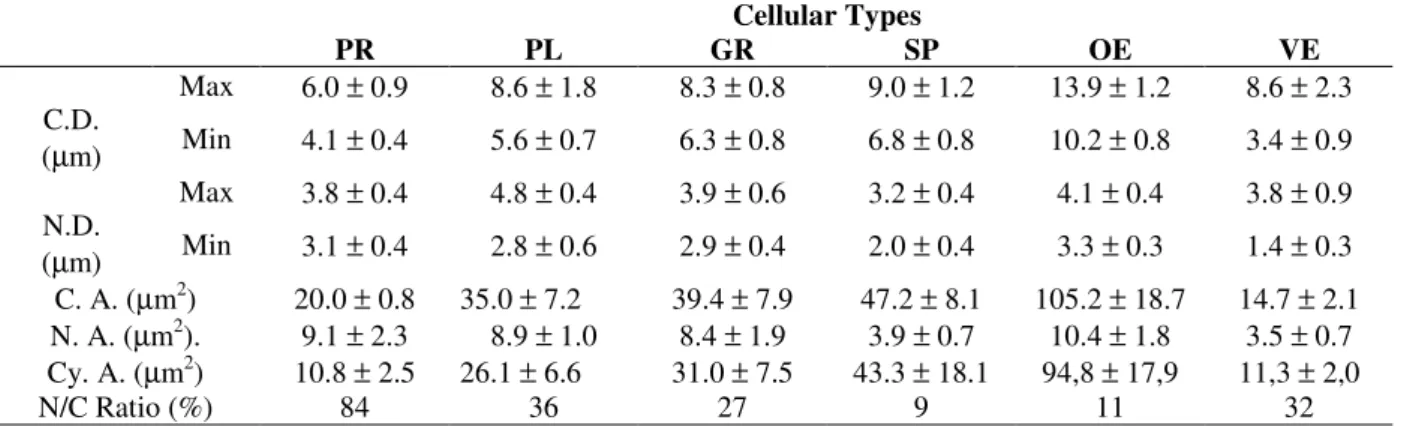
Table 2 - The morphometric analysis of the different hemocyte types found in the hemolymph of the D. saccharalis larvae.
Cellular Types
PR PL GR SP OE VE
Max 6.0 ± 0.9 8.6 ± 1.8 8.3 ± 0.8 9.0 ± 1.2 13.9 ± 1.2 8.6 ± 2.3 C.D.
(µm) Min 4.1 ± 0.4 5.6 ± 0.7 6.3 ± 0.8 6.8 ± 0.8 10.2 ± 0.8 3.4 ± 0.9 Max 3.8 ± 0.4 4.8 ± 0.4 3.9 ± 0.6 3.2 ± 0.4 4.1 ± 0.4 3.8 ± 0.9 N.D.
(µm) Min 3.1 ± 0.4 2.8 ± 0.6 2.9 ± 0.4 2.0 ± 0.4 3.3 ± 0.3 1.4 ± 0.3 C. A. (µm2) 20.0 ± 0.8 35.0 ± 7.2 39.4 ± 7.9 47.2 ± 8.1 105.2 ± 18.7 14.7 ± 2.1 N. A. (µm2). 9.1 ± 2.3 8.9 ± 1.0 8.4 ± 1.9 3.9 ± 0.7 10.4 ± 1.8 3.5 ± 0.7 Cy. A. (µm2) 10.8 ± 2.5 26.1 ± 6.6 31.0 ± 7.5 43.3 ± 18.1 94,8 ± 17,9 11,3 ± 2,0
N/C Ratio (%) 84 36 27 9 11 32
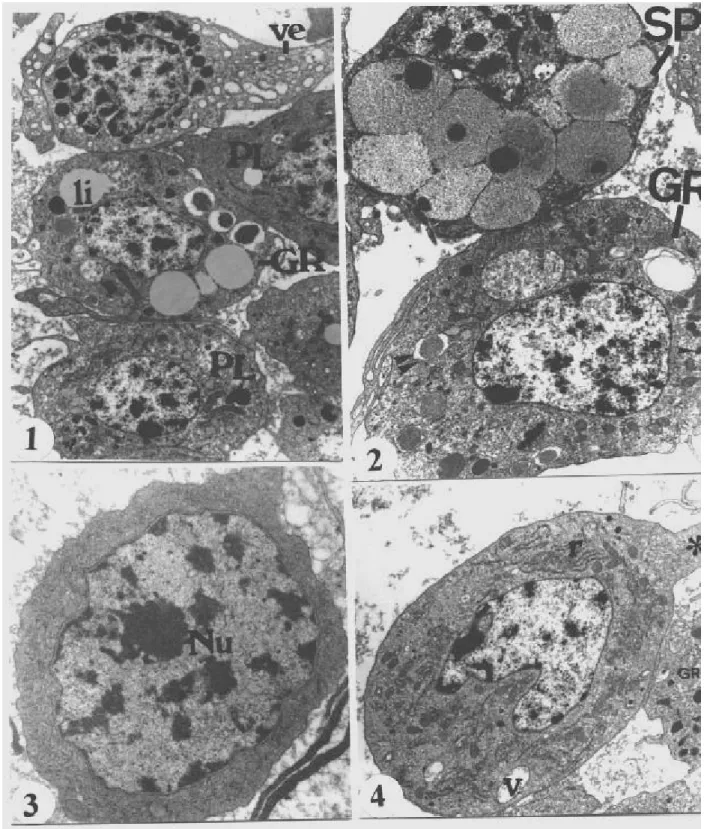
Figure 1 - General aspect of hemocytes from 23 day-old larvae, plasmatocytes (PL), vermicytes (VE), granulocytes (GR), li (lipids drop). X 7800.
Figure 2 - General aspect of spherulocytes (SP) from 23 day-old larvae. Cytoplasm full of spherules, flocculent content and electron dense core. Granulocytes (GR) showing structured granules (two arrow tips) and electron -dense granules (arrow tip). X 9400.
Figure 3 - Prohemocytes (PR) from 17 day-old larvae. Centralized and large nucleus, evident nucleoli (Nu), cytoplasm poor in organelles, cellular surface without projections. X18900.
Figure 5 - Vermiform cell from 23 day-old larvae (empty arrow) between oenocytoids (OE) and granulocytes (GR). Dense heterogeneous granules and translucid vesicles. Golgi complex (curved arrow) and mitochondria and ring-shaped cells in the OE. X 14000.
Figure 6 - General aspect of oenocytoids from 23 day-old larvae. Evident nucleoli (Nu), electron-dense cytosol, scarce mitochondria and vacuoles variable in content and size. Lipids drop (LI). X 10000.
DISCUSSION
Among the cellular types observed in Diatraea saccharalis (Fabricius) larvae, PR, PL, GR, SP and OE presented ultrastructural morphological features similar to those found in other Lepidoptera species (Akai and Sato, 1973; Lai-Fook, 1973; Beaulaton and Monpeyssin, 1976; Raina, 1976; Beeman et al., 1983; Essawy et al., 1985; Wago and Kitano, 1985; Saxena, 1992; Ribeiro et al., 1996), except Wago and Kitano (1985), who recognized only three types of hemocytes present in the hemolymph of Pieris rapae crucivora (Boisduval). In addition, the hemocytes of D. saccharalis described in this study presented, for each type of hemocyte, and in different insect species, morphometric values within the numerical intervals reported by Gupta (1985) which were numerically very close to the data obtained by Raina (1976).
The identification of VE cell as a distinct cellular type has not been pointed out in many of the insects studied, being included as a functional variant of PL. Although some authors have reported behavioral differences in this cell, which distinguished them from the PL, they were classified as PL in Prodenia (Jones, 1959) and in
Calpodes ethlius (Stoll) (Lai-Fook, 1973). Kurihara et al. (1992), analyzed hemocytes of
Spodoptera litura (Fabricius) by phase contrast microscopy and were able to find adhesive character of forms, different from the PL. They proposed that these cells and the so-called coagulocytes (CO) were of the same cellular kind, naming them Podocytes (PO).
In the D. saccharalis larvae, these cells showed quite individualized morphological characteristics, which permitted us to distinguish them as a cellular type and not only a transitional, a variant of the PL functional state. Our morphological description for this cellular type was similar to Ribeiro et al. (1996).
AD has not been considered a cellular type in the present work since, ultrastructurally, they showed similar morphology to that of GR, except in the large amount of lipid droplets in the cytoplasm. The word “adipohemocyte” has been applied to cells of different morphologies, being functional variants of other cellular types with lipid droplets, according to Lea and Gilbert (1966). Thus, the use of synonymies for these different cells which accumulated lipid, as observed in GR of various insects (Price and Ratcliffe, 1974; Kaaya and
Ratcliffe, 1982; Gupta, 1985; Ratcliffe et al. 1985, Fenoglio et al. 1993, among others), including the
D. saccharalis larvae, was not recommended as it generated controversies, which made the characterization of insect hemocytes even more difficult.
The identification of cells with intermediate features between PR-PL and between PL-GR suggested a possible cell differentiation of those hemocyte types in the circulation. The decrease in the number of PR and PL, simultaneously with the increase of PL and GR, respectively, suggested that a transformation of PR in PL and of PL in GR took place, as reported by Gupta (1979, 1985) and Ratcliffe et al. (1985).
Although the results of this study referred to the presence of transitional stages among types of hemocytes, they did not reach any conclusion the differentiation of the blood cells. Probably PR was an undifferentiated cell that would be transformed in PL, which then would give origin to the GR. No morphological transitional stage among any other type of the D. saccharalis hemocytes (ES, OE and VE) was found, which led us to believe that the cellular types had a different origin from that mentioned earlier. Further studies are needed to contribute to this discussion.
Results of this work may help in the investigation of the role played by the different cell types on the defense mechanisms of the sugarcane borer. They may also contribute to the improvement of the techniques used in the control of this plague, mainly the biological control that takes place when the parasitoid deceives successfully the defense system of the host.
ACKNOWLEDGEMENTS
The authors are grateful to Nivalde Basso, Maria H Moreno and Maria Euleda Perez, for the technical support. This work was partially supported by CPG/UEL and CNPq.
RESUMO
granulócitos (GR), esferulócitos (ES), oenocitói-des (OE) e vermiformes (VE). O PR foi o menor tipo celular; apresentando um núcleo grande, citoplasma com poucas organelas e superfície lisa e homogênea. O PL era polimórfico e abundante, com citoplasma rico em organelas e superfície celular com várias projeções citoplasmáticas. O GR era abundante, apresentando dois tipos de grânulos envoltos por membrana (denso e estru-turado), glicogênio, gotas de lipídio e projeções citoplasmáticas filopodiais. O ES era uma célula grande, com citoplasma carregado de esférulas intracitoplasmáticas. O OE foi o maior tipo de hemócito encontrado, apresentando citoplasma grande, homogêneo e escassas organelas. O VE era discóide e apresentou grânulos elétrons-densos.
REFERENCES
Akai, H.and Sato, S. (1973), Ultrastructure of the larval hemocytes of the silkworm, Bombyx mori L. (Lepidoptera: Bombycidae). International Journal of Insect Morphology and Embryology, 2, 207-231. Beaulaton, J. and Monpeyssin, M. (1976),
Ultrastructure et cytochimie des hémocytes d' Antheraea pernyi (Guér) (Lepidoptera, Attacidae) au cours du cinquième âge larvaire. Journal of Ultrastructural Research, 55, 143-156.
Beeman, S. C.; Wilson, M. E.; Bulla, L. A. et al., (1983),Structural characterization of the haemocytes of Plodia interpunctella. Journal of Morphology, 175, 1-16.
Brehélin, M. and Zachary, D. (1986), Insect haemocytes: a new classification to rule out the controversy. In: Brehélin, M. (ed.). ‘Immunity invertebrates, cells, molecules and defense reactions’. Heidelberg: Spring Verlag. pp. 37-48.
Brookman, J. L.; Ratcliffe, N. A. and Rowley, A. F. (1989), Optimization of a monolayer phagocytosis and its applications for studying the role of prophenoloxidase system in the wax moth, Galleria mellonella. Journal of Insect Physiology, 34, 337-346. Essawy, M. A.; Maleville, A. and Brehélin, M. (1985),
The hemocytes of Heliothis armigera. Ultrastructure, function, and evolution in the course of larval development. Journal of Morphology, 186, 225-264. Falleiros, A. M. F. and Gregório, E. A. (1995),
Hemócitos fagocitários em larvas de Diatraea saccharalis (Fabricius) (Lepidoptera, Pyralidae). Revista Brasileira de Zoologia, 12, 751-758.
Fenoglio, C.; Bernardini, P. and Gervaso, M. V. (1993), Cytochemical characterization of the hemocytes of Leucophaea maderae (Dictyoptera: Blaberoidea). Journal of Morphology, 218, 115-126.
Gupta, A. P. (1979), Hemocytes types, their structures, synonymies, interrelationships, and taxonomic significance. In: Gupta, A. P. (ed.). ‘Insect hemocytes: development, forms, functions and techniques’. Cambridge: Cambridge University Press. pp. 85-127.
Gupta, A. P. (1985), Cellular elements in the hemolymph In: ‘Comprehensive insect physiology biochemistry and pharmacology’. Kerkut, G.A., Gilbert, L. I. Eds. Oxford: Pergamon Press. pp. 402-444.
Hensley, S. D. and Hammond Jr., A. M. (1968), Laboratory techniques for rearing the sugarcane borer on an artificial diet. Journal of Economic Entomology, 61, 1742-1743.
Hung, S. Y. and Boucias, D. G. (1996), Phenoloxidase activity in hemolymph of naive and Beauveria bassiana infected Spodoptera exigua larvae. Journal of Invertebrate Pathology, 67, 35-40.
Jones, J. C. (1959), A phase contrast study of the blood cells in Prodenia larvae (Order Lepidoptera). Quarterly Journal of Microscopical Science, 100, 17-23.
Jones, J. C. (1979), Pathways and pitfalls in the classification and study of insect hemocytes. In: Gupta, A. P. (ed.). ‘Insect hemocytes: development, forms, functions and techniques’. Cambridge: Cambridge University Press. pp. 279-300.
Kaaya, G. P. and Ratcliffe, N. A. (1982), Comparative study of hemocytes and associated cells of some medically important dipterans. Journal of Morphology, 173, 351-365.
Kurihara, Y.; Shimazu, T. and Wago, H. (1992), Classification of hemocytes in the common cutworm, Spodoptera litura (Lepidoptera: Noctuidae) I. Phase Microscopic Study. Applied Entomology and Zoology, 27, 225-235.
Lackie, A. M. (1986), Evasion of insect immunity by helminthes larvae. Symposium Zoological Society of London, 56, 161-178.
Lai-Fook, J. (1973), The structure of the haemocytes of
Calpodes ethlius (Lepidoptera). Journal of
Morphology, 118, 79-104.
Lea, M. S. and Gilbert, L. (1966), The hemocytes of Hyalophora cecropia (Lepidoptera). Journal of Morphology, 118, 197-216.
Leonard, C.; Söderhäll, K. and Ratcliffe, N. A. (1985). Studies of pro-phenoloxidase and protease of Blaberus craniifer haemocytes. Insect Biochemistry, 15, 803-810
Price, C. D. and Ratcliffe, N. A. (1974), A reappraisal of insect haemocyte classification by the examination of blood from fifteen insect orders. Zoologische Zellforsch, 147, 537-549.
Raina, A. K. (1976), Ultrastructure of the larval hemocytes of the pink bollworm, Pectinophora gossypiella (Saunders) (Lepidoptera: Gelechiidae). International Journal of Insect Morphology and Embryology, 5, 187-195.
Ratcliffe, N. A.; Rowley, A. F. and Fitzgerald, S. W. (1985), Invertebrate immunity: basic concepts and recent advances. International Review of Cytology, 97, 183-349.
Ratcliffe, N. A. and Rowley, A. F. (1987), Insect responses to parasites and other pathogens. In: Jouslby, E. J. L. (ed.). ‘Imune responses in parasitic
infections: immunology, immulopathology and
immunoprophylaxis, protozoa, arthropods and invertebrate’. Florida: C. R. C. Press. pp. 123-254. Ribeiro, C.; Simões, N. and Brehélin M. (1996), Insect
immunity: the haemocytes of the armyworm Mythimna unipuncta (Lepidoptera: Noctuidae) and their role in defence reactions. In vivo and in vitro studies. Journal of Insect Physiology, 42, 815-822.
Saxena, B. P. (1992), Comparative study of three lepidopterans by light and scanning electron microscopy. Acta Entomologica Bohemoslovaca, 89, 323-329.
Wago, H. and Kitano, H. (1985), Morphological and functional characterization of the larval haemocytes of the cabbage white butterfly Pieris rapae crucivora. Applied Entomology and Zoology, 20, 1-7.