Printed version ISSN 0001-3765 / Online version ISSN 1678-2690 http://dx.doi.org/10.1590/0001-3765201620150574
www.scielo.br/aabc
Neuroprotective effect of
Portulaca oleracea
extracts against
6-hydroxydopamine-induced lesion of dopaminergic neurons
Waleska B. Martins1
, sheyla a. rodrigues1
, hataMy k. silva1
, CaMila g. dantas1
, WaldeCy de luCCa Júnior2, lauro Xavier Filho1,Juliana C. Cardoso1 and Margarete Z. goMes1
1
Instituto de Tecnologia e Pesquisa/ITP, Av. Murilo Dantas, 300, Farolândia, 49032-490 Aracaju, SE, Brasil 2
Universidade Federal de Sergipe, Cidade Universitária Prof. José Aloísio de Campos, Av. Marechal Rondon, s/n, Jardim Rosa Elze, 49100-000 São Cristóvão, SE, Brasil
Manuscript received on August 3, 2015; accepted for publication on November 27, 2015
aBstraCt
The Portulaca oleracea L. (Portulacaceae) is a cosmopolitan species with a wide range of biological activities, including antioxidant and neuroprotective actions. We investigated the effects of P. oleracea extracts in a 6-hydroxydopamine rat model of Parkinson’s disease, a debilitating disorder without effective
treatments. Chemical profiles of aqueous and ethanolic extracts of whole plant were analyzed by thin layer chromatography and the antioxidant activity was assessed by 2,2-diphenyl-1-picrilhidrazila method. Male Wistar rats received intrastriatal 6-hydroxydopamine and were treated with vehicle or extracts (oral,
200 and 400 mg/kg) daily for two weeks. The behavioral open field test was conducted at days 1 and 15.
Immunohistochemical analysis was performed 4 weeks after surgery to quantify tyrosine-hydroxylase cell counts in the substantia nigra pars compacta. Extracts presented antioxidant activity in concentrations
above 300 µg/kg. The chromatographic analysis revealed the presence of Levodopa, alkaloids, flavonoids,
saponins, tannins, terpenoids and polysaccharides. Both extracts improved motor recovery 15 days after lesion and protected from tyrosine-hydroxylase cell loss after 4 weeks, but these effects were more evident for the aqueous extract. Because the dopamine precursor is present, in addition to antioxidant compounds and neuroprotective effects, P. oleracea can be considered as potential strategy for treating Parkinson’s disease.
key words: Parkinson Disease, Antioxidant Response Elements, Levodopa, Purslane.
Correspondence to: Margarete Zanardo Gomes E-mail: margarete_zanardo@itp.org.br
introduCtion
Parkinson’s disease (PD) is a neurodegenerative
illness characterized by a progressive loss of
dopaminergic neurons in the substantia nigra
pars compacta (SNc). The consequent dopamine
depletion in the striatum is clinically related to
movement and cognitive disorders (Wichmann and Dostrovsky 2011). Its etiology is not fully understood, however, mechanisms of neuronal degeneration include mitochondrial dysfunction, altered proteasomal and lysosomal proteolysis,
glial mediated inflammation and oxidative stress
Currently, there is no available therapy to stop or slow neurodegeneration.
Portulaca oleracea L. (P. oleracea) is a com-monly found species and a medicinal food for human consumption. This plant, also called purs-lane, contains minerals, proteins, carbohydrates,
β-carotene, vitamins and fatty acids (Uddin et al.
2012, 2014). Among the bioactive components and biological activities assigned to the P. oleracea, the presence of catecholamines (Chen et al. 2003)
and its antioxidant and anti-inflammatory actions
(Dkhil et al. 2011, Yue et al. 2015) deserve to be highlighted.
Neuropharmacological actions of the P. ol-eracea extracts were previously reported in rodent models. Effects included the reduction of the lo-comotor activity, the increase in the onset time of pentylenetetrazole-induced convulsions in mice, the opioid mediated anti-nociceptive and muscle re-laxant activities in rats (Radhakrishnan et al. 2001).
Biological activities also include anti-inflammatory
actions (Chan et al. 2000, Lee et al. 2012).
It has been shown that P. oleracea protects neurons against hypoxia injury (Wanyin et al. 2012) and D-galactose induced toxicity in vivo (Hongxing et al. 2007). The treatment with betacyanins from P. oleracea improved cognitive deficits and attenuat -ed oxidative damage induc-ed by D-galactose in the brains of senescent mice (Wang and Yang 2010). Moreover, the P. oleracea extracts decreased apop-tosis and oxidative-stress-induced neurodegenera-tion caused by the pesticide rotenone (Al-Quraishy et al. 2012, Abdel Moneim 2013).
These neuroprotective actions and the exis-tence of dopamine and noradrenaline in extracts suggest that P. oleracea may be a potential can-didate for the treatment of Parkinson’s disease. Thus, the aim of this work was to evaluate the neu-roprotective effects of the ethanolic and aqueous
P. oleracea extracts, given orally, in animals with 6-hydroxydopamine-induced lesion of the nigros-triatal pathway, an animal model for the study of
progressive degeneration in PD. Since ethanolic and aqueous extracts are commonly studied, they
were also analyzed regarding chemical profiles and
antioxidant activity.
Materials and Methods
Plant Material
The plant material was collected in August 2012, from the greenhouse for medicinal plants at the campus of the Tiradentes University, located in Aracaju – Sergipe, Northeast of Brazil. The plant
was identified by Dr. Ana Paula do Nascimento
Prata and a voucher specimen (no. 20379) has been deposited at the Herbarium of the Federal University of Sergipe. The whole plant was used for the preparation of the extracts. The fresh vegetal material was manually chopped in pieces about 2 cm.
PreParationofthe extracts
The aqueous extract was prepared as previously reported (Hozayen et al. 2011). The vegetal material was boiled in distilled water (80 ºC, 1:1, m: v) for 60 min and then mashed with a blender apparatus (3 cycles of 15 sec). The mixture was coated (2 mm pores strainer) and lyophilized under vacuum.
The plant material was extracted with 100% ethanol (100 mL: 10 g) at room temperature, in a dark and closed container for one week. The extract
was centrifuged and filtered through a Whatman number 3 filter and then concentrated using a rotary
evaporator. The resultant extract was lyophilized to produce a powder (Wanyin et al. 2012).
PhytocheMical analysis
as a positive control and the samples were diluted in hydrochloric acid (Dighe et al. 2008). The phytochemical screening was carried out using standard procedures that have been previously described (Harborne 1998): the frothing test was used for the detection of saponins, the ferric chloride test was used for the detection of tannins, the Dragendorff precipitation assay was used for the detection of alkaloids, Mg-HCl was used for the
detection of flavonoids, Liebermann-Burchard and
Salkowski reactions was used for the detection of triterpenes and lugol was used for the detection of polysaccharides.
QualitativeanD Quantitative 2,2-DiPhenyl -1-PicrilhiDrazila (DPPH) free raDical scavenging activity assay
The extracts were analyzed by TLC in a plate eluted in ethyl acetate and ethanol (50%:50%). After drying, a 0.4 mmol/L solution of DPPH in MeOH was added. The antioxidant activity was determined by the presence of yellow spots on a purple background (Souza et al. 2007).
The quantitative assays were performed accord-ing to the method previously described (Djouossi et al. 2015). Samples of the extracts and the positive control (trolox) were prepared in methanol (5 mg/ mL) and diluted in the concentrations of 50, 100,
200, 300, 400 and 550 μg/mL. Measurement of
the absorbance in the reactive mixtures (0.3 mL of sample solution with 2.7 mL DPPH solution in a 40
μg/mL concentration) was made on a 515 nm, after
40 minutes incubation at room temperature, pro-tected from light. Absorbance values were convert-ed in percentage of the antioxidant activity and the
antioxidant efficacy was stabilized by linear regres
-sion analysis (p<0.05 confidence interval). Results
were expressed through µmol trolox/g-equivalent
antioxidant capacity and the sufficient necessary
sample concentration to scavenge 50% of DPPH radical was also estimated.
Biological assay
Animals
Forty-eight adult male Wistar rats weighting 200– 250 g were housed in a temperature controlled room (at approximately 25ºC), under 12-h light/ dark cycle, with free access to food and water. The experiments were carried out according to the rules of the local Animal Use and Care Committee (ap-proval number 141110) and the Brazilian National Council for the Control of Animal Experimentation (CONCEA). Every effort was made to minimize animal suffering and to keep a number of animals used to a minimum.
Experimental design
After surgical procedures for the intrastriatal injection of 6-hydroxydopamine (6-OHDA) or saline, the animals were divided into groups (n = 6 per group) that received, via oral administration, vehicle (2% Tween 80 in saline), aqueous extract (AEPO) or ethanolic extract (EEPO) of P. oleracea, at 200 or 400 mg/kg for 15 days, consecutively. Doses were chosen based on previous work
(Wanyin et al. 2012). At the first and fifteenth day after surgery, an open field test was performed in
order to evaluate the motor abilities of the animals.
The animals were sacrificed at 28 days following
the beginning of the treatments and their brains were extracted and processed for the histological analysis.
6-OHDA microinjections
The heads of the rats were fixed on a Kopf ste
-reotaxic instrument under general anesthesia (ket-amine/xylazine, 90 and 15 mg/kg i.p.). A midline skin incision was made, exposing the skull for
sub-sequent drilling. The neurotoxin, 6-OHDA (20 μg in 4 μl dissolved in 0.9% saline containing 0.02
mg/mL of ascorbic acid; RBI-Sigma) was injected
into the right striatum with a 10 μl Hamilton sy
anterior, 3.0 mm lateral (right side) from Bregma, and 5.0 mm ventral to the surface of the skull, with
the tooth-bar set at −3.0 mm (Paxinos and Watson
1998). The microinjections were performed in a
rate of 1 μl/min with an infusion pump (Insight,
BR) and the needle was left in place for an
addi-tional 180 s to prevent reflux before being slowly
retracted. The movement of an air bubble inside the PE-10 polyethylene tubing connecting the
mi-cro-syringe with the needle confirmed drug flow.
Sham operated (control) animals were submitted to the same procedure but received ascorbate saline (0.02% ascorbic acid) instead of the neurotoxin.
The open field test
The open-field test was used to assess spontaneous
locomotor activity. The following parameters evaluated during 5 mins: locomotion or crossings (number of line crosses) and rearings (the number of times the rat stood on its hind legs) (Whimbey and
Denenberg 1967). The open field was made of white
colored wood, and it consisted of a quadrilateral with an area of 4,830.25 cm2 and walls that were
34.5 cm high, with the base subdivided into sixteen quadrants, visibly marked by black lines.
Histological analysis
Animals were euthanized in a CO2 chamber and
their brains extracted, fixed in formalin and paraf
-fin-embedded in preparation of subsequent immu
-nohistochemical procedures. Fifteen micrometer serial, sections were cut within a microtome
(Lei-ca). Neuroanatomical sites were identified using
a rat brain atlas (Paxinos and Watson 1998). The sections were taken from -5.4 mm from Bregma to -6.0 mm). The brain sections were stained for tyrosine-hydroxylase (TH), a marker for dopami-nergic neurons.
Immunohistochemistry of tyrosine-hydroxylase (TH)
For TH immunohistochemistry, the antigen recov-ery was carried out in citrate buffer (pH 6) by using
a microwave (3 cycles of 5 min). The endogenous peroxidase was blocked (0.3 % H2O2) and tissue sections were incubated 18 h with the primary antibody (rabbit anti TH: 1/1000, Pel-Freez Bio-logicals). Sections were processed by the strepta-vidin-biotin immunoperoxidase method (strepta-vidin HRP Kit, Dako) and immunopositive cells were visualized by addition of the chromogen 3, 3-diaminobenzidine (DAB; Sigma, 1 m g/mL) and hydrogen peroxide (0.2%). The tissue was always washed in phosphate-buffered saline between pro-cedures. Immunopositive cells were revealed by a brown reaction product. The sections were mount-ed onto gelatin-coatmount-ed glass slides, dehydratmount-ed in ethanol, cleared in xylene, and cover-slipped for microscopic observations. In all experiments, tis-sues, from every group were always processed in the same assay (Douhou et al. 2002).
Image analysis
Semi-quantitative analysis of midbrain dopami-nergic cells was performed on six sections from 3 different animals. To ensure that neurons were not counted twice, counts were made using every sixth section (i.e., separated by 225 µm). All the
histo-logical quantification was performed with experi
Statistical analysis
Results from antioxidant activity and histological counts were expressed as percentage of controls,
while the results from the open field are expressed
as the mean ± SEM. Effects of the treatments and time on behavioral tests were analyzed by two way repeated measures (MANOVA). In the case of
significant interaction it was followed by one-way
analysis of variance (ANOVA) and Tukey post-test. The histological and antioxidant evaluations were submitted to ANOVA followed by Tukey post-test.
Values of p<0.05 were considered significant. All
statistical analyses were performed using SPSS for Windows (version 19.0).
results
The TLC analysis revealed the presence of three fluorescent bands, observed on the UV light at the same position. Each one corresponded to the AEPO, EEPO and levodopa positive control. In the phytochemical analysis, booth extracts showed positive results for the search of alkaloids,
flavonoids, tannins, saponins, polysaccharides and
terpenoids. Also, the qualitative evaluation of the extracts by TLC showed the existence of substances with antioxidant activity on both extracts.
Both AEPO and EEPO were able to sequester DPPH radicals. However, the EEPO showed
antioxidant activity higher than 70% at the 500 μg/
mL, while it was 52% for AEPO. The amount of extract in the samples necessary to decrease the initial concentration of the DPPH in 50% was
162.62 ± 16.37 μg/mL for EEPO and 463.92 ± 40.88 μg/mL for AEPO (Fig. 1). The statistical analysis (ANOVA) revealed significant differences in both
the AEPO [F5,30=126.2, p<0.05] and the EEPO
[F5,35 =96.21, p<0.05]: For the AEPO analysis, each
increment in concentration (i.e., from 50, 100, 200,
300, 400, and 500 µg/mL) produced a significant
increase in its respective antioxidant activity, except that no further increase in antioxidant activity was observed from 400 to 500 µg/mL increase in concentration (Fig. 1a). Similarly, a stepwise increase in antioxidant activity was found for nearly every increase in concentration of EEPO (Fig. 1b).
The MANOVA analysis of the crossings and
rearing in the open field test revealed an interac
-tion between treatment and time factors and signifi -cant differences among treatments [F1,50 = 2403.84, p<0.0001 and F1,50=802.18, p<0.0001, respectively]. Analysis of the tests conducted 24h after 6-OHDA
administration showed significant decrease in lo
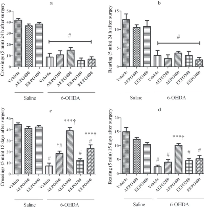
-comotion from all 6-OHDA groups compared to saline microinjections, independent of the treatment received orally (crossing: F9,59 = 45.28, p<0.0001; rearing: F9,59 = 14.15, p<0.0001, one-way ANOVA followed by Tukey post-test, Fig. 2).When the test
Figure 2 - Results from the open field test: Means of crossings (a and c) and rearing (b and d) at 24 h and 15 days after surgery, respectively. # indicates significant decrease from saline groups (p < 0.001), * indicates significant increase from 6-OHDA/vehicle with p < 0.05, *** indicates significant increase from 6-OHDA/vehicle with p < 0.0001 and (†) indicates significant difference
was carried out at 15 days after surgery (Fig. 2), a
significant reduction of crossing for animals treated
with 6-OHDA was still observed when compared with groups which received intracerebral saline, except for 6-OHDA/AEPO 400 mg/kg. Treatments with both extracts at higher doses (6-OHDA/ AEPO and 6-OHDA/EEPO 400 mg/kg) resulted
in significantly higher values when compared to
those obtained for the group 6-OHDA/vehicle and 6-OHDA/AEPO or 6-OHDA/EEPO 200 mg/kg. Moreover, statistical analysis revealed an increase in crossings for the 6-OHDA/AEPO 400 mg/kg compared with all other 6-OHDA groups (F9,59 = 44.52, p<0.0001, one-way ANOVA followed by Tukey post-test) reaching values similar to control groups.
The mean number of rearing was also reduced in the groups that received 6-OHDA/vehicle, 6-OHDA/AEPO (200 mg/kg), 6-OHDA/EEPO (200 mg/kg) and 6-OHDA/EEPO (400 mg/kg) when compared with the groups that received intracerebral saline in 15 days. The 6-OHDA/AEPO
at 400 mg/kg treatment significantly increases these
values compared with all other 6-OHDA groups (F9,59 = 17.00, p<0.0001). Regarding temporal analysis, both extracts of the P. oleracea (400 mg/ kg) promoted an increase in the number of crossings (AEPO: p = 0.001; EEPO: p = 0.02) and rearings (AEPO: p = 0.002; EEPO: p = 0.05, paired t test) from 24 hours to 15 days in the lesioned animals.
The extent of the denervation in the nigrostria-tal system induced by unilateral 6-OHDA microin-jection was also determined by TH immunohisto-chemistry (Figs. 3 and 4). The 6-OHDA promoted
significant decrease in the density of TH-positive
neurons in the SNc, relative to the saline-microin-jected counterparts (33% ± 2.31 remaining cells). The AEPO exerted protective action (200 mg/ kg: 60.67% ± 3.57), this effect was dose depen-dent (400 mg/kg: 78.17% ± 4.22). The administra-tion of EEPO at the lower dose did not modify the 6-OHDA action (37.67% ± 1.38), but the 400 mg/
kg dose promoted an increase the percentage of residual neurons (68.5% ± 6.06) similar to those observed in rats given the higher dosage of AEPO (Figs. 3 and 4, F4,29 = 25.71, p = 0.041).
disCussion
In this study, spontaneous motor activity in the open
field maze was performed 24 hours and 15 days
after the administration of 6-OHDA, in order to access both initial functional symptoms and more stabilized lesion signs (Meredith et al. 2008). The
6-OHDA injections induced a significant decrease
in locomotion at 24 hours, corroborating the find
-ings of other authors (Dauer and Przedborski 2003, Truong et al. 2006). After 15 days, these effects were attenuated in a dose dependent manner in the 6-OHDA-treated rats given AEPO and EEPO. Re-sults of immunohistochemistry are compatible with these observations, since the tyrosine-hydroxylase
Figure 3 - Percentages of tyrosine-hydroxylase positive cell density (TH+) in the substantia nigra pars compacta (SNc), 28 days after surgery, from saline microinjected rats. Different number of asterisks indicates statistical difference between groups (p< 0.05) while equal characters indicate similar results. AEPO: aqueous extract of
is the enzyme that catalyzes the rate-limiting step in the synthesis of catecholamines and a marker for dopaminergic reminiscent neurons (Daubner et al. 2011). Here, the administration of both extracts produced a nearly 70% increase in preservation of dopaminergic cells. It is in agreement with the re-ports that spontaneous locomotion, measured in an
open field, is impaired in a manner closely related
to striatal dopamine depletion after 6-OHDA in ro-dents (Kirik et al. 1998, Willard et al. 2015).
Our data indicate that P. oleracea contains bioactive compounds that are able to exert a pro-tective effect against degeneration of dopaminer-gic neurons. It has already been demonstrated in
the rotenone model of PD that aqueous extracts of
P. oleracea protects the rat brain from oxidative stress through a decrease in the levels of glutathi-one (Al-Quraishy et al. 2012). Also, the biochemi-cal changes, including an increase in the intercel-lular content of calcium and dopamine metabolites and decrease in the complex I activity, as well as apoptosis induced by rotenone in the rat striatum, were effectively counteracted by administration of
P. oleracea (Abdel Moneim et al. 2012, Abdel Mo-neim 2013).
As a catecholaminergic neurotoxin, 6-OHDA, when applied into rat striatum, induces a slow de-generative process in dopaminergic neurons. One Figure 4 - Photomicrographs of tyrosine-hydroxylase positive neurons (TH+) in the substantia nigra pars compacta (SNc) of rats, 28 days after surgery. a: saline microinjected and treated with vehicle; b: 6-hydroxydopamine microinjected and treated with vehicle; c and d: 6-hydroxydopamine microinjected and treated with aqueous extract (AEPO) or ethanolic extract (EEPO) of
of the underling mechanisms of cell death is oxi-dative stress, which has gained special attention because of its association with the reported H2O2 and free radical formation, decreased superoxide dismutase (SOD) activity, increased monoaminoxi-dase (MAO) activity and mitochondrial complex toxicity (Martínez-Cerdeño et al. 2010, Decressac et al. 2012).
Both aqueous (Hongxing et al. 2007, Al-Quraishy et al. 2012) and ethanolic (Wanyin et al. 2012) P. oleracea extracts have demonstrated protective effects against oxidative stress-related pathologies. For example, the oral administration
of EAPO significantly increased the activities of
antioxidant enzymes, such as SOD and decreased the production of MAO (Dkhil et al. 2011). In agreement with previous reports, our results from
the DPPH test confirm the antioxidant activity of
both types of P. oleracea extracts. The possible neuroprotection mechanism could be related to this antioxidant activity (Molyneux 2004, Chen et al. 2009), since both extracts in concentrations above 300µg/kg dose provided 50% of the antioxidant effects of ascorbic acid. It is interesting to note that
P. oleracea ethanolic crude extract did not present toxic effect on normal mice even at 500 mg/kg (Aljeboori et al. 2014).
The antioxidant action of P. oleracea extracts can be attributed to its constituents. It has been reported the presence of gallotannins, omega-3 fatty
acids, ascorbic acid, α-tocopherols, kaempferol,
quercetin, and apigenin (Yan et al. 2012). The analysis of the extracts showed the presence of
alkaloids, flavonoids, tannins, saponins, terpenoids
and polysaccharides. The same compounds were found in other studies concerning P. oleracea
chemical composition (Lu et al. 2009, Dong et al. 2010, Zhu et al. 2010, Uddin et al. 2012, Tian et al. 2014, Liang et al. 2014). The consumption of
foods rich in flavonoids is associated with several beneficial effects for human health and can reduce
the risk of developing PD in humans (Gao et al.
2012). Also, the alkaloids compounds betacyanins, soluble pigments found in P. oleracea, had neuroprotective effects in senescent mice, reducing the levels reactive oxygen species damage and lipid peroxidation (Wang and Yang, 2010).
In addition, the tannins demonstrated a strong antioxidant activity (Figueroa-Espinoza et al. 2015), polysaccharides of P. oleracea exhibited free radical scavenging activities and protective effect against oxidative damage (Chen et al. 2009), and terpenoids also exerted a neuroprotective role against neuronal hypoxia (Zhu et al. 2007). Also, previous work showed that AEPO can modulate the activity of acetylcholinesterase (Yang et al. 2012) and increase the monoamine contends due
its melatonin, omega-3 fatty acids, flavonoids and
phenolic compounds (Abdel Moneim et al. 2012).
The group of flavonoids possesses important
biological activities and kaempferol and apigenin have been found in ethanolic extracts of leaves and stems (Xu et al. 2006, Zhu et al. 2010). It may explain the higher antioxidant activity of EEPO. On the other hand, AEPO was more effective against 6-OHDA deleterious effects and it suggests that neuroprotection could have been mediated by a mechanism other than oxidative stress.
The TLC analysis revealed the occurrence of a fluorescent band at the same position of levodopa in both extracts. Catecholamines, such as dopamine, are synthesized from levodopa precursor by tyrosine-hydroxylase and have important roles as neurotransmitters in the central nervous system and in PD, once the dopamine depletion is the responsible for the hallmark symptoms of disease (Missale et al. 1998). The presence of cathecolamines such as dopamine has been reported in P. oleracea (Gandía-Herrero et al. 2009, Weng et al. 2005) as well as the presence of levodopa in P. oleracea and the ability of converting tyrosine to levodopa (Harris et al. 2012).
dyskinesia (Del Bel et al. 2014, Padovan-Neto et al. 2009), futures studies are needed to explore the potential of P. olearecea in dopamine replacement therapy. From our results, in addition to the antioxi-dant effects, the improvement in behavioral perfor-mance promoted by P. oleracea could be explained, at least in part, by some dopaminergic-like action.
In conclusion, the present results indicated that aqueous and ethanolic extracts of P. oleracea were
able to reverse the behavioral motor deficits and
neuronal loss induced by 6-OHDA and that these effects may be related to their antioxidant activ-ity and bioactive compounds. The P. oleracea is, therefore, an important target of study with thera-peutic potential for the treatment of neurodegenera-tive diseases.
aCknoWledgMents
The authors thank to the Fundação de Apoio à Pesquisa e à Inovação Tecnológica do Estado de Sergipe (FAPITEC-SE), Grant number 04/2011.
reFerenCes
ABDEL MONEIM AE. 2013. The neuroprotective effects of purslane (Portulaca oleracea) on rotenone-induced biochemical changes and apoptosis in brain of rat. CNS Neurol Disord Drug Targets 12: 830-841.
ABDEL MONEIM AE, NASR IA, DKHIL MA AND AL-QURAISHY S. 2012. Neuronal activities of Portulaca oleracea in adult rats. J Med Plants Res 6: 3162-3168. AL-QURAISHY S, DKHIL MA AND MONEIM AE. 2012.
Protective effects of Portulaca oleracea against rotenone mediated depletion of glutathione in the striatum of rats as an animal model of Parkinson’s disease. Pestic Biochem Physiol 103: 108-114.
ALJEBOORI KH, AL-RUBAI OH AND NAHI YY. 2014. Study of pathological effects of crude extract of Portulaca oleracea L. in the albino mice organs. Int J Technol Res Applications 2: 29-32.
CHAN K, ISLAM MW, KAMIL M, RADHAKRISHNAN R, ZAKARIA MN, HABIBULLAH M AND ATTAS A. 2000. The analgesic and anti-inflammatory effects of Portulaca oleracea L. subsp. Sativa (Haw.) Celak. J Ethnopharmacol 73: 445-451.
CHEN CJ, WANG WY, WANG XL, DONG LW, YUE YT, XIN HL, LING CQ AND LI M. 2009. Anti-hypoxic
activity of the ethanol extract from Portulaca oleracea in mice. J Ethnopharmacol 124: 246-250.
CHEN J, SHI YP AND LIU JY. 2003. Determination of noradrenaline and dopamine in Chinese herbal extracts from Portulaca oleracea L. by high-performance liquid chromatography. J Chromatogr A 1003: 127-132.
DAUBNER SC, LE T AND WANG S. 2011. Tyrosine hydrox-ylase and regulation of dopamine synthesis. Arch Biochem Biophys 508: 1-12.
DAUER W AND PRZEDBORSKI S. 2003. Parkinson’s disease: Mechanisms and models. Neuron 39: 889-909. DECRESSAC M, MATTSSON B AND BJöRKLUND A.
2012. Comparison of the behavioural and histological characteristics of the 6-OHDA and α-synuclein rat models of Parkinson’s disease. Exp Neurol 235: 306-315.
DEL BEL E, PADOVAN-NETO FE, SZAWKA RE, DA SILVA CA, RAISMAN-VOZARI R, ANSELMO-FRANCI J, ROMANO-DUTRA AC AND GUIMARAES FS. 2014. Counteraction by nitric oxide synthase inhibitor of neurochemical alterations of dopaminergic system in 6-OHDA-lesioned rats under L-DOPA treatment. Neurotox Res 25: 33-44.
DEXTER DT AND JENNER P. 2013. Parkinson disease: from pathology to molecular disease mechanisms. Free Radic Biol Med 62: 132-144.
DIGHE V, DHOTRE O, PAREKH G AND GURSALE A. 2008. Quantification of dopamine in Portulaca oleracea
Linn. by high-performance thin-layer chromatography. J Plan Chromatogr 21: 183-186.
DJOUOSSI MG, TAMOKOU JD, NGNOKAM D, KUIATE JR, TAPONDJOU LA, HARAKAT D AND VOUTQUENNE-NAZABADIOKO L. 2015. Antimicrobial and antioxidant flavonoids from the leaves of Oncoba spinosa Forssk. (Salicaceae). BMC Complement Altern Med 28: 15-134. DKHIL AM, MONEIM AE, AL-QURAISHY S AND SALEH
RA. 2011. Antioxidant effect of puslane (Portulaca oleracea) and its mechanism of action. J Med Plant Res 5: 1589-1563.
DONG CX, HAYASHI K, LEE JB AND HAYASHI T. 2010. Characterization of structures and antiviral effects of polysaccharides from Portulaca oleracea L. Chem Pharm Bull 58: 507-510.
DOUHOU A, DEBEIR T, MURER MG, DO L, DUFOUR N, BLANCHARD V, MOUSSAOUI S, BOHME GA, AGID Y AND RAISMAN-VOZARI R. 2002. Effect of chronic treatment with riluzole on the nigrostriatal dopaminergic system in weaver mutant mice. Exp Neurol 176: 247-253. F I G U E R O A - E S P I N O Z A M C , Z A F I M A H O VA A ,
ALVARADO PG, DUBREUCQA E AND PONCET- LEGRAND C. 2015. Grape seed and apple tannins: emul-sifying and antioxidant properties. Food Chem 178: 38-44. GANDíA-HERRERO F, JIMéNEZ-ATIéNZAR M,
GARCíA-CARMONA F. 2009. Fluorescence detection of tyrosinase activity on dopamine-betaxanthin purified from Portulaca oleracea (common purslane) flowers. J Agric Food Chem 57: 2523-2528.
GAO X, CASSIDY A, SCHWARZSCHILD MA, RIMM EB AND ASCHERIO A. 2012. Habitual intake of dietary flavonoids and risk of Parkinson disease. Neurology 78: 1138-1145.
GOMES MZ, RAISMAN-VOZARI R AND DEL BEL EA. 2008. A nitric oxide synthase inhibitor decreases 6-hy-droxydopamine effects on tyrosine hydroxylase and neu-ronal nitric oxide synthase in the rat nigrostriatal pathway. Brain Res 1203: 160-169.
HARBORNE JB. 1998. Phytochemical Methods: a guide to modern techniques of plant analysis. 3nd
ed., London, UK: Published by Chapman and Hall, 278 p.
HARRIS NN, JAVELLANA J, DAVIES KM, LEWIS DH, JAMESON PE, DEROLES SC, CALCOTT KE, GOULD KS AND SCHWINN KE. 2012. Betalain production is possible in anthocyanin-producing plant species given the presence of DOPA-dioxygenase and L-DOPA. BMC Plant Biol 12: 34.
HONGXING Z, NANCAI Y, GUOFU H, JIANBO S, YANXIA W, HANJU H, QIAN L, WEI M, YANDONG Y AND HAO H. 2007. Neuroprotective effects of purslane herb aquenous extracts against D-galactose induced neurotoxicity. Chem Biol Interact 170: 145-152.
HOZAYEN W, BASTAWY M AND ELSHAFEEY H. 2011. Effects of Aqueous Purslane (Portulaca Oleracea) Extract and Fish Oil on Gentamicin Nephrotoxicity in Albino Rats. Nat Sci 9: 47-62.
KIRIK D, ROSENBLAD C AND BJORKLUND A. 1998. Characterization of behavioral and neurodegenerative changes following partial lesions of the nigrostriatal dopa-mine system induced by intrastriatal 6-hydroxydopadopa-mine in the rat. Exp Neurol 152: 259-277.
LEE S, KIM JS, LEE YJ, KANG DG AND LEE HS. 2012. Anti-TNF-𝛼 activity of Portulaca oleracea in vascular endothelial cells. Int J Mol Sci 13: 5628-5644.
LIANG X, TIAN J, LI L, GAO J, ZHANG Q, GAO P AND SONG S. 2014. Rapid determination of eight bioactive al-kaloids in Portulaca oleracea L. by the optimal microwave extraction combined with positive-negative conversion multiple reaction monitor (+/−MRM) technology. Talanta 120: 167-172.
LU JR, HE TR AND PUTHETI R. 2009. Compounds of Purslane extracts and effects of antikinetic fatigue. J Med Plants Res 3: 506-510.
MARTíNEZ-CERDEñO V, NOCTOR SC, ESPINOSA A, ARIZA J, PARKER P, ORASJI S, DAADI MM, BANKIE-WICZ K, ALVAREZ-BUYLLA A AND KRIEGSTEIN AR. 2010. Embryonic MGE precursor cells grafted into adult rat striatum integrate and ameliorate motor symp-toms in 6-OHDA-lesioned rats. Cell Stem Cell 6: 238-250.
MEREDITH GE, SONSALLA P AND CHESSELET MF. 2008. Animal Models of Parkinson’s Disease Progression. Acta Neuropathol 115: 385-398.
MISSALE C, NASH S, ROBINSON S, JABER M AND CARON MG. 1998. Dopamine receptors: from structure to function. Physiol Rev 78: 189-225.
MOLYNEUX P. 2004. The use of the stable free radical diphenylpicrylhydrazyl (DPPH) for estimating antioxidant activity. J Sci Tech 26: 211-219.
PADOVAN-NETO FE, ECHEVERRY MB, TUMAS V AND DEL BEL EA. 2009 Nitric oxide synthase inhibition attenuates L-DOPA-induced dyskinesias in a rodent model of Parkinson’s disease. Neuroscience 159: 927-935. PAXINOS G AND WATSON C. 1998. The Rat Brain in
Ste-reotaxic Coordinates. 4th
ed., New York, USA: Academic Press, 120 p.
RADHAKRISHNAN R, ZAKARIA MN, ISLAM MW, CHEN HB, KAMIL M, CHAN K AND AL-ATTAS A. 2001. Neuropharmacological actions of Portulaca oleraceae L. v. sativa (Hawk). J Ethnopharmacol 76: 171-176.
SOUZA CM ET AL. 2007. Total phenols and antioxidant activity of five medicinal plants. Quim Nova 30: 351-355. TIAN JL, LIANG X, GAO PY, LI DQ, SUN Q, LI LZ AND SONG SJ. 2014. Two new alkaloids from Portulaca oleracea and their cytotoxic activities. J Asian Nat Prod Res 16: 259-264.
TRUONG L, ALLBUTT H, KASSIOU M AND HENDERSON JM. 2006. Developing a preclinical model of Parkinson’s disease: a study of behaviour in rats with graded 6-OHDA lesions. Behav Brain Res 169: 1-9.
UDDIN MK, JURAIMI AS, ALI ME AND ISMAIL MR. 2012. Evaluation of Antioxidant Properties and Mineral Composition of Purslane (Portulaca oleracea L.) at Different Growth Stages. Int J Mol Sci 13: 10257-10267. UDDIN MK, JURAIMI AS, HOSSAIN MS, NAHAR MA,
ALI ME AND RAHMAN MM. 2014. Purslaneweed (Portulaca oleracea): a prospective plant source of nutrition, omega-3 fatty acid, and antioxidant attributes. Sci World J. Available at: http://www.hindawi.com/ journals/tswj/2014/951019/. Accessed on April 15, 2015. Article ID 951019.
WANG CQ AND YANG GQ. 2010. Betacyanins from
Portulaca oleracea L. ameliorate cognition deficits and attenuate oxidative damage induced by D-galactose in the brains of senescent mice. Phytomedicine 17: 527-553. WANYIN W, LIWEI D, LIN J, HAILIANG X, CHANGQUAN
L AND MIN L. 2012. Ethanol extract of Portulaca oleracea
L. protects against hypoxia-induced neuro damage through modulating endogenous erythropoietin expression. J Nutr Biochem 23: 385-391.
capillary chromatography with amperometric detection. Se Pu 23: 18-21.
WHIMBEY AE AND DENENBERG VH. 1967. Two independent behavioral dimensions in open-field performance. J Comp Physiol Psychol 63: 500-504. WICHMANN T AND DOSTROVSKY JO. 2011. Pathological
basal ganglia activity in movement disorders. Neuroscience 198: 232-244.
WILLARD AM, BOUCHARD RS AND GITTIS AH. 2015. Differential degradation of motor deficits during gradual dopamine depletion with 6-hydroxydopamine in mice. Neuroscience 301: 254-267.
XU X, YU L AND CHEN G. 2006. Determination of flavonoids in Portulaca oleracea L. by capillary electrophoresis with electrochemical detection. J Pharm Biomed Anal 41: 493-499.
YAN J, SUN LR, ZHOU ZY, CHEN YC, ZHANG WM, DAI HF AND TAN JW. 2012. Homoisoflavonoids from
the-medicinal plant Portulaca oleracea. Phytochemistry 80: 37-41.
YANG Z, ZHANG D, REN J, YANG M AND SHUO L. 2012. Acetylcholinesterase inhibitory activity of the total alkaloid from traditional Chinese herbal medicine for treating Alzheimer’s disease. Med Chem Res 21: 734-738. YUE T, XIAOSA W, RUIRUI Q, WENCAI S, HAILIANG
X AND MIN L. 2015. The Effects of Portulaca oleracea
on Hypoxia-Induced Pulmonary Edema in Mice. High Alt Med Biol 16: 43-51.
ZHU H, WANG Y, LIU Y, XIA Y AND TANG T. 2010. Analysis of Flavonoids in Portulaca oleracea L. by UV-Vis Spectrophotometry with Comparative Study on Different Extraction Technologies. Food Anal Meth 3: 90-97.