Tese de Mestrado
Mestrado em Bioquímica Aplicada
Especialização em Biomedicina
Trabalho efetuado sob a orientação da
Doutora Susana Alexandra Rodrigues Chaves
Outubro 2013
Ana Rita Correia Pacheco
Nome: Ana Rita Correia Pacheco
Endereço eletrónico: aritacpacheco@hotmail.com Telefone: +351 914 341 505
Nº Cartão Cidadão: 13815474 Título da Tese de Mestrado:
The role of Cks proteins in apoptosis
Orientadora:
Doutora Susana Chaves
Instituição de acolhimento:
Centro de Biologia Molecular Ambiental (CBMA)
Ano de Conclusão: 2013 Designação do Mestrado:
Mestrado em Bioquímica Aplicada – Especialização em Biomedicina
1. É AUTORIZADA A REPRODUÇÃO INTEGRAL DESTA TESE, APENAS PARA EFEITOS DE INVESTIGAÇÃO, MEDIANTE DECLARAÇÃO ESCRITA DO INTERESSADO, QUE A TAL SE COMPROMETE.
Universidade do Minho, 31 de Outubro de 2013
____________________________________________ Ana Rita Correia Pacheco
iii
Agradecimentos
É com a escrita dos agradecimentos que me consciencializo que a escrita e todo o trabalho desenvolvido ao longo deste ano não me competem apenas a mim. E assim, quero agradecer:
Em primeiro lugar, á minha orientadora de tese, à Doutora Susana Chaves pela sua admirável orientação marcada pela sapiência, paciência, disponibilidade e palavra certa quando os resultados não eram os esperados. Obrigada Susana pela partilha de conhecimento e por toda a ajuda, aprendi muito contigo.
À professora Doutora Manuela Côrte-Real pela simpatia, disponibilidade, sabedoria e carinho.
Aos meus colegas de laboratório, que conseguiram transformar um local de trabalho num local mais divertido. Ao António, Rita, Andreia e Dário, por todo o apoio, simpatia e estarem sempre lá quando precisei, mesmo com as dúvidas mais banais. Aos restantes colegas, Natália, Bete, Vera, Tânia, Marco, Selma, Lisandra, Sara, Helena, Flávio, obrigada por todas as gargalhadas partilhadas e por conseguirem fazer de um dia menos bom, um dia melhor. Obrigada Pedrinho pelas imensas ofertas de alíquotas de células competentes ☺.
À D. Isabel e ao Sr. José por me colocarem sempre com um sorriso na cara por cada entrada na sala de lavagens.
Aos meus colegas e amigos de mestrado, em especial, à Luísa, Cris, Carina, Rosana, Marina, Natália, Bete, pelo acolhimento, simpatia, entreajuda, e pelos cafezinhos nos momentos mais difíceis, por tudo, por fazerem desta etapa, uma etapa menos penosa.
Aos meus amigos, aos de sempre e aos de agora, em especial, ao Joel, Sabrina, Ritinha, Carla, Rui, Maria Rita, Leandro, Flávia, Diana, Suzy, Ricardina, Andreia, pelas palavras amigas em dias mais cinzentos, alegria, apoio, companheirismo, incentivo, carinho e amizade constante. Por último, mas em primeiro, à minha família, em especial aos meus pais, Patrícia e irmão, que em todos os momentos, onde quer que eu esteja, me apoiam incondicionalmente, por tudo o que representam na minha vida, pelo amor, confiança e por me terem ensinado a ser quem sou.
Ao Afonso, pelo amor sincero e sorriso genuíno, por me ensinar a importância das pequenas coisas da vida.
iv
The role of Cks proteins in apoptosis Abstract
Cyclin-dependent kinase subunits (Cks) proteins are evolutionary conserved small proteins that interact with Cyclin-dependent kinase (Cdk) and are frequently overexpressed in several types of cancer, which is correlated with poor prognosis and aggressive behavior. Lower eukaryotes as budding yeast express one Cks protein (Cks1), whereas mammalian cells express two (Cks1 and Cks2).
Cks proteins were most frequently associated with a direct role in cell cycle regulation, but are also involved in efficient transcription of multiple genes. However, despite their similarity, Cks proteins can have specific functions. For instance, mammalian Cks1, but not Cks2, is involved in ubiquitination of p27, a cyclin-dependent kinase inhibitor. There is some indication that modulation of Cks protein levels can impact apoptosis in some mammalian cell lines, but there is a lack of focused research on this subject and the mechanism involved remains uncharacterized. A role of Cks proteins in apoptosis has therefore yet to be explored as a target in cancer therapy.
The yeast model has already demonstrated its potential for application in the apoptosis field, as evidenced by the increasing number of studies using yeast as a model for neurotoxicity and cancer. Most functions attributed to Cks proteins are conserved in yeast, and indeed mammalian Cks proteins can functionally substitute for yeast Cks1p. Our aim was therefore to determine whether modulation of Cks1p levels alters apoptotic signaling in the yeast Saccharomyces cerevisiae, which would be indicative of a conserved function in apoptosis. We found that deletion of yeast CKS1 led to significantly increased sensitivity to short-term exposure to the chemotherapeutic agent cisplatin, but not to the other DNA damaging agents - methyl methanesulfonate and 5-fluorouracil, nor to the general apoptosis inducers - acetic acid and hydrogen peroxide. This is a strong indication of a specific regulated role for Cks1p in DNA damage-induced cell death, independent from its role in cell cycle. Further studies to unravel its function, as well as the pathways involved, could therefore provide novel targets to exploit in the treatment of cancers where Cks proteins are up-regulated.
v
O papel das proteínas Cks na apoptose Resumo
Subunidades Kinase dependente de ciclina (Cks) são pequenas proteínas conservadas evolucionariamente, que interagem com kinases dependentes de ciclina (Cdks) e encontram-se sobreexpressas em diversos tipos de cancro, relacionado com mau prognóstico e comportamento agressivo. Eucariotas inferiores como a levedura expressam uma proteína Cks (Cks1), enquanto as células de mamífero expressam duas proteínas (Cks1 e Cks2).
Proteínas Cks estão mais associadas a um papel direto na regulação do ciclo celular, mas também estão envolvidas na transcrição eficiente de vários genes. Contudo, apesar das suas semelhanças, as proteínas Cks podem ter funções específicas. Por exemplo, a Cks1 de mamífero está envolvida na ubiquitinação do p27, um inibidor da kinase dependente de ciclina. Há algumas indicações que a modulação dos níveis de proteínas Cks podem ter impacto na apoptose em algumas linhas celulares de mamífero, mas há pouca investigação neste assunto e o mecanismo continua por caracterizar. Um papel das proteínas Cks na apoptose tem ainda que ser explorado para ser usado como alvo na terapia de cancro.
O modelo da levedura tem mostrado potencial para aplicação na área da apoptose, como é evidenciado pelo aumento do número de estudos a usarem a levedura como modelo para neurotoxicidade e cancro. Muitas funções atribuídas a proteínas Cks são conservadas em levedura, e de facto proteínas Cks de mamífero podem substituir funcionalmente a proteína Cks1p de levedura. O nosso objetivo foi determinar se a modulação dos níveis de Cks1p altera a sinalização apoptótica na levedura Saccharomyces cerevisiae, o que seria indicativo da conservação da função na apoptose. Determinamos que a deleção de CKS1 em levedura leva ao aumento significativo da sensibilidade a uma exposição curta à cisplatina, mas não a outros agentes danificadores de ADN, metanosulfato metil e 5-fluorouracil, nem a indutores gerais de apoptose, ácido acético e peróxido de hidrogénio. Isto é uma forte indicação para um papel de regulação específico de Cks1p em morte celular induzida por danos de ADN, independente do seu papel no ciclo celular. Estudos futuros para desvendar esta função, assim como as vias envolvidas, podem fornecer novos alvos para explorar no tratamento de cancros onde as proteínas Cks estão sobreexpressas.
vi Index Agradecimentos ... iii Abstract ... iv Resumo ... v Index ... vi Abbreviations ... viii INTRODUCTION ... 1 Cks proteins ... 2 Relevance in cancer ... 2
Structure and functions ... 2
Potential link to apoptosis ... 5
The importance of apoptosis in cancer therapy ... 6
Apoptosis regulation ... 7
Extrinsic apoptosis ... 7
Intrinsic apoptosis ... 9
Saccharomyces cerevisiae as model system to study apoptosis regulation ... 11
OBJECTIVES AND RESEARCH PLAN ... 13
MATERIALS AND METHODS ... 15
Reagents ... 16
Strains and plasmids ... 16
Fluorescence Microscopy ... 17
Growth conditions and treatments ... 17
vii
Colony-forming units (c.f.u.) ... 18
Spot assays ... 18
Chronic exposure to DNA damaging agents ... 18
Cell cycle analysis ... 18
Detection of apoptotic markers ... 19
Plasma membrane integrity ... 19
Production of ROS ... 19
RESULTS ... 20
Deletion of yeast CKS1 does not alter mitochondrial morphology ... 21
Deletion of yeast CKS1 does not alter sensitivity to the yeast apoptosis inducers acetic acid or hydrogen peroxide ... 22
Deletion of yeast CKS1 results in differential sensitivity to DNA damaging agents ... 24
Expression of yeast CKS1 from a multicopy plasmid does not alter sensitivity to cisplatin ... 32
Expression of yeast CKS1 from a multicopy plasmid does not alter sensitivity to hydroxyurea ... 34
DISCUSSION AND FUTURE PERSPECTIVES ... 38
viii
Abbreviations 5-FU – 5-Fluoururacil AA – Acetic Acid
AIF – Apoptosis Inducing Factor ANT – Adenine Nucleotide Translocator APAF1 – Apoptotic Proteasome Activating
Factor 1
ATP – Adenosine Triphosphate BER – Base Excision Repair
Bid – BH3-interacting domain death agonist c.f.u. – Colony-forming units
cDDP – cis-diamminedichloroplatinum (II) Cdk – Cyclin-dependent kinase
Cks – Cyclin-dependent kinase subunit CypD – Cycliphilin D
DHE – Dihydroethidium
DIC – Differential Interference Contrast DISC – Death Inducing Signaling Complex DMSO – Dimethyl Sulfoxide
DNA – Deoxyribonucleic acid FASL – FAZ ligand
FdUMP – 5-Fluorodeoxyuridine
monophosphate
GFP – Green Fluorescent Protein H2O2 – Hydrogen peroxide
HTRA2 – High Temperature Requirement
Protein A2
HU – Hydroxyurea
IAP – Inhibitor of Apoptosis Protein MMR – Mismatch Repair
MMS – Methyl methanesulphonate
MOMP – Mitochondrial Outer Membrane
Permeabilization
MPF – Cdk1/Cyclin B complex NER – Nucleotide Excision Repair PCD – Programmed Cell Death pI – Isoelectric point
PI – Propidium Iodide
PTP – Permeability Transition Pore RNA – Ribonucleic acid
ROS – Reactive Oxygen Species siRNA – small interfering RNA
SMAC/DIABLO – Second
Mitochondria-derived Activator of Caspases/Direct IAP-binding protein with low pI
tBid – Truncated Bid
TNF – Tumor Necrosis Factor TS – Thymidylate Synthase UV – Ultra-violet
VDAC – Voltage-dependent Anion Channel Δψm – Mitochondrial transmembrane
2
Cks proteins: Relevance in cancer
Cyclin-dependent kinase subunit (Cks) proteins are evolutionary conserved small (9-18 kDa) proteins [1-3]; lower eukaryotes express only one Cks1 protein, whereas mammals, and possibly other vertebrates, express two orthologs, Cks1 and Cks2 [2-6]. Cks proteins have been the subject of increased attention in recent years, since they are frequently overexpressed in several cancers, which is correlated with poor prognosis. Cks1 overexpression has been strongly associated with various clinical and pathological features that are commonly used to determine aggressive tumor behavior [7-9]. High expression of Cks1 may be involved in the pathogenesis of non-small cell lung carcinoma [10], oral squamous cell carcinoma [11], colorectal carcinoma [8, 12], gastric carcinoma [13], lung carcinoma [11] and breast cancer [14]. Overexpression of Cks1 correlates with the increased radiotherapy resistance of esophageal squamous cell carcinoma [15] and tumor stage and positive lymph node metastasis in breast cancer [14]. It has also been found that Cks2 is expressed at significantly higher levels in tumors with metastasis [16] and can be responsible for aggressive behavior of tumours [17]. Cks2 levels were also increased in cervical cancer [18], metastatic androgen-independent prostate cancer [19] and cholangiocarcinoma [17]. In addition, CKS2 has been proposed as a biomarker for the diagnosis and staging of bladder cancer [20] and melanocytic tumour progression [21]. Expression of Cks1 and Cks2 was elevated in prostate tumors and forced expression of both Cks1 and Cks2 in benign prostate tumor epithelial cells accelerated cell growth [22]. Therefore, Cks proteins may be considered as potential novel prognostic marker and target for the future development of specific therapeutic interventions. Targeting these proteins may be a promising therapeutic option for the treatment of cancers where they are up-regulated.
Cks proteins: Structure and functions
Cks proteins were originally identified through their ability to genetically suppress defective alleles of the cyclin-dependent kinase (Cdk) of fission and budding yeast [1-3], which pointed to a prominent role in the cell cycle.
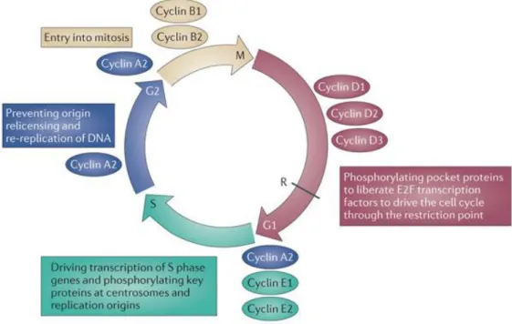
The cell-cycle machinery is distinguished by a series of coordinated events essential to ensure faithful DNA replication and segregation of replicated chromosomes into two separate cells. It consists of specific phases: the G1 phase, during which the cell prepares for DNA synthesis; the S phase, during which active replication of the chromosomes occurs; G2 gap
3
period, prior to chromosome segregation and cytokinesis in M phase (mitosis) (Figure 1) [23-24]. Cells in G1 can, before committing to DNA replication, enter a quiescent state, called G0 [25].
Progression through each cell-cycle phase and transition from one phase to the next are monitored by specific checkpoints, which maintain the correct order of events [25-26]. If these sensors detect aberrant or incomplete cell-cycle events, checkpoint pathways relay a signal that can lead to cell cycle arrest until the problem is resolved [27-28]. These checkpoints are governed by a tight relationship of specific Cdks [29], serine/threonine kinases that must bind to cyclin proteins to become active [30] and regulate the cell cycle division [1]. Cdks are required for the G1 to S phase cell cycle transition, initiation of DNA replication, the G2 to M phase cell cycle transition, and initiation of multiple mitotic events [31]. Cks proteins bind a subset of Cdks with high affinity at a position remote from the ATP and cyclin binding sites, functioning as activating partners. However, unlike cyclins, they are not required for the general activation of the Cdk activity [4], but seem to modulate substrate choice or the extent of phosphorylation [1].
Figure 1. Cell Cycle regulation in mammals. Cyclins D are synthesized in response to mitogenic signals and they direct bind CDK4 (and/or CDK6) to phosphorylate pocket proteins. CDK2–cyclin E functions at the G1/S transition to trigger chromosomal DNA replication and to initiate centrosome duplication. The longer-lived CDK2–cyclin A complex collaborates with CDK2–cyclin E to regulate DNA replication both positively and negatively thus that DNA is only replicated once. The final wave of CDK1–cyclin B activity reorganizes the cell for mitosis (Moore (2013) [26]).
4
Cks proteins are structurally and functionally conserved in eukaryotes, and share over 80% sequence identity [4, 22, 32-35]. The Cks structure consists of a four-stranded β-sheet capped at one end by two or three short α-helices and a highly conserved sequence, the so-called “hinge loop” that links the exchanged β-strand with the rest of the protein [5]. The Cdk-binding and phosphate-Cdk-binding surfaces are also conserved in Cks proteins. In contrast, the major structural differences are located on the two α helices and also at the carboxyl terminus, which generate a largely contiguous surface. It is likely that these regions perform Cks-specific functions [1, 3].
It has been shown that the yeast Cks1p and Cdk1 are involved in chromatin remodeling and play a role in gene expression [36], and that one function of Cdk1/Cks1p and the 19S subunit of the proteasome is to evict nucleosomes from chromatin in the context of gene induction [37]. A role for Cks1p in cyclin B degradation has been suggested in S. cerevisiae. Both genetic and biochemical data indicate that Cks1p is required for efficient proteasomal targeting of ubiquitinated cyclin B [33].
Despite being 87% similar, Cks proteins in mammalian can have specific functions [1, 3]. For example, Cks1 plays an important role in facilitating the ubiquitin-mediated proteolysis of p27, a cyclin-dependent kinase inhibitor, through interaction with Skp2 (Figure 2) [38]. Cks1 is part of the substrate-binding surface, and is necessary for efficient ubiquitination of T187-phosphorylated p27kip1 [11, 39]. Tissues from mice lacking Cks1 accumulate p27Kip1 and exhibit
proliferative defects. Accordingly, Cks1-/- mice exhibit a small body size, resembling the size
phenotype of Skp2-/- mice. Overexpression of Cks1, in general, is correlated with decreased p27
levels [40], which are associated with high aggressiveness, poor prognosis and aggressive behavior in a large variety of malignant tumors [13, 39]. However, Cks1 supports hepatocarcinogenesis independently of Skp2, indicating it can play a separate role in carcinogenesis [41]. In contrast, Cks2 seems to counter Cks1 and stabilize p27; absence of Cks2 results in increased cyclin A/Cdk2 activity, shortening of the cell cycle, and DNA damage [34]. Cks2 is also essential for the first metaphase/anaphase transition of meiosis [22, 35].
5
Cks proteins: Potential link to apoptosis
There is some indication that modulation of Cks proteins levels can impact apoptosis in select mammalian cell lines. Knockdown of Cks1 expression inhibited the growth of oral squamous cell cancer cells both in cultured cells and in vivo [11] and increased the activity of caspase 3, promoting apoptosis of breast cancer cells [42]. Inhibition of Cks1 by siRNA also induced accumulation of cells at the G2/M phase and apoptosis in Cks1-overexpressing lung cancer cells, but not in normal lung fibroblasts [6]. Down-regulation of Cks2 expression inhibited cell proliferation, colony formation and tumorigenesis by suppressing cell cycle progression and enhancing the susceptibility of cholangiocarcinoma cells to Bax-mediated mitochondrial caspase-dependent apoptosis [17]. In prostate tumor cells, knockdown of Cks2 expression induced apoptosis in vitro and compromised tumorigenic activity of the cells in vivo [22]. Additionally, knockdown of both Cks1 and Cks2 in malignant prostate tumor cells inhibited cell growth, anchorage-independent growth, and migration activity [22]. However, due to the lack of focused research on this subject, it is still not clear what role Cks proteins play in apoptosis, and especially if overexpression of Cks1 or Cks2 does indeed protect cells from undergoing apoptosis,
Figure 2. Skp2-dependent degradation of p27. After phosporylation of p27Kip1
at T187 by cyclin E/A-Cdk2, p27Kip1
is recognized by Skp2 and Cks1, which stimulates its targeting for ubiquitylation by the SCFSkp2
complex. The ubiquitylated p27Kip1
is then rapidly destroyed by the proteasome, allowing the activity of cyclin E/A-Cdk2 and progression to the S phase (Hershko (2008) [38]).
6
particularly in response to antitumor drugs. A role of Cks proteins in apoptosis has thus yet to be explored as a target in cancer therapy.
The importance of apoptosis in cancer therapy
Apoptosis is an important process in normal organ development and cellular regulation, playing a role in a variety of physiological and pathological conditions [43]. Apoptosis is also an important variable in cancer development, prevention and therapy [44]. It is well-established that a large number of anticancer agents induce apoptosis, and that disruption of apoptosis programs can reduce treatment sensitivity [45]. Indeed, abundant therapeutic opportunities have been uncovered by investigations of the fundamental mechanisms of apoptosis regulation and identification of the various cell survival genes that become de-regulated in tumors [46]. Since apoptotic programs can be manipulated to produce massive changes in cell death, apoptosis regulators are potential drug targets. Two observations suggest that such strategies are feasible. Firstly, most anti-apoptotic mutations act relatively upstream in the program, implying that tumor cells retain the machinery and latent potential for apoptosis. Secondly, tumor-specific alterations in apoptotic programs provide opportunities to target cell death in a selective manner. Several new strategies have been developed: for instance, apoptosis can be impaired by dominant oncogenes, and agents that inhibit their anti-apoptotic function can lead to a remarkable increase in cell death. In addition, hyperactivation of cell survival signaling may accompany tumor development, and these pathways are particularly exciting targets for small molecule inhibition. When apoptosis is lost by a recessive mutation, restoring the dysfunctional gene or activity can promote massive cell death. Indeed, strategies using this approach are currently in clinical trials. By enhancing the effects of apoptotic mutations, it is also possible to directly harness the pro-apoptotic forces produced by certain oncogenic mutations to selectively destroy tumor cells [45]. In past years, there has been an extraordinary increase in the understanding of apoptosis, and its contribution to cancer development and cancer therapy. It seems likely that rational strategies to manipulate cell suicide programs will produce new therapies that are less toxic and mutagenic than current treatments [45].
7
Apoptosis regulation
The term apoptosis stems from the Greek language, to describe a process reminiscent of the “dropping off” or “falling off” of petals from flowers, or leaves from trees [47]. Apoptosis is a type of programmed cell death (PCD), categorized as a type of cell death that is not accidental (necrosis), but a genetically controlled sequence of steps that lead to morphological and biochemical changes [48], involving the cytoplasm, nucleus and plasma membrane [49]. There are particular morphological changes in the apoptotic process: rounding of the cell, retraction of pseudopodes, reduction in cellular volume, condensation of chromatin, fragmentation of the nucleus, plasma membrane blebbing and maintenance of an intact plasma membrane until the late stages of death [44, 50-54]. In contrast, during accidental necrosis, cells first swell, and then the plasma membrane collapses and cells are rapidly lysed, resulting in damage to neighboring cells and a strong inflammatory response in the corresponding tissue [55].
The apoptotic process is mediated by two classical pathways: the extrinsic and intrinsic pathways, which are activated by the binding of ligands to death receptors, or by stress triggered by intrinsic factors, such as oncogenes, exposure to irradiation and other environmental stresses, respectively.
I. Extrinsic apoptosis
The term extrinsic apoptosis has been used to designate apoptotic cell death induced by extracellular stress signals that are received and propagated by specific transmembrane receptors. Extrinsic apoptosis can be initiated by the binding of lethal ligands, such as FAS/CD95 ligand (FASL/CD95L), tumor necrosis factor α (TNFα) and TNF ligand superfamily, to various death receptors. Alternatively, an extrinsic pro-apoptotic signal can be dispatched by the netrin receptors, which only exercise lethal functions when the concentration of their specific ligands is lower than a critical threshold level [56-57].
There are several transmembrane proteins that, at least under special circumstances, can transduce lethal signals in response to ligand binding. Most of these proteins have a double function, depending on the cellular context and triggering stimulus, and they can convey either pro-survival or pro-death signals [56]. A signaling pathway leading to extrinsic apoptosis is illustrated by FAS ligation (Figure 3). In the absence of FAS ligand (FASL), FAS subunits spontaneously assemble at the plasma membrane to produce trimers. Ligand binding stabilizes these trimers while at the same time inducing a conformational change that allows the assembly
8
of a dynamic multiprotein complex at the cytosolic tail of the receptor. The resulting supramolecular complex, which has been called death-inducing signaling complex (DISC), constitutes a platform that regulates the activation of caspase -8 (or -10) [49, 56, 58]. In some cell types, such as lymphocytes, so-called type I cells, active caspase -8 directly catalyzes the proteolytic maturation of caspase -3, triggering the executioner phase of caspase-dependent apoptosis in a mitochondrion-independent mode. In other cells, type II cells, including hepatocytes and pancreatic b cells, caspase -8 mediates the proteolytic cleavage of BH3-interacting domain death agonist (Bid), leading to the generation of a mitochondrion-permeabilizing fragment, known as truncated Bid (tBid). Therefore, while type I cells undergo extrinsic apoptosis irrespective of any contribution by mitochondria, type II cells give way to the activation of death receptors while showing signs of mitochondrial outer membrane permeabilization (MOMP), including the dissipation of mitochondrial transmembrane potential (Δψm) and the release of toxic proteins that are normally retained within the mitochondrial intermembrane space [56].
Figure 3. Extrinsic apoptosis. FASL: FAS ligand; FADD: FAS-associated protein with a death domain; cIAPs: cellular Inhibitor of Apoptosis Proteins; DISC: Death Inducing Signaling Complex; MOMP: Mitochondrial Outer Membrane Permeabilization; DAPKI: Death-associated Protein Kinase 1; PP2A: Protein Phosphatase 2A (Galluzzi et al (2012) [56]).
9
Extrinsic apoptosis features one of three major lethal signaling cascades: (i) death receptor signaling and activation of the caspase-8 (or -10) caspase-3 cascade; (ii) death receptor signaling and activation of the caspase-8 tBid Mitochondrial outer membrane permeabilization caspase-9 caspase-3 pathway; or (iii) ligand deprivation-induced dependence receptor signaling followed by (direct or mitochondrial outer membrane permeabilization-dependent) activation of the caspase-9 caspase-3 cascade. Extrinsic apoptosis is thus a caspase-dependent cell death process, and hence can be suppressed, at least theoretically, by pancaspase chemical inhibitors. [56].
II. Intrinsic apoptosis
Apoptosis can also be triggered by an excess of intracellular stress, including DNA damage, oxidative stress, cytosolic Ca2+ accumulation, excitotoxicity, related to glutamate receptor
overstimulation in the nervous system, accumulation of unfolded proteins in the endoplasmic reticulum and many others [56, 59]. Although the processes that trigger intrinsic apoptosis are highly heterogeneous as far as the initiating stimuli are concerned, they are all associated with a mitochondrion-centered control mechanism.
Intrinsic apoptosis is mediated by MOMP and thus is associated with generalized and irreversible Δψm dissipation, release of mitochondrial intermembrane space proteins into the
cytosol (and their possible relocalization to other subcellular compartments), and respiratory chain inhibition [56]. The connection between the extrinsic and intrinsic pathways and amplification of death signal is mediated by Bid, a pro-apoptotic Bcl-2 family member (Figure 4). Bid is cleaved by caspase-8 and when the truncated form is translocated into the mitochondria it acts to induce MOMP and release of pro-apoptotic proteins [60]. MOMP can occur due to the pore-forming activity of pro-apoptotic members of the Bcl-2 protein family such as Bak and Bax [61]. Bcl-2 and Bcl-xL can directly induce changes in conformation of the proteins Bax and Bak,
preventing their activation and oligomerization, and blocking the release of pro-apoptotic mitochondrial factors that lead to apoptosis [62]. Another model suggests a permeability transition pore (PTP) that is formed, which allows the passage of solutes and water into the mitochondria matrix, causing mitochondrial depolarization, uncoupling of oxidative phosphorylation and osmotic swelling. The precise localization and composition of the PTP has not been fully determined, but it appears to be localized at the site of contact between the inner and outer mitochondrial membranes and to contain as main components as the
voltage-10
dependent anion channel (VDAC), the adenine nucleotide translocator (ANT), and cyclophilin D (CypD) [63]. After MOMP, cytosolic cytochrome c participates with apoptotic proteasome activating factor 1 (APAF1), cytoplasmic adaptor protein, and dATP in the formation of the apoptosome, which triggers the caspase-9 caspase-3 proteolytic cascade (Figure 4) [64].
Independently of the precise biochemical and physical mechanisms through which it develops, irreversible mitochondrial outer membrane permeabilization affecting most mitochondria within a single cell has multiple lethal consequences: the dissipation of the Δψm,
with cessation of mitochondrial ATP synthesis and Δψm-dependent transport activities; the release
of pro-apoptotic proteins that circulate freely in the mitochondrial intermembrane space into the cytosol, like cytochrome c, apoptosis inducing factor (AIF), endonuclease G, direct IAP-binding protein with low pI (DIABLO, also known as second mitochondria-derived activator of caspases, SMAC) and high temperature requirement protein A2 (HTRA2); and the inhibition of the Figure 4. Intrinsic apoptosis. MOMP: Mitochondrial Outer Membrane Permeabilization; Δψm:
mitochondrial transmembrane potential; ROS: Reactive Oxygen Species; IMS: Mitochondrial Intermembrane Space; CYTC: cytochrome c; DIABLO: Direct IAP-binding protein with low pI; HTRA2: High Temperature Requirement Protein A2; IAP: Inhibitor of Apoptosis Protein; AIF: Apoptosis Inducing Factor; ENDOG: endonuclease G; IM: Mitochondrial Inner Membrane; OM: Mitochondrial Outer Membrane; PTPC: Permeability Transition Pore Complex (Galluzi et al (2012) [56]).
11
respiratory chain (favored by the loss of cytochrome c), eliciting or aggravating reactive oxygen species (ROS) overproduction and hence activating a feed-forward circuit for the amplification of the apoptotic signal [56, 65-66].
Saccharomyces cerevisiae as model system to study apoptosis regulation
Like mammalian cells, yeast cells can trigger apoptosis showing characteristic markers, such as DNA fragmentation and chromatin condensation, externalization of phosphatidylserine to the outer leaflet of the plasma membrane and cytochrome c release from mitochondria [67]. Accumulating evidence points toward the phylogenetic conservation of the core machinery and the core regulators of cell death between yeast and mammals. This entails the possibility of using yeast as a research tool that can provide new hints to elucidation of cell death pathways [68].
An apoptotic phenotype in yeast was first described in a cdc28 temperature-sensitive mutant (Figure 5) [67]. When incubated above the restrictive temperature, cdc48 mutant cells showed an apoptotic phenotype characterized by phosphatidylserine exposure, DNA damage, chromatin condensation and fragmentation, release of cytochrome c and ROS production [69-70]. Since then, CDC48, an essential gene that encodes an AAA-ATPase localized in the endoplasmic reticulum and necessary for vesicle trafficking/translocation of ubiquitinated proteins from the endoplasmic reticulum to the proteasome for degradation [67], has been confirmed as a regulator of mammalian apoptosis, with anti-apoptotic functions, particularly apparent in neuronal pathology [71]. Various other yeast orthologues of vital apoptotic regulators have been identified, such as the caspase orthologue YCA1 (Figure 5) [72]. Yca1p belongs to the family of metacaspases that are found in fungi, plants and protists [68], but not in organisms containing “classical” caspases, and might represent an alternative form of caspase that has developed from the same ancient ancestor as human caspases [67]. Several studies also identified yeast orthologs of several other members of the mammalian apoptotic machinery, including AIF [73], inhibitor of apoptosis protein (IAP) [74], the apoptotic serine protease HTRA2/Omi [75] and endonuclease G [76].
12
Regulators such as Apaf-1 and most members of the Bcl-2 family proteins seem to be absent in yeast. Until now, only a putative yeast BH3-only protein was identified. Ybh3p translocates to the mitochondria and is capable of mediating the mitochondrial pathway of apoptosis [77]. However, heterologous expression of Bax in yeast leads to apoptotic cell death that can be prevented by heterologous expression of anti-apoptotic Bcl-2 and Bcl-xL, suggesting
the function of Bcl-2 family proteins is potentially conserved in yeast [78].
Figure 5. The apoptotic yeast cell. Red question marks indicate pathways that are known in mammals but not in yeast thus far (Madeo et al (2004) [67]).
OBJECTIVES
AND
RESEARCH
PLAN
14
The major objective of this work is to determine whether Cks proteins have a specific role in apoptosis that could contribute to tumorigenesis and/or resistance of Cks-overexpressing tumors to treatment or prevention of cancer. The yeast apoptotic model has already demonstrated its potential for application in the human system, as evidenced by the increasing number of studies using yeast as a model for neurotoxicity and cancer. Since most functions attributed to Cks proteins are also conserved in yeast, and mammalian Cks proteins can functionally substitute for yeast Cks1p, we aimed to use S. cerevisiae as a model system to determine:
I: Whether deletion of CKS1 alters apoptotic signaling in yeast;
MATERIALS
AND
16
Reagents
Yeast nitrogen base, bactopeptone and tryptone were purchased from Difco Laboratories and yeast extract from Cultimed. The carbon source used was glucose (Fisher). All aminoacids were purchased from Formedium. 5-Fluorouracil (5-FU) (Sigma) was dissolved in dimethyl sulfoxide (DMSO) and keep at 4ºC. Cisplatin (Sigma) was stored in aliquots of up to 2mg in the dark and dissolved in the medium immediately prior to use. Hydroxyurea (Formedium) was dissolved in the medium immediately before use.
Strains and plasmids
All Saccharomyces cerevisiae strains used in this study and respective phenotypes are listed in table 1. Plasmids YEp13 and YEp13-CKS1 [79], and pYX232-mtGFP [80], expressing a GFP tagged with a mitochondrial targeting sequence, have been previously described.
All plasmids were amplified by transforming 100 ng of DNA into Escherichia coli DH5α chemically competent cells by transformation using standard procedures (reviewed in [81]) and selected on Luria Bertani (LB) medium [LB: 1 % (w/v) tryptone, 0,5 % (w/v) yeast extract, 1 % (w/v) NaCl and 2 % (w/v) agar] supplemented with 100 µg/mL ampicillin. One colony was inoculated in LB-Amp media, cultures were grown overnight, and DNA was extracted using the GenElute Plasmid Miniprep kit according to manufacturer’s instructions (Sigma).
BF264-15D (15D) and cks1Δ cells were transformed with plasmid YEp13, YEp13-CKS1 or pYX232-mtGFP using the lithium acetate method [82]. Transformants were selected on Synthetic Complete (SC) medium [SC: 0.17 % (w/v) Yeast nitrogen base without aminoacids and ammonium sulfate, 0.5 % (w/v) ammonium sulfate, 0.14 % (w/v), dropout mixture lacking histidine, leucine, tryptophan and uracil, 0.008 % (w/v) histidine, 0.04 % (w/v) leucine, 0.008 % (w/v) Tryptophan and 0.008 % (w/v) uracil] lacking the appropriate aminoacids plus 2 % (w/v) of carbon source and 1,5 % agar.
17
Table 1. S. cerevisiae strains used in this work
Fluorescence Microscopy
Cells were harvested by centrifugation and resuspended in 1x PBS (80 mM Na2HPO4, 20
mM NAH2PO4 and 100 mM NaCl). Cells were visualized in a Leica Microsystems DM5000B
epifluorescence microscope using appropriate filter settings with a 100x oil immersion objective. Images were acquired with a Leica DCF350FX digital camera and processed with LAS AF Leica Microsystems software.
Growth conditions and treatments
S. cerevisiae strains 15D and cks1Δ were grown overnight in rich medium [YPD: 2 % (w/v) bactopeptone, 1 % (w/v) yeast extract, 2 % (w/v) glucose] at 30ºC, 200 rpm. Cells were collected by centrifugation at OD
600 0,5-0,8 and resuspended in fresh YPD medium (pH 3.0 set
with HCl in the case of acetic acid treatments) with or without 120 mM acetic acid (AA), 0,025 % methyl methanesulphonate (MMS), 0,2 mg/mL cis-diamminedichloroplatinum (II) (cDDP) or 2 % hydrogen peroxide (H2O2) (in this case cells were diluted to an OD600 ~ 0,2) for up to 180 min, at
30ºC, 200 rpm. Growth conditions and treatments of transformed strains were performed in the same manner, but using SC medium lacking the appropriate aminoacid instead of YPD.
Strain Phenotype Reference
BF264-15D Mat a, leu2, trp1, ade1, his3 [83]
cks1Δ BF264-15D cks1Δ :: KanMX4 [84]
15D YEp13 BF264-15D harboring YEp13 This study
15D YEp13-CKS1 BF264-15D harboring YEp13-CKS1 This study
cks1Δ YEp13 cks1Δ harboring YEp13 This study
cks1Δ YEp13-CKS1 cks1Δ harboring YEp13-CKS1 This study
15D pYX232-mtGFP BF264-15D harboring pYX232-mtGFP This study
18
Viability assays
I. Colony-forming units (c.f.u.)
At specific time points, cells were collected by centrifugation, ressuspended in water, diluted and plated on YPDA plates [YPDA: 2 % (w/v) bactopeptone, 1 % (w/v) yeast extract, 2 % (w/v) glucose and 1,5 % (w/v) agar]. After 2 days of incubation at 30ºC, colony-forming units (c.f.u.) were counted. OD
600 was measured for each sample and c.f.u. at each time point was
normalized to the OD
600. Percentages of viability were calculated in relation to time 0
(corresponding to 100 % of viability). Statistical analysis and cell viability quantification were performed with GraphPad Prism 6 software.
II. Spot assays
For semi-quantitative viability assays, 4 µL of cell suspensions and of ten-fold serial dilutions (10-1 to 10-4) were spotted onto YPDA plates. After 2 days of incubation at 30ºC, plates
were photographed using a ChemiDoc XRS (BioRad) and Quantity One® software (BioRad).
III. Chronic exposure to DNA damaging agents
Strains were grown overnight in liquid medium, cells were collected by centrifugation at OD
600 0,5-0,8 and resuspended in water to an OD of 0,5. 4 µL of cells suspensions and of ten-fold
serial dilutions (10-1 to 10-4) were spotted onto YPDA or SCGLU (in case of exposure to 5-FU or
cDDP) plates, containing different concentrations of HU (0, 200, 400 and 600 mM), MMS (0, 0075 and 0,015 %), 5-FU (0, 200 and 500 µg/mL) or cDDP (0, 50 and 100 µg/mL). After 2 days of incubation at 30ºC, plates were photographed using a ChemiDoc XRS (BioRad) and Quantity One® software (BioRad).
Cell cycle analysis
For cell cycle analysis, cells were collected, fixed in 70 % (v/v) ethanol and stored at 4ºC. Then, cells were washed and resuspended in 50 mM sodium citrate (pH 7.5). RNAse A (Sigma-Aldrich) and proteinase K (NZTech) were added to a final concentration of 0,25 mg/mL and 1 mg/mL, respectively, and samples incubated overnight at 37ºC. Afterwards, cells were centrifuged and resuspended in 50 mM sodium citrate buffer (pH 7.5) containing 1 µM of sytox green (Molecular probes) and kept overnight in the dark at 4ºC. Samples were sonicated briefly
19
(3 impulses 30 %, 1-2 sec each) prior to analysis in an Epics® XL™ flow cytometer (Beckman
Coulter), with excitation and emission wavelengths of 497nm and 520nm, respectively (FL-1 channel). Analysis of the results was performed using FlowJo 7.6 software (Tree Star, Inc.).
Detection of apoptotic markers
I. Plasma membrane integrity
50 µL of cells were collected at specific time points, centrifuged and resuspended in 500 µL 1x PBS. Propidium iodide (PI) (Sigma) was added to a final concentration of 2 µg/mL, and samples incubated for 10 minutes in the dark. Fluorescence was detected in an Epics® XL™ flow
cytometer (Beckman Coulter). Cells with red fluorescence (FL-3 channel (488/620nm)) were considered to contain plasma membrane disruption.
II. Production of ROS
50 µL of cells were collected at specific time points, diluted into 450 µL 1x PBS and incubated with 5 µg/mL dihydroethidium (DHE) (Molecular Probes), for detection of superoxide anion, for 30 min in the dark. Fluorescence was measured in an Epics® XL™ flow cytometer
(Beckman Coulter). Cells with red fluorescence (FL-3 channel (488/620nm)) were considered to contain superoxide anion.
21
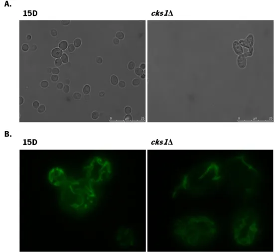
Deletion of yeast CKS1 does not alter mitochondrial morphology
It has previously been reported that depletion of Cks1/2 in murine embryonic fibroblasts led to a significant increased in the number of fragmented mitochondria, which could affect a mitochondrial-mediated apoptotic pathway [85]. To evaluate whether the absence of yeast CKS1 altered mitochondria morphology, wild-type strain 15D and strain cks1Δ were transformed with a plasmid expressing a mitochondrial green fluorescent protein and visualized by fluorescence microscopy. As shown in Figure 6, even though cks1Δ cells had an “aberrant” cellular morphology, the tubular mitochondrial network was visible in both strains and no difference in mitochondrial morphology was found.
Figure 6. Morphology of S. cerevisiae 15D and cks1Δ cells. Strains 15D and cks1Δ were transformed with pYX232-mtGFP and visualized by fluorescence microscopy (A) DIC, (B) GFP fluorescence.
22
Deletion of yeast CKS1 does not alter sensitivity to the yeast apoptosis inducers acetic acid or hydrogen peroxide
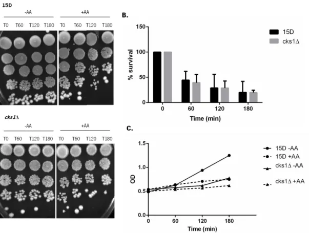
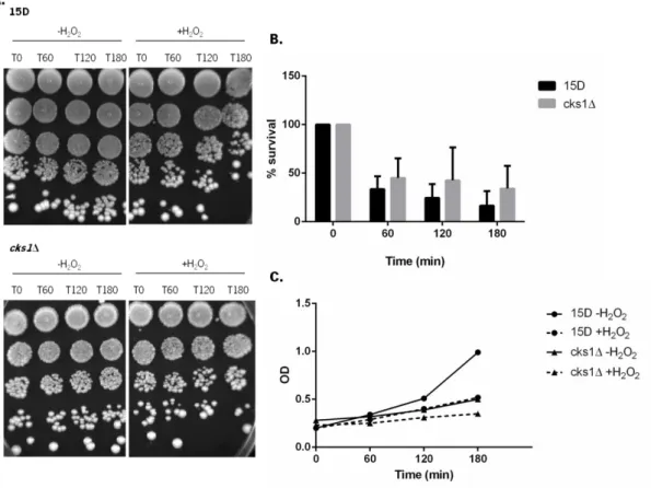
In order to determine whether CKS1 could play a general role in the apoptotic process, we first tested if absence of Cks1p affected viability of S. cerevisiae cells exposed to the most commonly used yeast apoptosis inducers, acetic acid and hydrogen peroxide.
Acetic acid is a weak acid that can be formed as an end sub-product of alcoholic fermentation by S. cerevisiae [86]. After acetic acid enters the cell, it dissociates (when the intracellular pH is higher than the extracellular pH), compromising cell viability [87], leading to the intracellular acidification [88] and induction of apoptosis [89]. Exposure of S. cerevisiae to low doses to acetic acid at pH 3.0 results in cell death with features of mammalian apoptosis. Cells exhibit chromatin condensation, exposure of phosphatidylserine and DNA strand breaks [89]. Like in mammalian cells, yeast apoptosis induced by acetic acid was linked to mitochondria. It was shown that acetic acid can lead to the release of cytochrome c, ROS accumulation, transient hyperpolarization of mitochondria followed by depolarization, decrease of mitochondrial respiration associated with decrease in cytochrome oxidase activity [90] and mitochondrial ultrastructural changes, namely decrease of cristae number, formation of myelinic bodies and swelling [91].
Hydrogen peroxide is a reactive oxygen species described as an apoptotic stimulus at low doses [92], and is known to induce apoptosis both in S. cerevisiae and in mammalian cells [93]. High concentrations of hydrogen peroxide result in cell death associated with disintegration of intracellular structures but without the phenotypic markers of apoptosis [92]. Apoptotic cell death induced by hydrogen peroxide in mammalian cells, promotes decrease in the intracellular superoxide anion (O2
-) concentration, reduction and acidification of the intracellular melieu,
activation of caspases [94], namely caspase-9 and -3, release of cytochrome c [95], among others. In yeast, apoptosis induced by hydrogen peroxide is accompanied by accumulation of ROS [92], phosphatidylserine exposure [96], cytochrome c release [96], DNA fragmentation [92], and plasma membrane vesicles reminiscent of blebbing observed in mammalian cells [96].
Strain 15D and cks1Δ were exposed to apoptosis-inducing concentrations of acetic acid and hydrogen peroxide for up 180 min, at 30ºC, and viability assessed by semi-quantitative spot assay and c.f.u. counts.
23
Figure 7. Sensitivity of wild-type strain 15D and deleted strain cks1Δ to AA. Exponential cultures of 15D and cks1Δ strains grown in YPD medium at 30ºC, were transferred to fresh YPD medium (pH 3) with (+AA) or without (-AA) 120 mM AA. Cells were grown for 180 minutes at 30°C. Samples were taken after 0, 60, 120 and 180 min. (A) Serial dilutions (1:10) were spotted onto YPD plates and incubated for 2 days at 30ºC. (B) For c.f.u. measurements, dilutions were plated on YPD plates, incubated for 2 days at 30°C and colonies counted. Values represent means and standard deviations of 3 independent experiments. (C) OD
24
There were no differences in sensitivity of the wild-type strain 15D and the strain cks1Δ to acetic acid (Figure 7B) or hydrogen peroxide (Figure 8B), indicating that Cks1p is not involved in a general stress response.
Deletion of yeast CKS1 results in differential sensitivity to DNA damaging agents
Our global aim is to determine whether Cks proteins have a role in apoptosis contributing to tumorigenesis and/or resistance of Cks-overexpressing tumors to treatment regimens. We therefore sought to determine whether Cks1p plays a role in the DNA damage response, as DNA damaging agents are among the most commonly used anti-cancer agents.
Figure 8. Sensitivity of wild-type strain 15D and deleted strain cks1Δ to H2O2. Exponential cultures of 15D and cks1Δ strains grown in YPD medium at 30ºC, were transferred to fresh YPD medium with (+H2O2) or without (-H2O2) 2 % H2O2. Cells were grown for 180 minutes at 30°C. Samples
were taken after 0, 60, 120 and 180 min. (A) Serial dilutions (1:10) were spotted onto YPD plates and incubated for 2 days at 30ºC. (B) For c.f.u. measurements, dilutions were plated on YPD plates, incubated for 2 days at 30°C and colonies counted. Values represent means and standard deviations of 3 independent experiments. (C) OD
25
It had previously been reported that deletion of yeast CKS1 renders cells more sensitive to growth in the presence of hydroxyurea, but not to UV irradiation [84]. We next tested whether cks1Δ mutants were sensitive to other DNA damaging agents.
The DNA alkylating agent methyl methanesulfonate causes base mispairing and replication blocks [97-98] and induces both inter-chromosomal and intra-chromosomal recombination [97]. Exposure to MMS causes a checkpoint-independent reduction in the rate of replication fork progression, likely due to a physical impediment of fork progression caused by alkylated DNA or some intermediate in lesion processing [98]. Strains 15D and cks1Δ were exposed to MMS for up to 180 min, at 30ºC and viability assessed by semi-quantitative spot assay and c.f.u. counts.
cks1Δ cells were only mildly more sensitive to transient MMS exposure than the wild-type strain, though there were no significant differences (Figure 9B). However, growth of the cks1Δ Figure 9. Sensitivity of wild-type strain 15D and deleted strain cks1Δ to MMS. Exponential cultures of 15D and cks1Δ strains grown in YPD medium at 30ºC, were transferred to fresh YPD medium with (+MMS) or without (-MMS) 0,025 % MMS. Cells were grown for 180 minutes at 30°C. Samples were taken after 0, 60, 120 and 180 min. (A) Serial dilutions (1:10) were spotted onto YPD plates and incubated for 2 days at 30ºC. (B) For c.f.u. measurements, dilutions were plated on YPD plates, incubated for 2 days at 30°C and colonies counted. Values represent means and standard deviations of 3 independent experiments. (C) OD600of the cultures. (D) Serial dilutions (1:10) were spotted onto YPD plates containing 0, 0,0075 and 0,015 % MMS and incubated for 2 days at 30°C.
26
strain on plates containing MMS was severely impaired (Figure 9D), indicating Cks1p is important for the ability of cells to survive chronic exposure to MMS, but not to overcome its short-term effects.
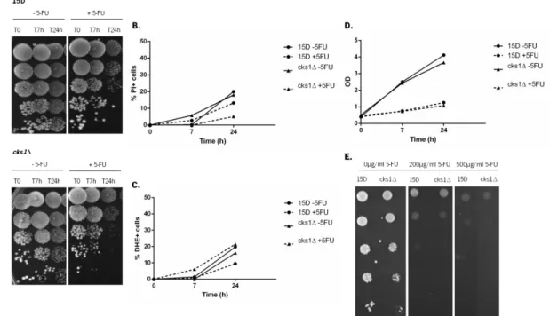
5-Fluorouracil is a pyrimidine analog antimetabolite commonly used in cancer treatment [99-102]. The cytotoxic mechanism of 5-FU occurs through inhibition of thymidylate synthase (TS), an enzyme involved in nucleotide synthesis [99], as well as through incorporation of fluoronucleotides into DNA and RNA, and disruption of RNA processing [101-102]. Since yeast lacks thymidine kinase, and therefore is unable to convert 5-FU into FdUMP, and inhibit TS, only the latter processes occur [103]. In order to determine whether yeast Cks1p affects sensitivity to 5-FU, strains 15D and cks1Δ were exposed to 5-FU for up to 24 hours, at 30ºC, and viability assessed by semi-quantitative spot assay.
Figure 10. Sensitivity of wild-type strain 15D and deleted strain cks1Δ to 5-FU. Exponential cultures of 15D and cks1Δ strains grown in SC medium at 30ºC, were transferred to fresh SC medium with (+5FU) or without (-5FU) 10 mM 5-FU. Cells were grown for 24 hours at 30°C. Samples were taken after 0, 7 and 24 hours. (A) Serial dilutions (1:10) were spotted onto YPD plates and incubated for 2 days at 30ºC. (B) Samples were incubated with PI (5 µg/mL) and fluorescence measured by flow cytometry. Increase of cells with PI staining is shown. (C) Samples are stained with DHE (5 µg/mL) and the fluorescence measured by flow cytometry. Increase of cells stained with DHE is shown. (D) OD
600of the cultures. (E) Serial dilutions (1:10) were spotted onto SC plates containing 0, 200 and 500
27
As seen in figure 10, there was no difference in the sensitivity of wild-type strain 15D and strain cks1Δ to 5-FU. Exposure to 5-FU did not lead to an increase in the number of cells stained with DHE or PI in comparison with non-treated cells (Figure 10B and 10C), indicating it does not cause an accumulation of ROS, typical of mitochondria-mediated apoptosis, but also does not lead to loss of plasma membrane integrity, typical of necrosis. Further studies will be required to characterize the mechanism mediating 5-FU-induced cell death in yeast, and confirm whether it is an active process. Nonetheless, there was no difference in the sensitivity of both strains to long-term exposure to 5-FU (Figure 10E), indicating CKS1 does not play a role in the cellular response to 5-FU.
Cisplatin is a platinum-based chemotherapy drug with activity against a wide spectrum of tumors [104-108]. It acts by forming a platinum complex inside the cell which binds to DNA and forms adducts, leading to inter-strand and intra-strand DNA cross-links, as well as DNA-protein cross-links [107, 109-111]. However, nuclear DNA is not the only target of cisplatin. It also binds to mitochondrial DNA, interacts with phospholipids and phosphatidylserine in membranes, disrupts the cytoskeleton and affects the polymerization of actin [112]. When bound to DNA, cisplatin inhibits DNA replication and chain elongation [113-114]. Cunha et al, showed that cisplatin induces an atypical programmed cell death pathway in S. cerevisiae, which is active, but mitochondria-independent, and that proteasome inhibition protects yeast cell from cisplatin-induce cell death [115]. In order to determine whether deletion of CKS1 affects sensitivity to cisplatin, wild-type strain 15D and cks1Δ strain were exposed to cisplatin for up to 180 min, at 30ºC and viability assessed by semi-quantitative spot assay and c.f.u. counts.
28
We found that the cks1Δ mutant strain is more sensitive to transient exposure to cisplatin than the wild-type strain 15D, which was more significant at time 60 and 120 min, and also to growth on plates containing cisplatin (Figure 11B, 11D), demonstrating that Cks1p is involved in the cellular resistance to cisplatin. We therefore proceeded to explore whether there was any alteration in apoptotic markers.
Cell membrane damage is considered a marker of necrotic cell death. Therefore, we tested if cisplatin-induced cell death in this strain background is accompanied by loss of membrane integrity. For this purpose, yeast cells were stained with PI and the fluorescence was evaluated by flow cytometry. As described for W303 cells [115], wild-type 15D and cks1Δ cells remained impermeable to PI after 180 minutes of exposure to cisplatin (Figure 12A), even Figure 11. Sensitivity of wild-type strain 15D and deleted strain cks1Δ to cisplatin. Exponential cultures of 15D and cks1Δ strains grown in YPD medium at 30ºC, were transferred to fresh YPD medium with (+cDDP) or without (-cDDP) 0,2 mg/mL cisplatin. Cells were grown for 180 minutes at 30°C. Samples were taken after 0, 60, 120 and 180 min. (A) Serial dilutions (1:10) were spotted onto YPD plates and incubated for 2 days at 30ºC. (B) For c.f.u. measurements, dilutions were plated on YPD plates, incubated for 2 days at 30°C and colonies counted. Values represent means and standard deviations of 3 independent experiments (**p<0,005). (C) OD600 of the cultures. (D) Serial
dilutions (1:10) were spotted onto SC plates containing 0, 50 and 100 µg/mL cDDP and incubated for 2 days at 30°C.
29
though there was a significant loss of cell viability under the same conditions (Figure 11B). This indicates cisplatin-induced yeast cell death occurs without loss of membrane integrity, supporting the hypothesis that cisplatin-induced death is an active process in both strains, and that the increased cell death observed in the cks1Δ mutant is not necrotic in nature.
ROS have been widely recognized as crucial cell death regulators and have been connected to many of the known apoptotic pathways in yeast [69, 116]. Huang et al (2003) showed that DNA damaging agents such as γ-irradiation and cisplatin induce apoptosis in Jurkat cells by increased ROS formation, leading to the generation of hydrogen peroxide and superoxide anion [117]. However, it has previously been reported that exposure of yeast W303 cells to cisplatin did not result in an increased percentage of cells stained with DHE, which detects the accumulation of intracellular superoxide anion [115]. In order to test the involvement of ROS in cisplatin-induced death in the 15D background, ROS levels were determined by flow cytometry using DHE.
We could not detect a significant increase in the percentage of cells stained with DHE after exposure to cisplatin (Figure 12B), indicating that, under our experimental conditions and in the 15D strain background, cisplatin-induced death is also not mediated by intracellular Figure 12. Plasma membrane integrity and ROS accumulation of wild-type strain 15D and mutant strain cks1Δ exposed to cisplatin. Exponential cultures of 15D and cks1Δ strains grown in YPD medium at 30ºC, were transferred to fresh YPD medium with (+cDDP) or without (-cDDP) cisplatin. Cells were grown for 180 minutes at 30°C. Samples were taken after 0, 60, 120 and 180 min. (A) Incubated with PI (5 µg/mL) and fluorescence measured by flow cytometry. Increase of cells with PI staining is shown. (B) Stained with DHE (5 µg/mL) and the fluorescence measured by flow cytometry. Increase of cells stained with DHE is shown. Values represent means and standard deviations of 3 independent experiments.
30
superoxide anion formation, and that the increased sensitivity of the cks1Δ strain to cisplatin is not likely a result of increased ROS levels.
Sorensen et al (1990) and Grossman et al (1999) reported that DNA cross-links caused by cisplatin do not inhibit S-phase but cause a G2/M arrest in S. cerevisiae cells [113, 118]. Cunha et al (2013) showed that cisplatin-treated cells had an increased percentage of cells with sub-G0/G1 DNA content than untreated cells, especially after recovery in cisplatin-free media [115]. We therefore next tested whether the increased sensitivity of cks1Δ cells to cisplatin was due to higher levels of DNA fragmentation/degradation. For cell cycle analysis, cells were untreated or treated with cisplatin for 240 min, fixed and stained with sytox green, and DNA content was analyzed by flow cytometry. Representative histograms are shown in Figure 13A, the cell cycle distribution is shown in Figure 13B, and the quantification of the percentage of cells with sub G0/G1 DNA content after 240 min of cisplatin exposure and an additional 4 h recovery period are shown in Figure 13C.
31
In agreement with published data, we also observed that exposure to cisplatin leads to an accumulation of cells in G2/M in both the wild-type strain 15D and strain cks1Δ (Figure 13A and 13B). The percentage of cells in the sub G0/G1 region was also higher in cisplatin-treated cells than in cells grown in cisplatin-free media, both after 240 min of treatment and after a 4 h recovery in YPD (Figure 13C). The percentage of 15D and cks1Δ cells in sub G0/G1 was similar Figure 13. The effect of cisplatin on cell cycle distribution. Exponential cultures of 15D and
cks1Δ strains grown in YPD medium at 30ºC, were transferred to fresh YPD medium with (+cDDP) or without (-cDDP) 0,2 mg/mL cisplatin. Cells were grown for 240 minutes at 30°C. Samples were taken after 0 and 240 min, stained with sytox green (1 µM) and the fluorescence measured by flow cytometry. (A) Representative histograms. (B) Bars indicated the frequence (%) of cells in each cell cycle phase. (C) Cells were washed and resuspended in fresh medium without cisplatin and grown an additional 4h at 30ºC. The percentage of cells with Sub G0/G1 DNA content was measured by flow cytometry. Values represent means and standard deviations of 3 independent experiments.
32
after the treatment period, but slightly lower in cks1Δ cells after recovery, despite higher levels of cell death (Figure 11B). This difference was statistically significant, showing that the higher sensitivity of cks1Δ cells to cisplatin is not due to increased DNA fragmentation/degradation.
Expression of yeast CKS1 from a multicopy plasmid does not alter sensitivity to cisplatin
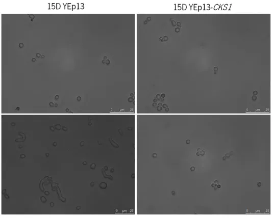
Since deletion of CKS1 resulted in higher sensitivity to cisplatin, it is conceivable that overexpression could result in higher resistance, potentially causing the observed higher resistance of Cks-overexpressing tumours to therapy. In order to characterize the phenotype of overexpression of Cks1p in yeast, strains 15D and cks1Δ were transformed with a multi-copy plasmid expressing Cks1 (YEp13-CKS1) and respective empty vector control (YEp13). As expected, the “aberrant” morphology of the cks1Δ strain (cks1Δ YEp13) was reversed by expressing Cks1p from the YEp13 vector (cks1Δ YEp13-CKS1) (Figure 14).
Figure 14. Morphology of S. cerevisiae strains 15D and cks1Δ expressing YEp13 and YEp13-CKS1.
33
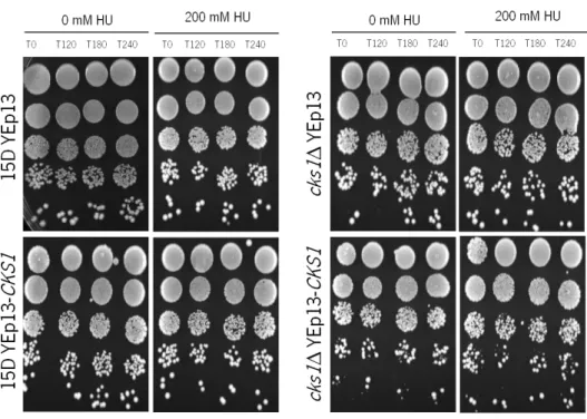
To test whether overexpression of Cks1p resulted in increased resistance to cisplatin, all strains were exposed to cisplatin for up 180 min, at 30ºC and viability assessed by semi-quantitative spot assay and c.f.u. counts.
There was no difference in the viability of 15D cells transformed with the empty vector and expressing YEp13-CKS1. However, expression of Cks1p in cks1Δ reverted the sensitivity of this strain to that of the wild-type strain. This indicates that either overexpression of Cks1p does Figure 15. Sensitivity of S. cerevisiae strain 15D and cks1Δ expressing YEp13 and YEp13-CKS1. Exponential cultures of 15D YEp13, 15D YEp13-CKS1, cks1Δ YEp13 and cks1Δ YEp13-CKS1 strains grown in SCGLU-Leu medium at 30ºC, were transferred to fresh SCGLU-Leu medium without (-cDDP) or with (+cDDP) 0,2 mg/mL cDDP. Cells were grown for 180 min at 30°C. Samples were taken after 0, 60, 120 and 180 min. (A) Serial dilutions (1:10) were spotted onto YPD plates and incubated for 2 days at 30ºC. (B) For c.f.u. measurements, dilutions were plated on YPD plates, incubated for 2 days at 30°C and colonies counted. Values represent means and standard deviations of 3 independent experiments (* p<0,05; **p<0,005). (C) OD600 of the cultures.
34
not have a phenotype or that expression of Cks1p from the YEp13 vector is not sufficient to induce an overexpression phenotype.
Expression of yeast CKS1 from a multicopy plasmid does not alter sensitivity to hydroxyurea
Hydroxyurea is a DNA replication inhibitor that represses both the elongation and initiation phases of replication [119]. HU inhibits ribonucleoside diphosphate reductase, thereby blocking DNA synthesis and repair [120-122]. HU slows down or inhibits S-phase progression and can compromise genetic integrity by increasing the rate of recombination [119]. HU also induces the generation of reactive oxygen species. The production of ROS induces oxidative stress, adversely affecting cellular metabolism and leading to cell cycle arrest and cell death [123].
It had previously been descrived that overexpression of human Cks1 or Cks2 in human mammary epithelial and breast cancer-derived cell leads to override of the intra–S-phase checkpoint that blocks DNA replication in response to replication stress [35]. To determine whether a similar phenotype could be observed in yeast, 15D YEp13, 15D YEp13-CKS1, cks1Δ YEp13 and cks1Δ YEp13-CKS1 strains were exposed to hydroxyurea for up to 240 min, at 30ºC. Cells were stained with sytox green, and DNA content was analyzed by flow cytometry. Representative histograms are shown in figure 16.
35
As expected, we observed that exposure to HU leads to an accumulation of cells in G1/S. There were no significant differences in the cell cycle profiles of the 15D YEp13 and 15D YEp13-CKS1 strains. Strain cks1Δ YEp13 still accumulated in G1/S, though the peak was broader, likely reflecting the “abnormal” morphology and cell cycle defects of this strain. Expressing YEp13-CKS1 in the cks1Δ strain reverted this phenotype, and the cell cycle profiles were similar Figure 16. The effect of hydroxyurea on cell cycle distribution. Exponential cultures of 15D YEp13, 15D YEp13-CKS1, cks1Δ YEp13 and cks1Δ YEp13-CKS1 strains grown in SCGLU-Leu medium at 30ºC were transferred to fresh medium without or with (+HU) hydroxyurea. Samples were collected after 0 and 240 min at 30ºC, stained with sytox green (1 µM) and the fluorescence measured by flow cytometry.
36
to those of the 15D strain. These results also indicate that either overexpression of Cks1p does not bypass the HU-induced cell cycle arrest in yeast or that expression of Cks1p from the YEp13 vector is not sufficient to induce an overexpression phenotype.
We next determined whether there were any differences in the viability of strains exposed to HU. Strains 15D YEp13, 15D YEp13-CKS1, cks1Δ YEp13 and cks1Δ YEp13-CKS1 were exposed to hydroxyurea for up to 240 min, at 30ºC and viability assessed by semi-quantitative spot assay.
There was no cell death observed even when exposing cells to a HU concentration as high as 600 mM for 240 min (data not shown), indicating under these experimental conditions HU was cytostatic but not cytotoxic.
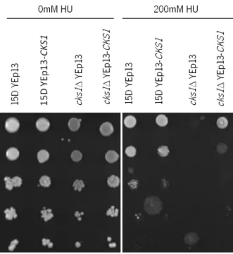
Strains 15D YEp13, 15D YEp13-CKS1, cks1Δ YEp13 and cks1Δ YEp13-CKS1 were then spotted onto plates containing HU to evaluate their survival after long-term HU exposure (Figure 18).
Figure 17. Sensibility of S. cerevisiae strains to HU. Exponential cultures of 15D YEp13, 15D YEp13-CKS1, cks1Δ YEp13 and cks1Δ YEp13-CKS1 strains grown in SCGLU-Leu medium at 30ºC were transferred to fresh medium without or with 200 mM HU. Samples were taken after 0, 120, 180 and 240 min. Serial dilutions (1:10) were spotted onto YPD plates and incubated for 2 days at 30ºC.
37
It has previously been described that cks1Δ cells are more sensitive to growth in the presence of HU [84]. We found that the viability of strain 15D expressing YEp13-CKS1 and strain 15D with the empty vector was identical, and, as expected, strain cks1Δ YEp13 was hyper-sensitive to HU. Expression of Cks1p (cks1Δ YEp13-CKS1) only partially reverted this phenotype, indicating that the protein levels of Cks1p when expressed from the YEp13 plasmid are low. This suggests that expression of Cks1p from the YEp13 plasmid is not sufficient to induce an overexpression phenotype, and that another expression system would be required to express high levels of Cks1p.
Figure 18. Chronic exposure of S. cerevisiae strains to HU. Exponential cultures of 15D YEp13, 15D YEp13-CKS1, cks1Δ YEp13 and cks1Δ YEp13-CKS1 strains grown in SCGLU-Leu medium at 30ºC. Serial dilutions (1:10) were spotted onto SCGLU-Leu plates containing 200mM HU and incubated for 2 days at 30°C.

![Figure 5. The apoptotic yeast cell. Red question marks indicate pathways that are known in mammals but not in yeast thus far (Madeo et al (2004) [67])](https://thumb-eu.123doks.com/thumbv2/123dok_br/17753711.834680/20.892.156.735.107.517/figure-apoptotic-yeast-question-indicate-pathways-mammals-madeo.webp)