Establishing the
Mechanism of
Glutaredoxin
Sodiq Odewale Waheed
Dissertação de Mestrado apresentada à
Faculdade de Ciências da Universidade do Porto
Química
2018
E stabli sh ing the M ec ha nism of G lutar ed ox in S o d iq O d ew ale W aheed FCUP UV EM 2018 2.º CICLOMechanism of
Glutaredoxin
Sodiq Odewale Waheed
Mestrado em Química
Departamento de Química e Bioquímica
2018
Orientadora
Maria João Ramos, Professora Catedrática, Faculdade de Ciências da
Universidade do Porto
Coorientadores
Sérgio F. Sousa, Investigador, Faculdade de Ciências da Universidade
do Porto
Iñaki Tuñón, Full Professor, Faculty of Chemistry, University of Valencia,
Spain
ACKNOWLEDGEMENTS
First and foremost, I am heartily grateful to the Erasmus Mundus Programme of the European Master in Theoretical Chemistry and Computational Modelling (EMTCCM) with the support of the European Union for the Erasmus Mundus grant they awarded me for this Master Programme.
I am extremely indebted to my supervisor – Professor Maria João Ramos who despite her hectic schedule gave all the needed guidance and help necessary for the successful completion of this thesis and the Master programme in general. My earnest desire is that God will continue to keep you at his right hand.
My profound gratitude also goes to Professor Pedro A. Fernandes, Dr. Sérgio F. Sousa and Dr. Rui P.P. Neves for their innovative ideas and support from the beginning to the end of this research. May the good Lord bless you all.
I would like to render my sincere appreciations to all the Professors in the Department of Chemistry and Biochemistry, FCUP and all the Professors during the Intensive Course part of the Master at the University of Valencia, Spain, as well as during the TCCM winter School at the University of Toulouse III - Paul Sabatier, France, who have in one way or the other imparted in me the skills needed for a competitive edge in life.
I wish to also express my gratitude to Professor Iñaki Tuñón’s research group at the Faculty of Chemistry, University of Valencia, Spain for the privilege that was given to me to carry out part of my research project in their group. I am highly thankful to Professor Iñaki Tuñón, Kirill Zinovjev and Carlos Ramos-Guzmán.
To my amiable and loving family miles away, thanks so much for your interminable love, support, prayers, and encouragements.
Finally, I thank God for His unfeigned love and unprecedented faithfulness during this Master programme.
ABSTRACT
Glutaredoxin (Grx) is a redox enzyme and it belongs to thioredoxin-family with Cys-Pro-Tyr-Cys active site motif. This enzyme catalyzes the reduction of glutathione disulfide (GSSG) to glutathione (GSH) through the dithiol mechanism that includes a Grx-S-S-G mixed disulfide intermediate between the enzyme (Grx) and the substrate (GSSG). Glutaredoxin functions as both electron donors and as regulators of cellular function in response to oxidative stress. Additionally, it functions in sulfur assimilation, dehydroascorbate reduction, and the regulation of cellular differentiation, transcription and apoptosis. Grx and other redox enzymes play a very crucial role in the protection of dopaminergic neurons. The loss of dopaminergic neurons results in Parkinson’s disease; hence, the study of this enzyme becomes important.
The work presented in this thesis contains the outcomes of the studies of the catalytic mechanism of the reduction of glutathione disulfide (GSSG) by glutaredoxin (Grx) using the ONIOM extrapolative QM/MM scheme as implemented in Gaussian09 package. The whole thesis contains four chapters. Chapter one presents an introductory information about the enzyme, its characteristic reaction; thiol-disulfide exchange, and the catalytic mechanism of the enzyme. Chapter two dwells on discussing the computational methods as applied to biological systems. This chapter contains four sections: (i) Quantum Mechanics (QM) Methods, (ii) Molecular Mechanics (MM) Methods, (iii) Hybrid Methods with emphasis on QM/MM Methods, and (iv) Computational Enzymatic Catalysis. Chapter three describes the studies that were carried out with the results obtained for the two mechanistic steps of the reaction as well as the conclusions drawn from each step. Lastly, chapter four outlines the general conclusions from the whole work.
Keywords: Glutaredoxin, quantum mechanics, thioredoxin-family, glutathione (GSH), molecular
mechanics, enzymatic catalysis, oxidized glutathione (GSSG), thiol-disulfide exchange, ONIOM QM/MM, dithiol mechanism.
RESUMO
A glutaredoxina (Grx) é uma enzima redox e pertence à família das tiorredoxinas com o motivo activo Cys-Pro-Tyr-Cys. Esta enzima catalisa a redução de dissulfeto de glutationa (GSSG) a glutationa (GSH) através do mecanismo de ditiol que inclui um intermediário dissulfeto de Grx-S-S-G entre a enzima (Grx) e o substrato (GSSG). A glutaredoxina funciona como dadora de eletrões e como reguladora da função celular em resposta ao stressoxidativo. Além disso, funciona na assimilação de enxofre, na redução do desidroascorbato e na regulação da diferenciação celular, transcrição e apoptose. O grx e outras enzimas redox desempenham um papel crucial na proteção dos neurônios dopaminérgicos. A perda de neurônios dopaminérgicos resulta na doença de Parkinson, tornando o estudo desta enzima importante.
O trabalho apresentado nesta tese contém os resultados dos estudos do mecanismo catalítico da redução da glutationa (GSSG), por glutarredoxina (Grx) usando o esquema ONIOM extrapolativo QM / MM como implementado no pacote Gaussian09. A tese contém no seu todo quatro capítulos.O capítulo um apresenta informação introdutória sobre a enzimae a sua reação característica; a permuta de tiol-dissulfureto, e o mecanismo catalítico da enzima. O capítulo dois inclui a discussão dos métodos computacionais e a sua aplicação a sistemas biológicos. Este capítulo contém quatro secções: (i) Métodos de Mecânica Quântica (QM), (ii) Métodos de Mecânica Molecular (MM), (iii) Métodos Híbridos com ênfase nos Métodos QM / MM, e (iv) Catálise Computacional Enzimática. O capítulotrês descreve os estudos que foram efetuados com os resultados obtidos para os dois passos mecanísticos da reação, bem como as conclusões retiradas a partir de cada etapa. Por fim, o capítulo quatro descreve as conclusões gerais de todo o trabalho.
Palavras-chave: Glutaredoxina, mecânica quântica, tiorredoxina-família, glutationa (GSH),
mecânica molecular, catálise enzimática, glutationa oxidada (GSSG), permuta de tiol-dissulfureto, ONIOM QM / MM, mecanismo ditiol.
TABLE OF CONTENTS
Acknowledgements………i Abstract………ii Resumo………iii Table of Contents………...iv Index of Figures………..vii Index of Tables………viii List of Abbreviations………ixAmino Acids Abbreviations………xi
Chapter One – Biological Problem 1.0 Introduction………..1
1.1 Glutaredoxins………...2
1.2 Structure of Glutaredoxins……….3
1.3 Catalytic Mechanism of Glutaredoxins……….6
1.3.1 Thiol-Disulfide Exchange Reaction……….6
1.3.2 Thiol-Disulfide Exchange Catalytic Mechanism of Glutaredoxins………..7
1.4 Major Doubts………..11
Chapter Two – Methods 2.0 Introduction……….12
2.1 Quantum Methods……….13
2.1.1 The Schrodinger Equation………..13
2.1.2 The Ab initio Methods………..14
2.1.3 Semi-Empirical Methods……….15
2.1.4 Density Functional Theory………..16
2.1.5 Exchange Correlation Functionals………18
2.1.6 Basis Sets……….19
2.2 Molecular Mechanics……….20
2.2.1 Force Fields………..20
2.2.1.2 Angle Bending………21
2.2.1.3 Torsional Terms……….21
2.2.1.4 Van der Waals Terms………21
2.2.1.5 Electrostatic Terms………22
2.2.2 Protein Molecular Mechanical Force Fields……….22
2.2.2.1 CHARMM Force Fields………..23
2.2.2.2 OPLS Force Fields………..23
2.2.2.3 AMBER Force Fields………..23
2.3 Hybrid Methods………..24 2.3.1 QM/MM Methods………..25 2.3.2 Types of QM/MM Schemes………26 2.3.2.1 Additive QM/MM Methods………26 2.3.2.2 Subtractive QM/MM Methods………26 2.3.3 ONIOM Method………27
2.3.3.1 Capping Bonds at the QM/MM Boundary………..28
2.3.3.1.1 The Link Atom Approach……….28
2.3.3.1.2 The Frozen Orbital Approach……….29
2.3.3.2 Handling the non-bonded Couplings Terms………...30
2.3.3.2.1 Mechanical Embedding………...30
2.3.3.2.2 Electrostatic Embedding……….31
2.3.3.2.3 Polarized Embedding………..31
2.4 Computational Enzymatic Catalysis………31
Chapter Three – Results and Discussion 3.0 Introduction……….33
3.1 Study of the Step One………34
3.1.1 Model Preparation………...35
3.1.1.1 Modelling Glutathione Disulfide (GSSG) into the Human Glutaredoxin……..35
3.1.1.2 Building the QM/MM Model……….37
3.1.1.3 Determination of the Catalytic Mechanism………...39
3.1.2 Results………40
3.1.4 Conclusions from the First Reaction Step………47
3.2 Study of the Step Two………...48
3.2.1 Determination of Second Step Mechanism………49
3.2.2 Results……….50
3.2.3 Analysis and Discussion of Results……….53
3.2.4 Conclusions from the Second Reaction Step………57
Chapter Four – Conclusions 4.0 Conclusions……….58
INDEX OF FIGURES
Fig. 1 – Glutaredoxin Structures from bacteria [28] and human [161]………..5
Fig. 2 – Catalytic Mechanism Scheme for the reduction of disulfide with Grx [26,28,29,33,57,63,64]………..8
Fig. 3 – Thiol-Disulfide Exchange related Structural Changes [29]………..10
Fig. 4 – Jacob’s ladder for DFT Functionals [106]………..19
Fig. 5 – Illustration of the QM/MM Method [84]………25
Fig. 6 – Different Methods to cap the QM region [137]………..30
Fig.7 – Catalytic Mechanism Scheme for the reduction of disulfide with Grx [26,28,29,33,57,63,64]……….34
Fig. 8 – First Step Reaction showing the formation of Grx-S-S-G mixed disulfide intermediate.35 Fig. 9 – Alignment result of the active site of the human glutaredoxin and the bacterial glutaredoxin………...36
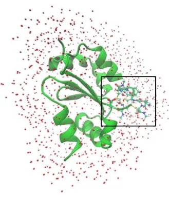
Fig. 10 – The optimized hGrx:GSSG complex and the water molecules as red dots………38
Fig. 11 – The DFT layer that contained the C24P25Y26C27 motif, Asp48 sidechain, important part of GSSG and water molecules………...38
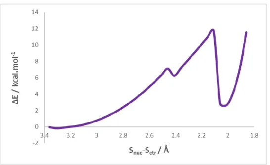
Fig. 12 – The scan plot for the model with 99 atoms in the DFT layer……….41
Fig. 13 – The scan plot for the model with 125 atoms in the DFT layer………..41
Fig. 14 – The scan plot for the model with 137 atoms in the DFT layer………..42
Fig. 15 – The plot for the reverse scan of the intermediate from Fig. 14……….42
Fig. 16 – The reaction states for the nucleophilic attack of the Cys24-thiolate to the GSSG-disulfide to form the mixed GSSG-disulfide intermediate………46
Fig. 17 – Thermochemical Profile for the First thiol-disulfide exchange………..46
Fig. 18 – Second Step Reaction showing the formation of the oxidized Grx (GrxS2) and the reduced GSH……….48
Fig. 19 – The scan plot for the deprotonation of Cys27-thiol by Asp48 via a bridging water molecule……….50
Fig. 20 – Structure and Scan plot for the model with GSHlg removed from the QM layer………51
Fig. 21 – Structure and Scan plot for the model with GSHlg protonated and retained in the QM layer………51
Fig. 22 – The scan plot for the intramolecular oxidation of the Grx-SSG intermediate………….52
Fig. 23 – The MD simulation plot of the Grx-SSG intermediate showing how the distance between the thiolates of GSHox and GSHlg changed with time during the simulation………52
Fig. 24 – The reaction states for the intramolecular oxidation of the Cys27 deprotonated mixed disulfide intermediate to form the oxidized Grx and the reduced GSH………56 Fig. 25 – Thermochemical Profile for the intramolecular oxidation (Second thiol-disulfide
exchange)………..56
INDEX OF TABLE
LIST OF ABBREVIATIONS
AM1 Austin Model 1
AMBER Assisted Model Building with Energy Refinement CHARMM Chemistry at HARvard Macromolecular Mechanics CPMD Car-Parrinello Molecular Dynamics
CPU Central Processing Unit
DDT Dithiothreitol
DFT Density Functional Theory DsbA Disulfide bond formation A EVB Empirical Valence Bond GAFF General Amber Force Field
GGA Generalized Gradient Approximation GHO Generalized Hybrid Orbital
GR Glutathione Reductase
Grx Glutaredoxin
GSH Glutathione
GSSG Glutathione Disulfide GTO Gaussian-type orbital HED Hydroxy ethyl disulfide
HF Hartree Fock
hGrx Human Glutaredoxin
HIV Human Immunodeficiency Virus
IMOMM Integrated Molecular Orbital and Molecular Mechanics IMOMO Integrated Molecular Orbital and Molecular Orbital INT Reaction Intermediates
kDa kilo Dalton
LDA Local Density Approximation LSCF Localized Self Consistent Field
MD Molecular Dynamics
MNDO Modified Neglect of Diatomic Differential Overlap NADPH Nicotinamide adenine dinucleotide phosphate NMR Nuclear Magnetic Resonance
NPT Isobaric-isothermal ensemble
NVT Canonical ensemble
ONIOM Our own N-layered Integrated Molecular Orbital and Molecular Mechanics
OPLS Optimized Potential Liquid Simulation OPLS-AA OPLS- All Atoms
OPLS-UA OPLS-United Atoms
P Product
PD Parkinson’s Disease
PDB Protein Data Bank
PDI Protein Disulfide Isomerase PES Potential Energy Surface PM3 Parametric Model number 3 PM6 Parametric Model number 6
QM Quantum Mechanics
QM/MM Quantum Mechanics/Molecular Mechanics
R Reactant
RNR Ribonucleotide Reductase
SCC-DFTB Self consistent charge Density Functional tight binding
SP Single Point
TS Transition State
STO Slater-type orbital
TIP3P Transferable Intermolecular Potential 3-Point
Trx Thioredoxin
AMINO ACIDS ABBREVIATIONS
Ala Alanine Arg Arginine Asn Asparagine Asp Aspartate Cys Cysteine Gln Glutamine Glu Glutamate Gly Glycine His Histidine Ile Isoleucine Leu Leucine Lys Lysine Met Methionine Phe Phenylalanine Pro Proline Ser Serine Thr Threonine Trp Tryptophan Tyr Tyrosine Val ValineChapter One – Biological Problem
1.0 Introduction
The chemistry of cysteine is the key to the enzymatic mechanism of the thiol-disulfide oxidoreductases of the thioredoxin subfamily, such as thioredoxins (Trxs), glutaredoxins (Grxs), protein disulfide isomerase (PDI), and DsbA [1]. These enzymes are ubiquitous and share similar architecture, known as the thioredoxin fold [2-3]. Trx, Grx, PDI, and DsbA active sites contain a -Cys-X-X-Cys- motif, where the N-terminal cysteine thiol has a pKa that is significantly
lower than that of a free cysteine [4-11]. Conversely, the pKa of the C-terminal active site
cysteine thiol is usually more basic than that of a free cysteine [1]. The understanding of the thiol-disulfide reaction is important because it is involved in a number of biological processes ranging from protein folding and stability to regulation of gene expression and catalytic activity [5-7].
Depending on the protein under study, measured values of the pKa of the N-terminal
active site cysteine range from ~7.1 in Trx [7,12,13] to ~3.2 in DsbA [9,14]. The thiol sulfur reactivity is highly dependent on the pKa of the thiol, as a lower pKa affects both nucleophilicity
and leaving group ability of the associated thiolate [15,16]. This is well illustrated in DsbA and its mutants, where a lower pKa of the N-terminal cysteine of the -Cys-X-X-Cys- motif is associated
with a more oxidative protein [14,17,18]. Although, other factors influence the redox properties of the -Cys-X-X-Cys- motif, the pKa of the N-terminal cysteine clearly contributes to these
properties [19]. In addition, the nature of the -X-X- residues also has crucial effects on the N-terminal cysteine thiol pKa and is mostly understood in terms of the number of hydrogen bonds
that the -X-X- residues can provide to stabilize the thiolate charge [1]. The replacement of a Tyr in the -Cys-Pro-Tyr-Cys- motif of Grx (or a His in the -Cys-Pro-His-Cys- motif of DsbA) by a Pro in its Trx counterpart (Cys-Gly-Pro-Cys-) allows the backbone of Grx (DsbA) to donate one more hydrogen bond to the thiolate sulfur than in Trx, which lowers the pKa of the N-terminal
active site cysteine in Grx (DsbA) more than in Trx [20,21]. The His of the -Cys-Pro-His-Cys- motif in DsbA can also form a hydrogen bond with the thiolate sulfur [22], which may also stabilize the thiolate anion in DsbA more than in Grx [20]. The thiol of the C-terminal cysteine is another group that can form a hydrogen bond with the thiolate [13]. The reduced Trx [23] and the reduced DsbA [22] X-ray crystal structures are compatible with such a hydrogen bond. The
direct evidence that supports this hydrogen bond is provided by the NMR experiments [10] and molecular dynamics (MD) simulations [20] using glutaredoxin 3 (Grx3) from Escherichia coli.
1.1 Glutaredoxins
The Glutaredoxin system was initially described by Holmgren in 1976 as an alternate electron donor to ribonucleotide reductase (RNR) in Escherichia coli mutants lacking thioredoxin [24]. A few years later, Grx was shown to also have GSH-disulfide transhydrogenase activity; thus, functioning as general thiol-disulfide oxidoreductases [25]. In the end, it became apparent that thioltransferases and glutaredoxins purified from different organisms were highly similar and were actually the same enzymes just named differently [5,11].
Glutaredoxins are small proteins, usually around 9-15 kDa, that exist in a large number of isoforms in basically all glutathione (GSH) containing life forms [26]. According to their active site motif, Grxs can be divided in two major categories: the dithiol Grxs with active site motif Cys-X-X-Cys and the monothiol Grxs with active site motif Cys-X-X-Ser [27,28]. The latter is subdivided into single- and multi-domain monothiol Grxs [27,28]. Dithiol and single-domain monothiol Grxs are present in all forms of life such as bacteria, plants, and vertebrates, while multi-domain Grxs were until now not yet identified in bacteria [29]. In addition, another division of the Grx family is based on a cysteine residue at the second position of the active site motif (Cys-Cys-X-Cys/Ser). These Grxs family, called CC-type Grxs, are exclusively found in land plants, where they control plant physiology, like flower development [30]. Dithiol Grxs are thiol-disulfide oxidoreductases that have high specificity for the glutathione moiety [31]. These Grxs are kept reduced by GSH, and the resulting oxidized form of GSH, glutathione disulfide (GSSG) is reduced by glutathione reductase (GR) with electrons from NADPH [31,32]. The classical dithiol Grxs efficiently reduce some protein disulfides like that in Escherichia coli ribonucleotide reductase with a dithiol mechanism [26]. However, the major action of Grxs appears to be to reduce protein-GSH mixed-disulfides using a mechanism that requires only the N-terminal active site residue, that is, a monothiol mechanism [33].
Grxs have been suggested to be involved in numerous functions, both as electron donors and as regulators of cellular function in response to oxidative stress, as demonstrated by the ability of yeast glutaredoxins to directly reduce hydroperoxides with catalytic rate comparable to glutathione peroxidase [34], and the sensitivity of the yeast mutants lacking Grx to oxidative stress [35]. Additionally, they function in sulfur assimilation [36,37],
dehydroascorbate reduction [38], and the regulation of cellular differentiation [39], transcription [40-42], and apoptosis [43-45]. The human Grx2 can also catalyze the reduction of hydroperoxides in vitro, but a reasonable high concentration of the enzyme is needed [46]. Furthermore, human Grx1 has been shown to be able to scavenge glutathione-thiyl radicals [47]. Grxs also mediate the recovery from oxidative damage by reducing protein-GSH mixed disulfides and restoring the protein thiol homeostasis in brain mitochondria [48], or human lens epithelial cells [49].
A study demonstrating the critical role of the glutaredoxin system in protecting macrophages from cell death induced by oxidized low density lipoprotein, was used to show how Grxs protect cells against apoptosis [50]. In addition, glutaredoxins have been detected within HIV-1 and have been proven to regulate the activity of glutathionylated HIV-1 protease in vitro [51]. Monothiol Grxs from E. coli, yeast and vertebrates including humans are crucially involved in iron-sulfur cluster biosynthesis and regulation of iron homeostasis [52].
The study of this enzyme becomes important, as it has been shown with other redox enzymes, to play a very crucial role in the protection of dopaminergic neurons, thus contributing to maintain neuronal cell viability. The loss of dopaminergic neurons results in the noxious disease called Parkinson’s disease (PD).
1.2 Structure of Glutaredoxins
Glutaredoxins have been extensively studied using both NMR spectroscopy and X-ray crystallography. There are over one hundred (100) structures of Grxs from different organisms, like Homo sapiens, E. coli, Saccharomyces cerevisiae, etc. in the protein database. Structurally, Grxs belong to the Trx fold family of proteins, and represent only a substructure or a domain in the other members of the family [2,3]. In bacterial glutaredoxins, the structure contains four stranded β-sheets that are surrounded by three α-helices (Fig. 1A), while other Grx types from yeast, human etc. do consist of four stranded β-sheets surrounded by five α-helices (Fig. 1B). The central β-sheet is composed of three antiparallel strands (b1,b3,b4) with b2 parallel to the adjacent b1 (Fig. 1). Furthermore, all the Trx oxidoreductase family of proteins shares similar Cys-X-X-Cys active site motifs that are located on the loop connecting the first β-sheet (b1) and the first α-helix (a1). The N-terminal cysteine residue in the Grx active site is similar to Trxs, surface-exposed with low pKa, below that of the free cysteine, while the C-terminal cysteine is buried in the molecule and has much higher pKa value [28].
Human glutaredoxin consists of a four-stranded mixed β-sheet composed of residues 15 to 19, 43 to 47, 72 to 75 and 78 to 81, and five α-helices composed of residues 4 to 9, 24 to 34, 54 to 65, 83 to 91 and 94 to 100 [53] in contrast to the three α-helices that are observed in E. coli Grx-1. The two additional helices in hGrx relative to its E. coli counterpart are located at the N and C termini of the protein [53]. The loop connecting α-helix 2 to β-strand 2 (α1 to β2 in E. coli) is longer in the human protein. It was reported that a hydrogen bond interaction between the NH of Lys39 and the CO of Ala104 serves to hold α-helix 5 in contact with the extended loop between α-helix 2 to β-strand 2 [53]; thus, enhances the interaction between the extended loop and α-helix 5 [53]. The helix containing the active site is one turn shorter in the human protein [53]. Also, α-helix 3 in hGrx is one turn longer and positioned differently than the counterpart E. coli Grx-1. This has been reported to result in the narrowing of the groove into which the RNR substrate has been shown to bind in E. coli Grx-1 [54]. The β-sheet forms the central core of the protein with helices 1 and 3 located on one side of the sheet and helices 2, 4, and 5 located on the other side [53]. Human Grx (hGrx) needs to be structurally characterized because it is a basic protein and longer than its E. coli counterpart. Furthermore, human Grxs have three additional cysteine residues (Cys8, Cys79 and Cys 83) in addition to those that are present in the active site [53]. These additional cysteines are proposed to play a regulatory role because their oxidation leads to inactivation in the case of the studied calf thymus Grx [55]. The exact role of these additional cysteine residues is yet to be established [53]. However, possible functional importance was suggested for them based on their spatial location and local environment [53]. Cys8 which is located in the N-terminal helix and solvent-exposed, decreases the lifetime of hGrx samples considerably with substantial aggregation of the protein if it is not maintained in a fully reduced state [53]. The second additional cysteine residue (Cys79) is also solvent-exposed and is located in β-strand 4, providing a site for oligomerization. Oligomerization of hGrx with disulfide cross-linking helps in the regulation of hGrx [53]. The last cysteine residue, Cys83 is located at the N-terminal of α-helix 4 and is solvent-exposed too [53]. Cys83 is located in a region of positive electrostatic potential, which is indicative that it will have a low pKa value and that likely will be active in redox reactions. The thiol would play a key role in the regulation of the protein or in one or more known activities of the protein [53].
A
B
Fig.1: Glutaredoxins structures from bacteria (A) [28] and other from human (B) [161]. The notation * in A and B represent the point where the Cys-Pro-Tyr-Cys active site motif is located in the enzyme.
1.3 Catalytic Mechanism of Glutaredoxins
The catalytic mechanism of Grxs has been reported to follow the thiol-disulfide exchange reaction, characteristic of the thioredoxin oxidoreductases family [10,26,28-33,56-61,64].
1.3.1 Thiol-Disulfide Exchange Reaction
The thiol-disulfide exchange reaction is a biological fundamental process [56]. This reaction is reversible and reaches an equilibrium based on the initial concentrations of the reactants and products, governed by the redox potentials of the thiol-disulfide couples involved [57]. Thiol-disulfide reaction plays a very crucial role in protein folding and stability [56], and the folding rate is usually limited by this reaction for proteins containing disulfide bonds [56]. The reaction can be represented by the set of chemical equations below [56]:
R-SH + H2O ⇌ R-S- + H3O+ (1)
R-S- + R´-SS-R´ ⇌ R-SS-R´ + R´-S- (2)
R-SS-R´ + R-S-⇌ R-SS-R + R´-S- (3)
R´-S- + H
3O+⇌ R´-SH + H2O (4)
The overall reaction is:
2 R-SH + R´-SS-R´ ⇌ R-SS-R + 2 R´-SH (5)
In the above overall reaction, a reduced thiol (R-SH) is exchanged with a disulfide (R´-SS-R´), leading to formation of a new disulfide (R-SS-R) and a new thiol (R´-SH). Although, the reaction is a redox reaction, it occurs as a nucleophilic displacement, with the thiol nucleophile attacking the electrophile disulfide. The rate of this reaction depends on the nucleophilicity of the thiol reactant, the reactivity of the central sulfur atom being attacked and the stability of the leaving group thiol product [56,57].
The overall reaction of Grx mediated by thiol-disulfide exchange follows a ping-pong mechanism [58]. Two different interconnected mechanisms have been described, the dithiol and the monothiol mechanism. In the dithiol mechanism, both active site cysteine residues of the Grxs are used to catalyze the reversible reduction of the disulfide while the monothiol mechanism utilizes only the N-terminal active site cysteine and reduces mixed disulfides formed between GSH and proteins or other small thiol compounds. Monothiol Grxs are not able to
perform the reduction of disulfide because of the lack of the C-terminal active site cysteine (buried cysteine) [29].
The thiol-disulfide exchange activity of Grxs can be determined by a limited set of assays. The experimentalists use these set of assays to distinguish between the monothiol and dithiol mechanisms in the laboratory [29]. The Grx deglutathionylation activity is usually measured using an artificial non-specific substrate HED (β-hydroxyethyl disulfide) in a spectrophotometric coupled assay measuring the consumption of NADPH by GR as described by Holmgren [25]. In addition, other substrates like cysteine-glutathione mixed disulfides or radiolabelled glutathionylated proteins are used for measuring the activity either coupled with NADPH or by radioactive determinations [17,58]. The disulfide reduction activity is measured using a ribonucleotide reductase (RNR) assay [25]. This assay measures the formation of 3
H-dCDP from 3H-CDP by RNR using electrons provided by GSH, GR and NADPH or with all the
three replaced with dithiothreitol (DDT) [25].
1.3.2 Thiol-Disulfide Exchange Catalytic Mechanism of Glutaredoxin
Glutaredoxin like other thioredoxin-like enzyme catalyzes the reduction of oxidized GSH (GSSG) to a reduced GSH (GSH) [59] via the dithiol mechanism that includes a mixed disulfide between Grx and substrate [60,61]. This mechanism is often regarded as ping-pong mechanism [58,62,63]. The catalytic mechanism scheme is shown in Fig. 2.Fig. 2: Catalytic Mechanism Scheme for the reduction of disulfide with Grx [26,28,29,33,57,63,64].
As shown above, the mechanism involves two basic steps. In the first, the nucleophilic sulfur atom of the N-terminal active site cysteine is used to attack the central sulfur atom of the substrate disulfide bond. The cysteine has low pKa value, solvent accessible, and it is primed to attack the disulfide bond of the oxidized substrate [57], while the C-terminal cysteine is inaccessible and buried [29]. Due to the low pKa value of the N-terminal active site cysteine when compared with the free cysteine, it is present as thiolate at physiological pH [11], and a larger fraction of the anionic thiolate would be available to react. Most reactive thiols are those with a pKa close to the pH of the solution [56]. The thiol-disulfide exchange reaction rate constant is dependent on the nucleophilicity of the attacking reactant and the stability of the leaving group. Thiolates have been proven to be better nucleophiles than thiols, and this explains the catalytic efficiency of Trx family in thiol-disulfide exchange reaction at physiological pH [29]. The second step involves the use of the C-terminal cysteine (buried cysteine) to reduce
the mixed disulfide intermediate that is formed in the first step. This step releases a reduced substrate and an oxidized protein with disulfide bond between the two active site cysteines [29].
The abstraction or deprotonation of the hydrogen from Cys-SnucH in the first step may be
due to the fact that the nucleophilic cysteine (Cys-SnucH) side chain is more exposed to the
solvent and its interaction with the solvent can trigger the loss of proton to give the thiolate that was used to attack the central sulfur of the disulfide bond [64]. However, even though the way the proton from Cys-SburH is lost is yet to be fully understood [64,67], the presence of specific
acidic groups like aspartate and glutamate near the cysteine are key for deprotonation [65,66]. This hypothesis may fail in cases in which the distance between the carboxylate of the Asp or Glu is more than 6 Å from the buried cysteine sulfur atom [64]. Based on this drawback, an alternative hypothesis of deprotonation using the anionic thiolate formed from the substrate after the first step has been suggested [67]. In a recent study of a similar enzyme protein disulfide isomerase (PDI) by Neves et al., the hypothesis that the deprotonation occurs through a solvent-mediated proton transfer to the glutamate residue Glu-47 was suggested. [68].
The optimal orientation for the nucleophilic attack of the N-terminal active site cysteine involves an angle of 180 ° between the nucleophilic active site cysteine and the two sulfur atoms of the substrate (Fig. 3). This orientation results in a shortened projection of the disulfide bond of the substrate. Studies have revealed that changes of both the disulfide bond length and the dihedral angle are important prerequisites to increase efficiency of a nucleophilic attack [29,56,69].
Fig. 3: Thiol-Disulfide exchange related structural changes [29].
The stabilization of the N-terminal cysteine anion thiolate has been proposed to arise from a variety of influences including the dipole moment of the active site helix [70-73], hydrogen bonding between active site thiols [74], hydrogen bonding between the N-terminal Cys sulfur atom and the amide proton of the C-terminal active site Cys [71,75-77], and electrostatic interactions between the thiolate anion and positively charged residues in the active site region [8,78]. Sun et al., [53] performed theoretical calculations to address the factors that contribute to the stabilization of the N-terminal Cysteine using QUANTA97. The results revealed the existence of hydrogen bond interactions in nine or more of the conformers between the thiolate anion and the NH of Tyr25, the NH of Cys26, and the SH of Cys26 [53]. It was also reported that the side chain of the ammonium group of Lys20 was within 3.0 Å of the N-terminal Cysteine sulfur atom, implying that this residue may also contribute to the local positive electrostatic potential [53]. This outcome is similar to what was also observed by Jao et al., in 2005, [79], but they went further to ascertain their conclusion by mutating Lys20 with leucine and glutamine. It was observed that the specificity of the Grx for glutathionyl was retained in both mutants, but their catalytic activity was lost due to higher pKa values for their N-terminal cysteine thiol moieties; thus, it was concluded that Lys20 contributed critically to the catalytic turnover [79].
The kinetic study of the human dithiol glutaredoxin as well as poplar GrxS12 shows that they have infinite kcat values, which are probably a common feature of enzymatically active Grxs
[57,62,63,80] and glutathione-dependent hydroperoxides [81,82]. The rate constant of mammalian glutaredoxins and poplar GrxS12 ranges from 2.5x10⁴ to 2x10⁶ M-1S-1; this supports
a rate limiting reduction half reaction [62,63,79,80].
1.4 Major Doubts
Researchers have done extensive work on glutaredoxins, as is well documented in the literature [25,33,38,43-48,51-55,57-63,76-81]. They have studied both the catalytic mechanism and the reaction kinetics of the protein and tried to analyze the factors that favor the active site motif anionic thiolate stabilization. In fact, some computational studies have analyzed the molecular basis for low pKa values at the N-terminal cysteine thiolate anion in hGrx and its role [79]. In addition, an explanation for the factors that are responsible for N-terminal cysteine thiolate anion stabilization was also put forward [79]. However, no computational work that specifically addresses the full catalytic mechanism of Grx was found in the literature; thus, this work is aimed at addressing this deficiency using human dithiol glutaredoxin. Hybrid quantum mechanics/molecular mechanics (QM/MM) methods were used in this study because they enable the study of large atomistic models, presenting results that involve the full enzyme [83]. In these methods, the QM region describes the part of the system where the reaction is taking place, that is, the active site, the substrate and other residues that are important for the reaction, while the MM part contains the rest of the enzyme [83].
Chapter Two – Methods
2.0 Introduction
This chapter focuses on giving the background information on the computational methods as applied to biological systems (enzymatic reaction mechanism). The chapter contains four sections: (1) Quantum Mechanics (QM) Methods, (2) Molecular Mechanics (MM) Methods, (3) Hybrid Methods; particularly, QM/MM methods, and (4) Computational Enzymatic Catalysis.
The chapter starts with QM methods, which are known to give accurate results and capable of being used to study chemical reactions involving breaking and forming of chemical bonds. Different QM methods like Ab initio, Semi-empirical and Density Functional Theory methods are addressed.
In the second section (section 2.2), the concept of molecular mechanics (MM) methods, force fields and protein molecular force fields like AMBER, CHARMM and OPLS are discussed.
The third section dwells on hybrid methods with emphasis on QM/MM methods. This discussion covers the following main themes: the basis of QM/MM method, its types with more focus on the subtractive scheme (ONIOM methodology). Also, capping bonds at the QM/MM boundary, using both the link atom and the frozen atom approaches, and handling the non-bonded coupling terms using mechanical embedding, electrostatic embedding and polarized embedding schemes are discussed in the last part of this section.
Finally, the last section, section 2.4 provides information on computational enzymatic catalysis.
2.1 Quantum Methods
The study of enzyme catalysis via computational means requires method(s) that is(are) capable of describing the cleavage and the forming of chemical bonds. To explain this, it is important to have quantum mechanical (also referred to as electronic structure or quantum chemical) methods that can accurately predict the structures and the energetics of reacting groups in large molecular systems [84]. Quantum mechanics provides the conceptual framework for understanding chemistry and the theoretical foundation for a computational methodology that models chemical compounds through their electronic structure [85].
2.1.1 The Schrödinger Equation
The basis of quantum mechanics is the Schrödinger equation, which can give a complete description of the electronic structure of a molecule. If the equation could be fully solved, all the information relating to a molecule could be determined [86].
Quantum mechanics describes molecules in terms of interactions among nuclei and electrons, and molecular geometry in terms of minimum energy arrangements of nuclei [87]. All quantum mechanical methods ultimately trace back to the Schrödinger equation, which for the special case of hydrogen atom (a single particle in three dimensions) may be solved exactly. [- ∇2 - ] Ψ(r) = EΨ (r) (6)
The quantity in the square brackets represents the kinetic and potential energy of an electron at a distance of r from a nucleus of charge, Z (1 for hydrogen atom). The electronic energy in atomic units is the E and Ψ is the wave function that describes the motion of the electron as fully as possible and it is a function of the electron coordinates. The wave functions for the hydrogen atom are the conversant s, p, d atomic orbitals.
The Schrödinger equation cannot be solved exactly for molecular systems containing more than one electron; thus, approximations are needed for many-electron systems. The Schrödinger equation to a multinuclear, multielectron system can be generalized to:
(7)
The QM methods that are of interest for this thesis are, depending on the approximation made: the ab initio, semi-empirical and density functional theory (DFT) methods.
2.1.2 The Ab Initio Methods
Ab initio translated from Latin means “from the beginning”. It refers to the fact that no
experimental data is used, and computations are based on quantum mechanics [88]. This is an approximate quantum mechanical calculation. The approximations made are usually mathematical approximations, such as using a simpler functional form for a function or finding an approximate solution to a differential equation [86].
The simplest and common ab initio calculation is the Hartree-Fock calculation, in which the mean field approximation is the main approximation. This means that the Coulombic electron-electron repulsion is not explicitly taken into account, however, its average effect is included in the calculation. This is a variational calculation, meaning that the approximate energies calculated are all equal to or greater than the exact energy. The approximation is the main flaw of HF as it ignores the tendency of electrons to avoid each other (electron correlation). This results in large errors that are associated with HF calculations and tend, with increasing basis size, to a limiting value called the Hartree-Fock limit [86].
In the Hartree-Fock method, the many-electron Schrödinger equation is broken into many simpler one-electron equations. Each one-electron equation is solved to yield a single electron wave function called an orbital, and an energy called an orbital energy. The orbital describes the behaviour of an electron in the net field of all the other electrons [86].
An additional issue that affects the accuracy of the computed results is the form chosen for the basis functions. The actual form of the single electronic molecular wave function (molecular orbital) is of course not known. The forms, used for the basis functions, can provide a better or worse approximation to the exact numerical single electron solution of the Hartree-Fock equation [86]. The basis functions used most often are combinations of either Slater type orbitals (exp (-ax)) or Gaussian type orbitals (exp (-ax2)), abbreviated STO and GTO
respectively. A molecular orbital is formed from a linear combinations of atomic orbitals, which are nothing more than linear combinations of basis functions with coefficients formed from the appropriate atomic HF calculations. Because of this approximation, most HF calculations give a computed energy greater than the Hartree- Fock limit [86].
Many correlated ab initio methods, such as those based on Møller-Plesset perturbation theory (MPn, where n is the order of correction), configuration interaction (CI) or coupled cluster
substantial improvement in accuracy over Hartree-Fock calculations, but have a much higher computational cost, which makes their application for systems with tens of atoms difficult [84,86].
The Quantum Monte Carlo (QMC) is another way of avoiding making the Hartree-Fock mistake. There are several types of QMC: variational, diffusion and green function Monte Carlo calculations. These methods work with an explicitly correlated wave function and evaluate integrals numerically using a Monte Carlo integration. These calculations can be very time consuming, but they could yield extremely accurate results [86].
The merit of ab-initio methods is that they eventually converge to the exact solution, once all of the approximations are made sufficiently small in magnitude. However, this convergence is not monotonic [86]. Sometimes, a more approximate calculation gives a better result for a given property, than a more elaborate calculation. The demerit of ab initio methods is that they are expensive. These methods often take enormous amounts of computer CPU time, memory and disk space. The HF method scales as N4, where N is the number of basis
functions, so a calculation twice as big takes 16 times as long to complete. Correlated calculations often scale much worse than this. In practice, extremely accurate solutions are only obtainable when the molecule contains a few electrons.
In general, ab initio calculations give very good qualitative results and can give increasingly accurate quantitative results as the molecules in question become smaller [89].
2.1.3 Semi-Empirical Methods
Semi-empirical methods are the simplest electronic structure theory methods. They are the least computationally intensive of the quantum mechanical methods, but they are also typically the least accurate, unless specifically parameterized for a particular property [84]. The semi-empirical methods start out from the ab initio formalism and a drastic assumption to speed up the calculations is introduced by neglecting many of the less important terms in the ab initio equations. To compensate for the errors from these approximations, empirical parameters are incorporated into the formalism and calibrated against reliable experimental or theoretical reference data [90].
Semi-empirical methods can be applied to larger systems than DFT or correlated Hartree-Fock methods (typically hundreds of atoms) [84]. They can also be used in molecular dynamics simulations [91]. Semi-empirical approaches include: Modified Neglect of Diatomic
Differential Overlap (MNDO), Austin Model 1 (AM1), Parametric Model 3 (PM3), self-consistent charge density functional tight-binding (SCC-DFTB) etc.
AM1 and PM3 are based on exactly the same model as MNDO, and they differ from MNDO only in the implementation: the effective atom-pair potential in the core-core repulsion function is represented by a more flexible function with several additional adjustable parameters [92-94]. MNDO, AM1 and PM3 employ an sp basis without d orbitals in their original implementation. Based on this, they cannot be applied to most transition metal compounds [84]. The PM3 method uses the same formalism and equations as the AM1 method, but the differences include: PM3 uses two Gaussian functions for the core repulsion function, instead of the variable number used by AM1; the numerical values of the parameters are different. The other differences are based on the methodology used during the parameterization. AM1 takes some of the parameter values from spectroscopic measurements, while PM3 treats them as optimizable values [85].
The SCC-DFTB methods are based on density functional theory and have been shown to provide geometries and relative energies that are comparable to DFT and ab initio calculations [95]. SCC-DFTB methods are popular in the studies on biochemistry and material science [96,97] and their conceptual origin and derivation is quite different from those of the conventional semi-empirical methods, although the implementations and the actual computational procedures share many similarities [98]. SCC-DFTB methods can be considered as semi-empirical methods on par with the traditional ones because the methods employ severe integral approximation and extensive parameterization [98].
2.1.4 Density Functional Theory
Density functional theory can offer accuracy approaching that of the correlated ab initio methods but at substantial lower computational expense [84,99]. The DFT basis is that the ground state energy of a molecule can be calculated from the knowledge of the electron density distribution [84,100]. According to quantum mechanics, all the information needed about a given system is contained in the wave function. A wave function is a complicated function, for an N electron system, as it depends on 4N variables, i.e. one spin and three spatial coordinates for every electron (for fixed nuclear positions). The electron density is the square of the wave function and each spin density only depends on three spatial coordinates, . The electron density does not depend on the number of electrons; hence, it is independent of the size of the
system [101]. The use of electron density in DFT speeds up the calculation by simplifying the problem, while the wave function complexity increases exponentially with the number of electrons [102]. The basis for DFT is the proof by Hohenberg and Kohn that the ground state electronic energy is determined by electron density [102]. Numerous approximate functionals like Thomas-Fermi model, Hohenberg-kohn, Kohn-Sham, and so on, have been developed based on a mixture of trial and error and known limiting features of the exact functional, but there is no systematic way to improve them for now [84].
The Thomas-Fermi model of atoms is the first theory in which the wave function is not used to describe an atom. Instead, the electron density is used. It started by introducing a homogeneous electron gas called ‘’jellium’’ with a uniform positively charge background. This electronic distribution (called the uniform electron gas) has a constant non-zero density [101]. The Thomas-Fermi approximate functional has a number of mathematical flaws, such as wrong asymptotic behaviour of the electrostatic potential. Furthermore, this model predicts that the chemical molecules are always unstable. Thus, it is not applicable to chemical problems. It usually underestimates the kinetic energy of atoms and molecules by about 10% and its main flaw is the poor representation of the kinetic energy and the electron-electron interaction [101,103].
Hohenberg and Kohn stated a theorem about the importance of electron density. The Hohenberg-Kohn theorem states that all ground state properties of a system are determined by the density of the system. In this case, the total ground state energy of a many-electron system is a functional of the density and once the electron density is known, the total energy of the system is also known. Although this theorem ascertains the existence of a functional relating the electron density and the energy of a system, it does not tell the form of such functional. One of the main goals of DFT methods is to search for functionals that are capable of linking these two quantities [104].
Kohn-Sham approach as developed by Kohn W., and Sham L., in 1965 [105] introduced atomic orbitals, which is the basis of the current DFT application. This approach yields a way to solve the Hohenberg-Kohn theorem for a set of interacting electrons, starting from a virtual system of noninteracting electrons having an overall ground state density equal to the density of some real system of chemical interest where electrons do interact [102]. The idea in the Kohn-Sham formalism is to split the kinetic energy into two parts, one that can be calculated exactly and that considers electron as noninteracting particles, and a small correction term that
accounts for electron-electron interaction. The Kohn-Sham model is closely related to the Hartree-Fock method, sharing identical formulas for the kinetic, electron-nuclear and Coulomb electron-electron energies [101,103].
2.1.5 Exchange Correlation Functionals
The exchange-correlation functional accounts for the difference in the kinetic energy between the noninteracting system and the real system and also the difference between the classical and quantum mechanical electron-electron repulsion [101].
The hierarchy of density functional approximation can be described with Jacob’s ladder (Fig. 4) [106]. The first introduced approximation was the local density approximation (LDA), and it is the simplest approach to represent the exchange-correlation function. LDA simplifies the electron-electron interactions by including the interaction between electrons and the charge density of the other electrons [107]. LDA is accurate despite its simplicity and it is mostly suitable for systems having slowly varying densities. Good results have been observed for several systems with relatively large density gradients [104]. On the other hand, LDA tends normally to underestimate atomic ground state energies and ionization energies, but it overestimates the binding energies [104]. The generalized gradient approximation (GGA) methods derive from the improvements of the local methods (LDA and LSDA). GGA functionals include the gradient, which makes the functional to contain their function of spin densities and their gradient [106,108]. GGA methods give better total energies [109], atomization energies [109-111], structural energy differences and energy barriers [112,113]. GGA methods give reliable results for hydrogen bridges, covalent, ionic, and metallic bonds, but they fail for van der Waals interactions [114,115]. Meta-GGA was developed based on GGA by including additional semi-local information beyond the first-order density gradient contained in the GGAs [104]. The hybrid density functional methods combine the conventional GGA method exchange-correlation with a percentage of Hartree-Fock or exact exchange [104]. These methods have become a popular choice in quantum chemistry and are now widely used [104].
Heaven Chemical Accuracy
Fig. 4: Jacob’s ladder for the DFT functionals [106].
2.1.6 Basis Sets
A basis set is a group of mathematical functions used to describe the shape of the orbitals in a molecule; each basis set has a different group of constants used in the wavefunction of the Schrödinger equation. The accuracy of a calculation is dependent on both the model and the type of basis set applied to it. There is a trade-off between accuracy and time. Larger basis sets will describe the orbitals more accurately but take longer to solve [89].
Basis sets for use in practical Hartree-Fock, density functional, Møller-Plesset and configuration interaction calculations make use of Gaussian-type functions. Gaussian functions are closely related to exponential functions, which are of the form of exact solutions to the one-electron hydrogen atom and comprise a polynomial in the Cartesian coordinates (x, y, z) followed by an exponential in r2. Several series of Gaussian basis sets now have received
widespread use and are thoroughly documented. Except for STO-3G and 3-21G, any of these basis sets can be supplemented with additional polarization functions and/or with diffuse functions. It should be noted that minimal (STO-3G) and split-valence (3-21G) basis sets, which lack polarization functions, are unsuitable for use with correlated models, in particular density functional, configuration interaction and Møller-Plesset models [116].
Random Phase Approximation
Hybrid
Meta-GGA
GGA
LDA
Unoccupied KS orbitals
E
xHartreeType equation here.
2.2 Molecular Mechanics
Molecular mechanics is the simplest and fastest way of evaluating molecular systems. It uses classical Newtonian mechanics to predict the energy of a molecule as a function of its conformation. In molecular mechanics, electrons are not explicitly included in the calculation, the energy of a system is calculated as a function of the nuclear position only. It can be used to supply the potential energy for molecular dynamics computations on large molecules, but they are not appropriate for bond cleavage and bond forming reactions. Molecular mechanics are faster than quantum mechanics and their computational requirements do not grow as fast with the size of a system.
Molecular mechanics is used in molecular dynamics to simulate the dynamic behaviour of molecules; in protein folding to predict the protein 3D structure from sequence; in protein-ligand docking to predict protein-ligand binding energy. Although molecular mechanics method is fast, it has many limitations: the method has limited precision that is significantly lower than QM calculations; it cannot be used to study chemical reactions that involves formation or cleavage of bonds; it requires parameterization.
2.2.1 Force Fields
Molecular mechanics uses force fields for the potential energy calculation between atoms. The force field energy is the sum of contributions due to covalent interactions and non-covalent interactions. A typical force field consists of bond stretching, angle bending, torsional rotation, van der Waals interaction and electrostatic interaction energy functions. Some force fields also include cross-terms. The final molecular mechanical energy, excluding cross-terms, is:
EMM = EStretching + EBending + ETorsion + EElectrostatic + EVdW (8)
2.2.1.1 Bond Stretching
Bond stretching accounts for the deformation energies of bond lengths with respect to their equilibrium values. It originates from molecular spectroscopy, where bond displacement near the equilibrium position can be described using a harmonic potential.
where Kb is the stretching force constant and is determined experimentally from Infra-red
spectroscopy or estimated from quantum calculations, b and bo are bond length and equilibrium
bond length respectively. The harmonic approximation can be used if atom bonding distances are near their equilibrium positions.
2.2.1.2 Angle Bending
The angle bending term describes deformation energies of the bond angles with respect to their equilibrium value. The energetic curve near the equilibrium angle can also be approximated by harmonic potential just like bond stretching. This approximation can also be used if bonding angles are near to their equilibrium positions.
(10)
The bending force constants Ka are also determined experimentally from Infra-red spectroscopy;
alternatively, quantum calculations can be performed.
2.2.1.3 Torsional Terms
Torsions describe the rotation of the angle between planes through two sets of three atoms that have two atoms in common (dihedral angle) [117]. The origin of this energy is through space steric and electronic interactions between non-bonded atoms. Torsions can also take purely quantum-mechanical effects, like hyperconjugation, into account. The hyperconjugation plays a crucial role in many biological phenomena, for example, in influencing glycine conformations [118]. Torsions are useful in sampling conformational states of molecules in order to find local and global energy minima [117].
(11)
2.2.1.4 Van der Waals Terms
Van der Waals term describes the van der Waals attractive and repulsive interatomic forces. This term is always approximated by the Lennard-Jones 12-6 potential, although other functions have also been proposed. The van der Waals term can be written as:
where Aij and Bij are the Lennard-Jones parameters and rij is the distance between atoms i and
j.
2.2.1.5 Electrostatic Terms
Electrostatic terms describe the Coulomb interactions between atoms with partial charges. These terms are always difficult to calculate because they do not fall off rapidly with distance, and long range electrostatic interactions are often important features of the system under study, especially for proteins. The electrostatic term can be written as:
where qi and qj are atomic charges, rij represents the distance between atoms i and j and is
the dielectric constant.
2.2.2 Protein Molecular Mechanical Force Fields
Force field development requires the determination of parameters that are described in bond stretching, angle bending, torsional terms, van der Waals and electrostatic terms equations above. The applicability of molecular mechanics to a biochemical system is limited by the way these parameters were derived and for atom types [117]. The parameters are usually derived from experimental data [117]. The equilibrium angles, bond lengths and van der Waals radii can be determined from crystal structures [117]. Quantum mechanics calculations are often used in determining torsional potential and atomic charges because they are difficult to determine [117]. The main drawback of many of the existing force fields are inaccurate electrostatics and torsional potentials [117]. Numerous types of force fields are available: all-atom force fields, where parameters are considered for every all-atom; united all-atom force fields, where aliphatic hydrogen atoms are implicitly represented; and the coarse-grained force fields, where group of atoms are treated as super atoms.
In the description and study of proteins, Assisted Model Building with Energy Refinement (AMBER), Chemistry at HARvard Macromolecular Mechanics (CHARMM) and Optimized Potential for Liquid Simulations (OPLS) are the most commonly used molecular force fields. These force fields have same thing in common, that is, the potential energy function is a function of pairs of atoms.
2.2.2.1 CHARMM Force Fields
The first CHARMM force field was developed in the early 1980s by Martin Karplus and his group at Harvard [119]. The CHARMM force fields, which are a prominent set of force fields for studying biological systems, use classical and quantum mechanical energy functions for different types of molecular system. They include parameters for lipids, carbohydrates, nucleic acids and proteins. The united-atom, CHARMM19, the all-atom CHARMM22 and the dihedral potential corrected variant CHARMM22/CMAP are the CHARMM force fields designed for proteins [120,121]. CHARMM19 parameters aimed to provide a balanced interaction between water-water and solute-water energies. In CHARMM19 parameters, the hydrogen atoms bonded to nitrogen and oxygen were explicitly represented; hydrogen bonded to carbon or sulfur were treated as extended atoms [122]. A general version of the CHARMM force field (CGenFF) also exists and it allows the treatment of drug-like compounds while maintaining compatibility with other CHARMM force fields [123].
2.2.2.2 OPLS Force Fields
The Optimized Potential for Liquid Simulations was developed by William L. Jorgensen and his co-workers. These force fields are parameterized to simulate organic liquids and the properties of the liquid states of water [122]. There are different varieties of OPLS, that is, a united-atom and all-atoms versions, OPLS-UA and OPLS-AA respectively [123]. The OPLS-AA force field uses similar parameters as the Amber force field for bond angles and stretching [124]. The torsional parameters are obtained using data from ab initio molecular orbital calculations [124].
2.2.2.3 AMBER Force Fields
AMBER force fields were developed by Peter Kollman’s group at the University of California, San Francisco for the calculations involving proteins and nucleic acids [125]. The AMBER force field contains values for the parameters of the corresponding equations like charges, force constants, equilibrium bond lengths and angles. The stretching and bending terms are calculated considering the harmonic approximation. The van der Waals interactions are approximated by the Lennard-Jones 12-6 formula, while the electrostatic interactions are calculated using the Coulomb formula. The torsional energies are determined by a Fourier series using the first six terms. The variations of AMBER force fields include: ff94, ff96, ff98,
ff99, ff99EP, ff02, ff02EP, ff03, ff10, ff12SB and ff14SB. The ff12SB reparameterizes backbone torsional angles, side chain torsions in selected amino acids, and incorporates improved backbone torsions in DNA and RNA [126]. ff14SB minimizes the dependency of protein side chain conformations on backbone conformations by including sidechain corrections and improves upon dihedrals in DNA and RNA. The General AMBER force field (GAFF) provides parameters for small organic molecules to facilitate simulations with drug-like molecules and small molecule ligands in conjunction with biomolecules [127]. Also, Rob Woods developed GLYCAM force fields (GLYCAM2000, GLYCAM04, GLYCAM04EP, GLYCAM06, GLYCAM06EP) for simulating carbohydrates. The AMBER energy function can be written as:
(14)
2.3 Hybrid Methods
The understanding of how enzymes facilitate chemical reactions is crucial, and atomistic simulations of enzymes can not only predict and establish their mechanisms but also provide valuable insight into the source of enzymatic rate enhancements [128]. To enable a sufficient balance in accuracy to describe chemical rearrangements and catalytic enhancement with the low computational cost needed to enable the inclusion of the full enzyme and/or sampling, a multilevel approach is employed [129-131]. Enzymes are too large to be described at any level of ab initio theory. At the same time, the available molecular mechanics force fields are not sufficiently flexible to model processes in which chemical bonds are cleaved and formed [132]. To overcome the limitations of a full quantum mechanical description on the one hand, and a full molecular mechanics treatment on the other hand, methods have been developed that treat a small part of the system at the level of quantum chemistry (QM), while retaining the computationally cheaper force field (MM) for the larger part [132]. This method is called a hybrid QM/MM. Also, hybrid QM/QM methods have been successfully used to study many enzymatic mechanisms [133-135]. In this method, a small part of the system is treated quantum mechanically using DFT or an ab initio method. This is often regarded as “high layer" and should include the parts of the system where bond cleavage and formation occurs. The larger non-reactive part, often named “low layer” comprises the remaining part of the system and it is always described with a simpler quantum mechanical method such as semi-empirical method,
AM1, PM3 or PM6. This hybrid QM/QM method is often used to study systems that contain between ~400 to ~700 atoms [136]. Although, both the QM/QM and the QM/MM methods have merits and demerits, a hybrid QM/MM methodology was used in this study owing to its popularity and its merit of being able to be used to model systems that contain the full enzyme.
2.3.1 QM/MM Methods
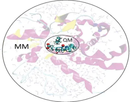
The hybrid QM/MM methodology was firstly introduced by Warshel and Levitt [130]. This methodology concurrently exploits the benefits of both the QM and the MM methods that were discussed in the last two sections. The region where the bond breaking and forming is taking place in the enzyme, along with selected catalytically relevant residues and water molecules, is treated with a QM method. The rest of the enzyme is treated with a MM force field as illustrated in fig. 5 below.
Fig. 5: Illustration of the QM/MM methodology. The system is divided into two layers: a small layer often regarded as the high layer, in which a chemical reaction occurs is treated with a QM method, while the rest of the enzyme and the surrounding solvent is modelled using MM [84].
The potential energy of the hybrid QM/MM comprises of three classes of interactions: interactions between atoms in the QM region, between atoms in the MM region and interactions between QM and MM atoms [84,132,136-139]. The QM and MM regions interactions are straightforward to describe using the QM and MM level respectively. However, the interactions between the two subsystems are more difficult to describe [132], and different approaches have
![Fig. 2: Catalytic Mechanism Scheme for the reduction of disulfide with Grx [26,28,29,33,57,63,64]](https://thumb-eu.123doks.com/thumbv2/123dok_br/16119727.1108799/22.918.188.754.205.643/fig-catalytic-mechanism-scheme-reduction-disulfide-grx.webp)
![Fig. 3: Thiol-Disulfide exchange related structural changes [29].](https://thumb-eu.123doks.com/thumbv2/123dok_br/16119727.1108799/24.918.161.429.145.520/fig-thiol-disulfide-exchange-related-structural-changes.webp)
![Fig. 4: Jacob’s ladder for the DFT functionals [106].](https://thumb-eu.123doks.com/thumbv2/123dok_br/16119727.1108799/33.918.215.608.117.495/fig-jacob-s-ladder-dft-functionals.webp)
![Fig. 6: Different methods to cap the QM region: link atoms (a) and frozen orbitals (b) and (c); b refers to LSCF orbitals and c to GHO orbitals [137]](https://thumb-eu.123doks.com/thumbv2/123dok_br/16119727.1108799/44.918.136.772.222.402/different-methods-region-frozen-orbitals-refers-orbitals-orbitals.webp)
![Fig. 7: Catalytic Mechanism Scheme for the reduction of GSSG-disulfide with Grx [26,28,29,33,57,63,64]](https://thumb-eu.123doks.com/thumbv2/123dok_br/16119727.1108799/48.918.166.862.123.625/fig-catalytic-mechanism-scheme-reduction-gssg-disulfide-grx.webp)