Mo
¨ ssbauer Characterization of Paracoccus denitrificans
Cytochrome c Peroxidase
FURTHER EVIDENCE FOR REDOX AND CALCIUM BINDING-INDUCED HEME-HEME INTERACTION*
(Received for publication, June 1, 1995, and in revised form, August 10, 1995) Susana Prazeres, Jose´ J. G. Moura, and Isabel Moura‡
From the Departamento de Quı´mica, Faculdade de Cieˆncias e Tecnologia, Universidade Nova de Lisboa, 2825 Monte de Caparica, Portugal
Raymond Gilmour, Celia F. Goodhew, and Graham W. Pettigrew‡
From the Department of Preclinical Veterinary Sciences, Royal (Dick) School of Veterinary Studies, University of Edinburgh, Summerhall, Edinburgh EH9 1QH, United Kingdom
Natarajan Ravi§, and Boi Hanh Huynh‡
From the Department of Physics, Emory University, Atlanta, Georgia 30322
Mo¨ ssbauer and electron paramagnetic resonance (EPR) spectroscopies were used to characterize the di-heme cytochrome c peroxidase from Paracoccus denitri-ficans (L.M.D. 52.44). The spectra of the oxidized enzyme show two distinct spectral components characteristic of low spin ferric hemes (S5 1/2), revealing different heme environments for the two heme groups. The Paracoccus peroxidase can be non-physiologically reduced by ascor-bate. Mo¨ ssbauer investigation of the ascorbate-reduced peroxidase shows that only one heme (the high potential heme) is reduced and that the reduced heme is diamag-netic (S5 0). The other heme (the low potential heme) remains oxidized, indicating that the enzyme is in a mixed valence, half-reduced state. The EPR spectrum of the half-reduced peroxidase, however, shows two low spin ferric species with gmax5 2.89 (species I) and gmax5
2.78 (species II). This EPR observation, together with the Mo¨ ssbauer result, suggests that both species are arising from the low potential heme. More interestingly, the spectroscopic properties of these two species are distinct from that of the low potential heme in the oxi-dized enzyme, providing evidence for heme-heme inter-action induced by the reduction of the high potential heme. Addition of calcium ions to the half-reduced en-zyme converts species II to species I. Since calcium has been found to promote peroxidase activity, species I may represent the active form of the peroxidatic heme.
A cytochrome c peroxidase has recently been isolated from
Para-coccus denitrificans (1), and initial characterization has been
per-formed using UV-visible, nuclear magnetic resonance (NMR) and electron paramagnetic resonance (EPR) spectroscopies (2–4). The
Paracoccus peroxidase is a periplasmic protein with a molecular
mass of 40 kDa (2). The physiological electron donor is most prob-ably the soluble cytochrome c550from the same organism (5). In many respects, this enzyme appears to be closely related to the peroxidase isolated from Pseudomonas aeruginosa, which has been extensively studied both spectroscopically and kinetically (6–16). Similar to the Pseudomonas enzyme (11), the Paracoccus peroxi-dase contains two c-type hemes with well separated midpoint redox potentials (2). For the high potential heme of the P. denitrificans enzyme, the redox potential was determined to be1176 mV. For the low potential heme, the redox potential was estimated to be within the range of2200 to 2100 mV (2). Based on the spectro-scopic and biochemical results (2), together with analogies drawn from the studies of P. aeruginosa peroxidase, it is concluded that the low potential heme is the peroxidatic center and the high potential heme functions as an electron transfer site.
Spectroscopic investigations of the Paracoccus peroxidase have revealed a complex activation mechanism that involves changes in the redox and spin state of the heme groups (2– 4). As purified, the peroxidase is in the oxidized form where both hemes are in the ferric state. At room temperature, the high potential heme is at least partly high spin (S5 5/2) and the low potential heme is low spin (S5 1/2). At low temperatures, both hemes are low spin exhibiting typical low spin ferric EPR signals (2). A NMR peak at 90 ppm, assignable to thee-CH3 group of an axial methionine ligand, was observed and sug-gested to be associated with the high potential heme, indicating that the high potential heme has a methionine-histidine axial coordination. The coordination of the low potential heme is not yet confirmed but has been proposed to be either bis-histidine or histidine-lysine by analogy to the Pseudomonas peroxidase. The oxidized form of the enzyme is catalytically inactive (17) and does not bind exogenous ligands such as cyanide. Addition of ascorbate reduces the high potential heme to the ferrous state, and a half-reduced mixed valence form of the enzyme is attained. The reduced high potential heme is low spin (S5 0) (4). This half-reduced form of the enzyme is catalytically active for the reduction of hydrogen peroxide (17) and readily binds exogenous ligands such as cyanide (2). Addition of sodium dithionite generates a fully reduced all ferrous form of the enzyme, which is a catalytically irrelevant species. Similar characteristics have also been found for other diheme bacterial peroxidases such as the peroxidases from P. aeruginosa (6 –16) and Pseudomonas stutzeri (18). In addition the Paracoccus * This work was supported by National Institutes of Health Grant
GM 47295 (to B. H. H.), a Wellcome Trust project grant (to G. W. P.), a Science and Engineering Research Council studentship (to R. G.), and the Junta Nacional de Investigac¸a˜o Cientifica e Tecnologica (Grants PBIC/QUI/1646/93 and STRDA/BIO/359/92 (to I. M.) and Grant STRDA/CEN/538/92 (to J. J. G. M.)). The costs of publication of this article were defrayed in part by the payment of page charges. This article must therefore be hereby marked “advertisement” in accordance with 18 U.S.C. Section 1734 solely to indicate this fact. To whom correspondence should be addressed.
§ Present address: Dept. of Chemistry, Carnegie-Mellon University, Pittsburgh, PA 15213.
‡ To whom correspondence should be addressed.
© 1995 by The American Society for Biochemistry and Molecular Biology, Inc. Printed in U.S.A.
24264
by guest on September 10, 2019
http://www.jbc.org/
enzyme was found to bind calcium ions (2, 3, 17). Other per-oxidases, such as lignin (19), manganese (20), and horseradish peroxidases (21–23), have also been found to contain bound calcium ions. Binding of calcium to the Paracoccus peroxidase promotes its activation (17) and appears to induce conforma-tional changes surrounding the immediate environment of the low potential heme (2, 3). In the presence of calcium, the low potential heme of the half-reduced enzyme becomes high spin at room temperature, suggesting a more open configuration for the heme site. The low potential heme is therefore ready for substrate binding. The binding of calcium also increases the midpoint redox potential of the high potential heme by approx-imately 50 mV, which should facilitate its reduction (2).
In this paper, a Mo¨ssbauer study of the P. denitrificans cytochrome c peroxidase is described. Both the oxidized and ascorbate-reduced forms of the enzyme were examined. Effects of the binding of calcium to the heme sites were investigated by both Mo¨ssbauer and EPR techniques. Intricate conformational changes induced by calcium binding to promote enzymatic ac-tivation are also suggested by this study.
MATERIALS AND METHODS
Cells of P. denitrificans (L.M.D. 52.44) were grown under low oxygen tension, and the periplasmic cytochrome c peroxidase was purified as de-scribed previously (1). The enzyme is isolated in the oxidized state. The growth medium was either enriched with the Mo¨ssbauer isotope57Fe (95%
plus enrichment) or contained natural abundance57Fe (2.2%). The protein
concentration of the purified enzyme solutions was determined by usinge 5 250 mM21cm21at 409 nm (oxidized enzyme) (1). The half-reduced form of the enzyme was achieved by anaerobic addition of a concentrated solution of sodium ascorbate. The enzyme was incubated under argon atmosphere for 60 min prior to calcium addition.
Mo¨ssbauer spectra of samples with a protein concentration of 1 mM
in 10 mMHepes (pH 6.4) and a volume of 350 – 400ml were recorded, either on a weak field Mo¨ssbauer spectrometer equipped with a Janis 8DT variable temperature cryostat or on a strong field Mo¨ssbauer spectrometer equipped with a Janis 12 CNDT/SC SuperVaritemp cry-ostat containing an 8-tesla superconducting magnet. Standard trans-mission Mo¨ssbauer measurements were made with a 50-mCi57Co(Rd)
source driven by a Doppler velocity transducer operating in the con-stant acceleration mode. The velocity scale was calibrated using room temperature spectra of a metallic iron foil, and the zero velocity of the Mo¨ssbauer spectra refers to the centroid of these spectra. EPR meas-urements were performed on a Bruker ER 200-SRC spectrometer with an Oxford Instrument ESR-9 continuous flow cryostat. For the EPR samples, the protein concentration was about 200mMin 10 mMHepes buffer (pH 6.4), and the volume was 200 –250ml.
RESULTS AND DISCUSSION
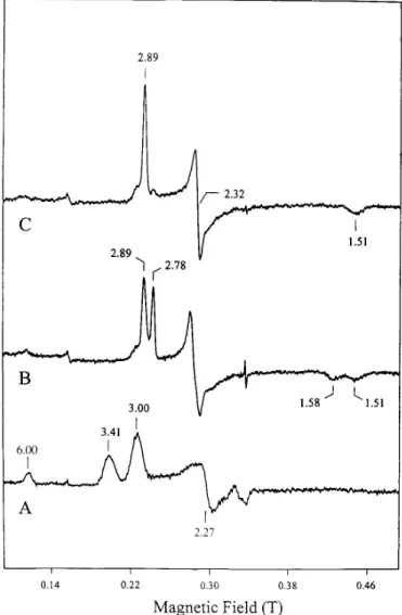
Fig. 1A shows the EPR spectrum of the oxidized cytochrome
c peroxidase from P. denitrificans recorded at 8 K. This
spec-trum is presented for the reason of clarity and for the purpose of comparison with those of the half-reduced state but is similar to the earlier published spectrum (2) in that two sets of reso-nances corresponding to the two c-type hemes were detected. The resonances at gmax5 3.00, gmed5 2.27, and gmin5 1.44 (not shown) were assigned to the low potential heme and the signal at gmax5 3.41 to the high potential heme (2). As with other low spin ferric hemes with a large gmax, the other two resonances for the high potential heme are not detected. It was assumed that gmedwas;2.0, and the corresponding gminwas estimated to be;0.6 according to the equation, gmax21 gmed2 1 gmin25 16 (24). The weak signal observed at g 5 6 repre-sents a minor high spin ferric heme component, possibly asso-ciated with the high potential heme, which is in a high spin-low spin equilibrium at room temperature (7).
Incubation of the oxidized sample with ascorbate for 60 min under an argon atmosphere yielded a half-reduced peroxidase, which exhibits the EPR spectrum shown in Fig. 1B.1In this
half-reduced enzyme, the high potential heme is reduced, as expected from its redox behavior and as supported by the absence of the gmax5 3.41 signal. Most interestingly, the low potential heme in this half-reduced enzyme displays two sets of EPR signals corresponding to two different species, both of which are distinct from that of the low potential heme in the oxidized enzyme. The observed g values are 2.89, 2.32, and 1.51 for species I and 2.78, 2.40, and 1.58 for species II. Also, these signals are sharp in comparison with that of the low potential heme in the oxidized enzyme. These observations indicate a subtle heme-heme interaction in which the reduction of the high potential heme induces a structural modification at and around the low potential heme. The relative proportion of spe-cies I and II was found to be dependent on the enzyme prepa-ration and is probably due to the different amounts of residual calcium bound to the purified enzyme (see below). For the sample shown in Fig. 1B, the relative concentration of species I to species II was estimated to be approximately 3 to 2 from the relative intensity of the peaks at gmax5 2.89 and 2.78, using the method of Aasa and Va¨nngård (25). Addition of calcium ions to this half-reduced state converts species II (gmax5 2.78)
1The spectra of Ref. 2 also contained a prominent peak at g max5 3.3,
which is absent in Fig. 1B. We are unsure of the basis for this, but it may be due to differences in enzyme preparations.
FIG. 1. EPR spectra of cytochrome c peroxidase from P. deni-trificans. A, oxidized enzyme; B, half-reduced enzyme (0.4 mM ascor-bate); C, half-reduced enzyme in the presence of 20 mMCa21.
Experi-mental conditions: temperature, 8 K; microwave frequency, 9.43 GHz; microwave power, 2 milliwatts; modulation amplitude, 1 mT; receiver gain, 83 104.
by guest on September 10, 2019
http://www.jbc.org/
into species I (gmax 5 2.89), resulting in an EPR spectrum showing a majority of species I (Fig. 1C). Consequently, both the reduction of the high potential heme and the binding of calcium ions to the protein have effects on the low potential heme. These effects could include protein conformation changes or changes in the coordination structure such as an increase in histidinate character of the proximal ligand, which is observed in some eukaryotic peroxidases (19, 23). It is inter-esting to note that, for peroxidases isolated from P. aeruginosa and P. stutzeri, the half-reduced enzymes display EPR spectra showing only one low spin ferric heme species. Either these proteins do not require Ca21or they bind Ca21so strongly that it is not lost during purification.
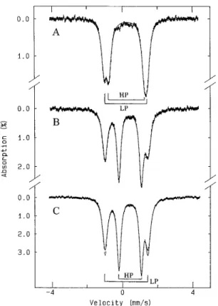
To further characterize the heme groups in the Paracoccus peroxidase, Mo¨ssbauer measurements were performed. We will first present the data recorded at 200 K, where the electronic relaxation time is fast in comparison with the nuclear preces-sion, resulting in the observation of quadrupole doublets for the heme irons regardless of their oxidation states. Under such
conditions, data analysis is simpler. Fig. 2 shows the 200 K spectra of the Paracoccus peroxidase in its oxidized (spectrum
A), ascorbate-reduced (spectrum B), and
ascorbate-reduced-plus-calcium (spectrum C) states. The oxidized peroxidase shows two partially resolved quadrupole doublets (Fig. 2A) corresponding to the two inequivalent c-type hemes. The data were least-squares fitted assuming two doublets with equal intensity and line width. For the half-reduced samples, the relative proportion of the two species of the low potential heme determined from the EPR data was used for the analysis of the Mo¨ssbauer data (Fig. 2, B and C). The results of the analysis are plotted as solid lines in Fig. 2, and the parameters obtained are listed in Table I. The good agreement between the data and the fits indicates that the Mo¨ssbauer data are consistent with the EPR findings. Also, this analysis allows unambiguous as-signments of the parameters to the two different hemes. For the high potential heme, the parameters (DEQ5 2.01 mm/s and d 5 0.24 mm/s for the oxidized S 5 1/2 state and DEQ5 1.27 mm/s and d 5 0.42 mm/s for the reduced S 5 0 state) are common for low spin heme compounds and are consistent with a methionine-histidine coordination (26 –32) (see also Table II). The quadrupole splittings for the low potential heme (2.45 mm/s in the oxidized enzyme and 2.46 mm/s in the calcium-bound enzyme), however, are atypically large (see Table II). The origin and significance of these unusually large values are currently unclear.
At 4.2 K, the electronic relaxation is slow and the oxidized peroxidase exhibits Mo¨ssbauer spectrum with magnetic hyper-fine structures as expected for low spin ferric heme compounds. Fig. 3 shows the Mo¨ssbauer spectra of the oxidized P.
denitri-ficans peroxidase recorded with the presence of a 50-mT2field applied parallel (spectrum A) and perpendicular (spectrum B) to the g-beam and with an 8-T parallel field (spectrum C). Similar to the high temperature data, these low temperature spectra can also be analyzed as a superposition of two compo-nents of equal intensity corresponding to the two low spin ferric hemes. The electronic g values have been determined from the EPR measurements, and the parameters,DEQandd, at 4.2 K can be extrapolated from the high temperature spectra. The only unknown parameters for the analysis of the low temper-ature data are the magnetic hyperfine coupling tensor A, which can be estimated from a ligand field theory commonly applied to low spin ferric heme complexes (33). In this theory the t2g orbitals are assumed to be well isolated from the egorbitals and are split by the tetragonal and rhombic crystal fields,D and V, respectively. The spin-orbit interaction mixes the t2gorbitals, resulting in three Krammers doublets. The observed g values and the low temperature Mo¨ssbauer spectra are originating from the ground doublet. If the g values of a low spin ferric compound are known, this theory can be used to estimate the corresponding A tensor (34) and the crystal field parametersD
2The abbreviations used are: mT, millitesla; T, tesla.
FIG. 2. High temperature Mo¨ ssbauer spectra of cytochrome c peroxidase from P. denitrificans. A, oxidized enzyme; B,
half-re-duced enzyme (6 mMascorbate); C, half-reduced enzyme in the presence of 17 mMCa21. The data were recorded at 200 K in the absence of an
applied field. The solid lines are least-squares fits of the experimental spectra (see text). The quadrupole doublets originating from the high potential (HP) and low potential (LP) hemes are indicated by brackets.
TABLE I
Least-squares fit parameters for the different oxidation states of the P. denitrificans cytochrome c peroxidase at 200 K
Oxidation state Heme group Spin DEQ d Ga Absorptionb
mm/s mm/s mm/s %
Oxidized High potential 1/2 2.016 0.03 0.246 0.02 0.33 50
Low potential 1/2 2.456 0.03 0.206 0.02 0.31 50
Half-reduced High potential 0 1.276 0.03 0.426 0.02 0.26 50
Low potential, I 1/2 2.466 0.03 0.256 0.02 0.32 30 Low potential, II 1/2 2.316 0.04 0.176 0.03 0.36 20 Half-reduced1 Ca21 High potential 0 1.276 0.03 0.436 0.02 0.25 50
Low potential, I 1/2 2.436 0.03 0.236 0.02 0.33 50 aFull width at half-maximum.
bFixed parameter.
by guest on September 10, 2019
http://www.jbc.org/
and V (33, 35, 36) in the units of the spin-orbit coupling con-stantl. Since the g values of the two hemes in the oxidized peroxidase are known from the EPR measurements, this theory was used to obtain initial guesses for the A values in the analysis of the low temperature Mo¨ssbauer spectra. These A values were then allowed to vary until a reasonable match between the theoretical simulations and experimental data was obtained. Results of such an analysis are shown as solid
lines in Fig. 3, and the parameters obtained are listed in Table
III along with the theoretical values. The good agreement be-tween the simulated and the experimental spectra indicates that the data support the presence of two distinct low spin ferric hemes in the oxidized peroxidase. The less than desirable fit at the outer absorption region is due to a distribution of the crystal field strength, which is not included in the analysis. Distributions ofD and V have generally been used to explain
the EPR and Mo¨ssbauer line shapes of low spin ferric complexes (37).
Since the crystal field parameters D and V can be readily determined from the EPR g values and since these parameters are expected to be affected by the ligands, EPR spectroscopy has been used for axial ligand assignment for proteins contain-ing low spin ferric protoheme IX. It was first demonstrated by Blumberg and Peisach (35) that proteins containing protoheme IX with the same axial ligands tend to haveD and V values that are clustered in a plot of the rhombic (V/l) versus the tetragonal (D/l) fields. This method of axial ligand assignment, however, was later found to contain ambiguities, particularly in the case of bis-histidine heme proteins (38, 39). Another method that has been proposed for heme axial ligand identification uses magnetic circular dichroism spectroscopy to measure the near infraredp to d charge transfer bands, the frequencies of which are sensitive to the axial ligation (38). In a study of 34 low spin ferric heme proteins, Gadsby and Thomson (38) established a linear correlation between the charge transfer transition en-ergy and the electronic hole enen-ergy of the t2gorbital (Eyz5 D/3 1 V/2). Consequently, Eyz can also be used for axial ligand assignment. Of the 34 heme proteins investigated, the Met/His ligation shows lower Eyz(1.5l 2 1.9l) than that of the His/His ligation (2.0l 2 2.3l). In Table III, the experimentally deter-mined and the theoretically estimated A values agree very well, suggesting that the theory used in our analysis describes the electronic properties of the hemes in the oxidized P.
denitrifi-cans peroxidase quite well. Using the theoretical values ofD
and V, the energy Eyzfor the high potential and the low poten-tial heme was found to be 1.15l and 1.91l, respectively. Earlier NMR measurements (2, 3) have established methionine liga-tion to the high potential heme, and the value 1.15l is therefore the smallest Eyzever reported for a Met/His ligation. Using the formula derived by Gadsby and Thomson (38), 1.15l for Eyz
FIG. 3. Low temperature Mo¨ ssbauer spectra of oxidized
cyto-chrome c peroxidase from P. denitrificans. The data were recorded
at 4.2 K with a 50-mT magnetic field applied parallel (A) and perpen-dicular (B) to theg-beam and an 8-T magnetic field applied parallel to theg-beam (C). The solid lines plotted over the experimental spectra are theoretical simulations using the parameters listed in Table III. The simulations of each spectral component in the presence of a 50-mT parallel field are also shown on top of spectrum A (solid line, high potential heme; dashed line, low potential heme).
TABLE II
Mo¨ssbauer parameters of some low spin ferric heme proteins with His/His or Met/His axial coordination
Ligands Protein (organism)a DEQ d G Temp.
(K) Ref.
mm/s mm/s mm/s
Met/His Cyt. c2(R. rubrum) 2.26 0.31 0.5 4.2 26
2.20 0.25 0.3 200 26
Cyt. c551(P. aeruginosa) 2.03 0.23 0.35 4.2 27
Cyt. c (Torula utilis) 2.14 0.21 200 28
Cyt. c (Tuna) 2.14 0.21 4.2 28
His/His HAO (N. europaer) 2.1 0.24 4.2 29
Cyt. b5(calf liver) 2.27 0.23 200 30
Cyt. c3(D. baculatus) 2.06 0.24 200 31
Nir (D. desulfuricans) 1.86–2.16 0.19–0.26 0.35 4.2 32
aCyt., cytochrome; HAO, hydroxylamine oxidoreductase; N, Nitrosomonas; D., Desulfovibrio; Nir, nitrite reductase.
TABLEIII
Crystal field and hyperfine parameters for the oxidized P. denitrificans cytochrome c peroxidase at 4.2 K
High potential heme Low potential heme Experiment Theory Experiment Theory
D/l 2.19 3.08 V/l 0.84 1.76 Eyz/l 1.15 1.91 gx 0.61 1.446 0.05 1.44 gy 2.00 2.276 0.01 2.27 gz 3.416 0.01 3.41 3.006 0.01 3.00 Axx/gnbn(T) 233.7 6 2.5 236.0 240.2 6 2.0 240.2 Ayy/gnbn(T) 128.5 6 1.0 130.0 119.3 6 1.0 119.3 Azz/gnbn(T) 178.7 6 0.3 180.2 153.4 6 0.5 153.4 DEQ(mm/s) 2.106 0.03 2.506 0.05 d (mm/s) 0.306 0.03 0.266 0.02 h 21.0 21.9 22.0 22.6 Line width (mm/s) 0.35 0.35 by guest on September 10, 2019 http://www.jbc.org/ Downloaded from
corresponds to a charge transfer energy in the order of 5300 cm21. It would be interesting to perform magnetic circular dichroism measurements to investigate whether the correla-tion between the Eyzand the charge transfer energy holds for the high potential heme. The value 1.91l obtained for the low potential heme falls into the range of Met/His axial ligation and is slightly outside of the range reported for bis-histidine heme proteins. It is, however, important to point out that axial ligand assignments using EPR and Mo¨ssbauer spectroscopy contain large uncertainties and that methionine ligation to the low potential heme is not supported by NMR measurements (3, 4). On the basis of their amino acid sequence work on the P.
aeruginosa enzyme, Ellfolk et al. (40) proposed that a
C-termi-nal domain contained a heme coordinated by a histidine and Met-254 (289 in P. denitrificans cytochrome c peroxidase num-bering) and an N-terminal domain contained a heme coordi-nated by a histidine and either His-240 (His-275 in P.
denitri-ficans cytochrome c peroxidase numbering) or a lysine.
Extension of sequence analysis to the enzyme from P.
denitri-ficans, the open reading frame f465 on the Escherichia coli
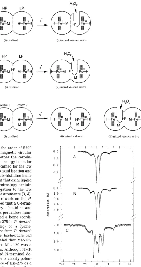
chromosome and the Mau G proteins3 revealed that Met-289 and His-275 were indeed conserved but also Met-129 was a conserved feature of the N-terminal domain. Although NMR evidence seems to preclude both the C- and N-terminal do-mains having methionine coordination, there is clearly poten-tial for this in the sequence, and the presence of His-275 as a possible ligand leads to a number of permutations of coordina-tion possibilities in the two redox states. Some of these possi-bilities are indicated in Fig. 4.
Fig. 5 shows the Mo¨ssbauer spectra of the half-reduced per-oxidase in the absence (spectrum A) and presence (spectra B and C) of calcium. The data were recorded at 4.2 K with a
3J. Van Beeumen, W. Hu, C. F. Goodhew, R. Gilmour, D. McGinnity,
N. Saunders, and G. W. Pettigrew, manuscript in preparation.
FIG. 5. Mo¨ ssbauer spectra of half-reduced Paracoccus
cyto-chrome c peroxidase in the absence (A) and presence (B and C) of added calcium. The data were recorded at 4.2 K with a 50-mT (A
and B) and an 8-T (C) magnetic field applied parallel to theg-beam. The solid lines are theoretical simulations using the parameters listed in Table III. In these simulations, the contributions from the oxidized and the reduced hemes are assumed to be equal. The relative proportions of species I and II for the oxidized low potential heme were taken from the EPR results. Diamagnetism was assumed for the reduced high poten-tial heme.
FIG. 4. Proposed models of the two
domains of P. denitrificans cyto-chrome c peroxidase. The cytocyto-chrome c
peroxidase is represented as a two-main structure. In model A, the heme do-main with His (H)-Met (M) coordination of the heme accepts the incoming electron and can therefore be considered high po-tential. Entry of the electron results in dissociation of His-275 (H*) from the heme of the second domain and allows access by hydrogen peroxide. HP, high po-tential; LP, low potential. In model B, both hemes are coordinated by methio-nine in the oxidized state. After reduction of one heme, the methionine of the second heme dissociates from the iron and allows access for hydrogen peroxide. In this model, His-275 (H*) is not a heme ligand but still plays a role as a distal catalytic residue. In model C, the entry of an elec-tron to center 1 causes a change in the properties of both hemes. Center 1 switches from high to low potential and reduces center 2, which becomes coordi-nated by a methionine. Center 1 itself becomes the peroxidatic center.
by guest on September 10, 2019
http://www.jbc.org/
50-mT (spectra A and B) and a 8-T magnetic field (spectrum C) applied parallel to theg-beam. These spectra clearly demon-strate that in the ascorbate-reduced peroxidase, one heme is reduced to a low spin ferrous (S 5 0) state and the other remains oxidized. The reduced heme (presumably, the high potential heme) displays a quadrupole doublet with parame-ters (DEQ5 1.23 6 0.03 mm/s and d 5 0.46 6 0.02 mm/s) typical of low spin ferrous hemes. Addition of calcium has no effect on this doublet. The oxidized heme (presumably, the low potential heme) exhibits a hyperfine split spectrum, which is distinguishable from that of the reduced heme. To analyze the spectral component of the oxidized heme, the contribution of the reduced heme was first removed, and the remaining spec-tral component was analyzed using the method described above. We began by analyzing the spectrum of the sample with calcium present, since it contains mostly species I (gmax 5 2.89). The parameters obtained for species I were then used in the analysis of the spectra of the sample without added calcium where both species I and II are present. The spectral data for species I and II are not sufficiently resolved to allow reliable determination of the hyperfine parameters for species II. The theoretical A values for species II and the concentration ratio of species I to species II obtained from the EPR measurements were used in the analysis. The results are listed in Table IV. The solid lines plotted in Fig. 5 are simulations using a 50/50 oxidized/reduced heme ratio. Good agreement is observed be-tween the experiments and the simulations, indicating that only one heme is reduced in these half-reduced protein samples with or without added calcium.
From these Mo¨ssbauer and EPR observations, the following conclusions can be drawn. The addition of ascorbate to the peroxidase reduces the high potential heme only. Reduction of the high potential heme affects the conformation surrounding the low potential heme, resulting in two low spin ferric states (species I and II) that are distinguishable from that in the oxidized form. The addition of Ca21 converts species II into species I. Since calcium has been found to promote the perox-idase activity, the low spin species I probably represents the peroxidatic heme in its active form.
In summary, Mo¨ssbauer and EPR spectroscopy have been used to characterize the two heme groups of P. denitrificans cytochrome c peroxidase in its oxidized and half-reduced forms. It was found that the spectroscopic properties of the peroxidatic heme can be affected by the reduction of the high potential heme and by the binding of calcium to the peroxidase, provid-ing further evidence for the long range heme-heme interactions observed in the bacterial cytochrome c peroxidases.
REFERENCES
1. Goodhew, C. F., Wilson, I. B. H., Hunter, D. J. B., and Pettigrew, G. W. (1990)
Biochem. J. 271, 707–712
2. Gilmour, R., Goodhew, C. F., Pettigrew, G. W., Prazeres, S., Moura, I., and Moura, J. J. G. (1993) Biochem. J. 294, 745–752
3. Prazeres, S., Moura, I., Moura, J. J. G., Gilmour, R., Goodhew, C. F., and Pettigrew, G. W. (1993) Magn. Reson. Chem. 31, S68 –S72
4. Prazeres, S., Moura, I., Gilmour, R., Pettigrew, G., Ravi, N., and Huynh, B. H. (1995) in Nuclear Magnetic Resonance of Paramagnetic Macromolecules (LaMar, G. N., ed) NATO ASI series, series c, Vol. 457, pp. 141–163, Kluwer Academic Publishers, Norwell, MA
5. Pettigrew, G. W. (1991) Biochim. Biophys. Acta 1058, 25–27
6. Foote, N., Turner, R., Brittain, T., and Greenwood, C. (1992) Biochem. J. 283, 839 – 843
7. Foote, N., Peterson, J., Gadsby, P. M. A., Greenwood, C., and Thomson, A. J. (1985) Biochem. J. 230, 227–237
8. Foote, N., Peterson, J., Gadsby, P. M. A., Greenwood, C., and Thomson, A. J. (1984) Biochem. J. 223, 369 –378
9. Ellfolk, N., Ro¨nnberg, M., Aasa, R., Va¨nngård, T., and Ångstro¨m, J. (1984)
Biochim. Biophys. Acta 791, 9 –14
10. Ellfolk, N., Ro¨nnberg, M., Aasa, R., Andre´asson, L.-E., and Va¨nngård, T. (1984) Biochim. Biophys. Acta 784, 62– 67
11. Ellfolk, N., Ro¨nnberg, M., Aasa, R., Andre´asson, L.-E., and Va¨nngård, T. (1983) Biochim. Biophys. Acta 743, 23–30
12. Aasa, R., Ellfolk, N., Ro¨nnberg, M., and Va¨nngård, T. (1981) Biochim. Biophys.
Acta 670, 170 –175
13. Ro¨nnberg, M., Araiso, T., Ellfolk, N., and Dunford, H. B. (1981) J. Biol. Chem.
256, 2471–2474
14. Ro¨nnberg, M., Araiso, T., Ellfolk, N., and Dunford, H. B. (1981) Arch. Biochem.
Biophys. 207, 197–204
15. Araiso, T., Ro¨nnberg, M., Dunford, H. B., and Ellfolk, N. (1980) FEBS Lett.
118, 99 –102
16. Ro¨nnberg, M., O¨ sterlund, K., and Ellfolk, N. (1980) Biochim. Biophys. Acta
626, 23–30
17. Gilmour, R., Goodhew, C. F., Pettigrew, G. W., Prazeres, S., Moura, J. J. G., and Moura, I. (1994) Biochem. J. 300, 907–914
18. Villalaı´n, J., Moura, I., Liu, M. C., Payne, W. J., LeGall, J., Xavier, A. V., and Moura, J. J. G. (1984) Eur. J. Biochem. 141, 305–312
19. Poulos, T. L., Edwards, S. L., Wariishi, H., and Gold, M. H. (1993) J. Biol.
Chem. 268, 4429 – 4440
20. Banci, L., Bertini, I., Bini, T., Tien, M., and Turano, P. (1993) Biochemstry 32, 5825–5831
21. Haschke, R. H., and Friedhoff, J. M. (1978) Biochem. Biophys. Res. Commun.
80, 1039 –1042
22. Morishima, I., Kurono, M., and Shiro, Y. (1986) J. Biol. Chem. 261, 9391–9399 23. Shiro, Y., Kurono, M., and Morishima, I. (1986) J. Biol. Chem. 261, 9382–9390 24. De Vries, S., and Albracht, S. P. J. (1979) Biochim. Biophys. Acta 546, 334 –340 25. Aasa, R., and Va¨nngård, T. (1975) J. Magn. Reson. 19, 308 –315
26. Huynh, B. H., Emptage, M. H., and Mu¨ nck, E. (1978) Biochim. Biophys. Acta
534, 295–306
27. Dwivedi, A., Toscano, W. A., Jr., and Debrunner, P. G. (1979) Biochim.
Biophys. Acta 576, 502–508
28. Lang, G., and Yonetani, T. (1968) J. Chem. Physiol. 49, 944 –950
29. Lipscomb, J. D., Andersson, K. K., Mu¨ nck, E., Kent, T. A., and Hooper, A. B. (1982) Biochemistry 21, 3973–3976
30. Mu¨ nck, E. (1978) Methods Enzymol. 54, 346 –379
31. Ravi, N., Moura, I., Costa, C., Teixeira, M., LeGall, J., Moura, J. J. G., and Huynh, B. H. (1992) Eur. J. Biochem. 204, 779 –782
32. Costa, C., Moura, J. J. G., Moura, I., Liu, M. Y., Peck, H. D., Jr., LeGall, J., Wang, Y., and Huynh, B. H. (1990) J. Biol. Chem. 265, 14382–14387 33. Griffith, J. S. (1957) Nature 180, 30 –31
34. Oosterhuis, W. T., and Lang, G. (1969) Physiol. Rev. 178, 439 – 456 35. Blumberg, W. E., and Peisach, J. (1971) Adv. Chem. Ser. 100, 271–291 36. Taylor, C. P. S. (1977) Biochim. Biophys. Acta 491, 137–149
37. Debrunner, P. (1990) in Iron Porphyrins (Lever, A. B. P., and Gray, H. B., eds) Part 3, pp. 139 –234, VCH Publishers, Inc., New York
38. Gadsby, P. M. A., and Thomson, A. J. (1990) J. Am. Chem. Soc. 112, 5003–5011 39. Walker, F. A., Huynh, B. H., Scheidt, W. R., and Osvath, S. R. (1986) J. Am.
Chem. Soc. 108, 5288 –5297
40. Ellfolk, N., Ro¨nnberg, M., and O¨ sterlund, K. (1991) Biochim. Biophys. Acta
1080, 68 –70 TABLE IV
Crystal field and hyperfine parameters for the half-reduced P. denitrificans cytochrome c peroxidase at 4.2 K
Low potential heme High potential heme (reduced (S5 0)) Species I Species II D/l 2.90 2.58 V/l 1.99 2.34 Eyz/l 1.96 2.03 gx 1.516 0.02 1.586 0.02 gy 2.326 0.01 2.406 0.01 gz 2.896 0.01 2.786 0.01 Axx/gnbn(T) 223.4 6 2.0 (241.2) a (242.4)a Ayy/gnbn(T) 12.2 6 1.0 (120.3) a (122.1)a Azz/gnbn(T) 143.4 6 0.5 (148.0) a (141.0)a DEQ(mm/s) 2.526 0.03 2.46 0.1 1.236 0.03 d (mm/s) 0.316 0.02 0.236 0.05 0.466 0.02 h 1.1 1.0 0 Line width (mm/s) 0.35 0.35 0.27
aThe numbers in the parentheses are the theoretical values.
by guest on September 10, 2019
http://www.jbc.org/
Graham W. Pettigrew, Natarajan Ravi and Boi Hanh Huynh
Susana Prazeres, José J. G. Moura, Isabel Moura, Raymond Gilmour, Celia F. Goodhew,
HEME-HEME INTERACTION
FURTHER EVIDENCE FOR REDOX AND CALCIUM BINDING-INDUCED
Peroxidase:
c
Cytochrome
Paracoccus denitrificans
Mössbauer Characterization of
doi: 10.1074/jbc.270.41.24264
1995, 270:24264-24269.
J. Biol. Chem.
http://www.jbc.org/content/270/41/24264
Access the most updated version of this article at
Alerts:
When a correction for this article is posted
•
When this article is cited
•
to choose from all of JBC's e-mail alerts
Click here
http://www.jbc.org/content/270/41/24264.full.html#ref-list-1
This article cites 39 references, 11 of which can be accessed free at
by guest on September 10, 2019
http://www.jbc.org/