Universidade de Lisboa
Faculdade de Ciências
Departamento de Biologia Animal
The role of the serosa in Immune defense of the
beetle Tribolium castaneum
Joana Pimenta Bernardes
Dissertação
Mestrado em Biologia Evolutiva e do
Desenvolvimento
Universidade de Lisboa
Faculdade de Ciências
Departamento de Biologia Animal
The role of the serosa in Immune defense of the
beetle Tribolium castaneum
Joana Pimenta Bernardes
Dissertação
Mestrado em Biologia Evolutiva e do
Desenvolvimento
Dissertation oriented by:
Professor Élio Sucena and Professor Maurijn van der Zee
Sumário
O modelo de investigação Tribolium castaneum tem sido amplamente utilizado em áreas como a biologia do desenvolvimento e evolução. Os trabalhos de investigação nesta espécie são importantes devido à sua relevância em termos económicos e de controlo de pragas. Esta espécie está inserida na ordem Coleopetera, a ordem com maior diversidade biológica conhecida. Este modelo é amplamente estudado pela sua facilidade de manutenção em condições laboratoriais; possui um ciclo de vida curto e alta fecundidade. T. castaneum possui bastantes ferramentas úteis para o seu estudo como por exemplo; o seu genoma encontra-se sequenciado (2008) e técnica de RNA de interferênciapode ser aplicada de forma estandartizada. Esta técnica permite uma análise rápida e simples da função do gene em estudo. Ao injectar fêmeas de Tribolium castaneum com RNA dupla cadeia (dsRNA) específico para um determinado gene resulta num knockdown ou inactivação do gene na prole da fêmea. Este efeito parental permite ao investigador fazer screenings funcionais até três genes em simultâneo.
Os embriões de T. castaneum possuem uma serosa que cobre por completo o embrião, esta serosa secreta a cutícula serosa, que se pensa ser uma inovação evolutiva. Esta tem como função a protecção do desenvolvimento dos insectos em ambiente terrestre. O desenvolvimento da serosa não se verifica em todas as classes de insectos, por exemplo, dípteros como Drosophila melanogaster apresenta somente um estado transiente em que o tecido conhecido como a amnioserosa cobre o embrião.
O desenvolvimento de tecido extra embrionário em insectos tem início na etapa de blastoderme, neste período uma porção da blastoderme anterior ou posterior especifica a serosa, que secreta uma cutícula quitinosa. Em T. castaneum, a serosa forma-se quando blastoderme posterior envolve o ovo ficando a blastoderme ventral interiorizada. A blastoderme ventral dá origem ao âmnio e ao embrião em gastrulação, o espaço entre o âmnio e o embrião forma a cavidade amniótica. Durante o fecho dorsal dá-se a fusão da serosa com o âmnio, formando um epitélio contínuo.
O gene que define a formação de tecido extra embrionário em insectos, tem por nome
zerknüllt(zen), este apresenta diferentes expressões e funções em insectos. Por exemplo, em
D. melanogaster na ausência da actividade do zen o embrião não forma qualquer tipo de
tecido extra embrionário. Em T. castaneum o gene zen sofreu uma duplicação seguida de um subfuncionalização produzindo dois loci (parálogos) Tc-zen1 e Tc-zen2. Nos embriões de T.
castaneum Tc-zen1 é um gene que actua na padronização antero-posterior, especificando a
serosa e Tc-zen2 actua mais tarde no desenvolvimento e inicia o fecho dorsal, este é também responsável pela correcta fusão do âmnio e da serosa. Ao fazer o knockdown de Tc-zen1 por RNAi, verifica-se a supressão do desenvolvimento da serosa e consequentemente da cutícula serosa, tendo o embrião apenas um epitélio extra embrionário simples constituído por células do âmnio. Os indivíduos Tc-zen1 RNAi, sem serosa, desenvolvem-se normalmente, apresentando uma fase transiente em que possuem uma cabeça comparativamente maior que o embrião selvagem com serosa. No entanto, se fizermos o knockdown por RNAi para o gene
Tc-zen2, os embriões não são viáveis e possuem problemas no fecho dorsal, apresentando um
fenótipo diferente do normal onde o embrião faz um ‘fecho ventral’ e fica totalmente invertido de fora para dentro. Os embriões de T. castaneum podem sobreviver sem a serosa apresentando apenas uma taxa de eclosão ligeiramente mais baixa quando comparado com o selvagem. O que nos leva a pensar; qual é afinal a função da serosa?
Outro gene que está intrinsecamente ligado à formação da serosa é o gene Chitin
advém não possui a cutícula serosa. Este fenótipo é viável apresentando apenas uma taxa de eclosão ligeiramente mais baixa.
O objectivo do nosso trabalho foi averiguar se a serosa tinha um papel na resposta imunitária inata; sendo que esta função pode actuar de duas maneiras: 1) a cutícula serosa pode funcionar como uma barreira física que previne a entrada de parasitas; ou 2) As células da serosa possuem a capacidade intrínseca na defesa do individuo, fazendo estas parte do sistema imunitário inato.
Estudos preliminares em T. castaneum revelam que a NFkB dorsal, um solicitador da resposta imunitária inata (Toll pathway), é altamente expresso na serosa. Outro estudo indica que a proteína Dorsal, um transactivador da Toll pathway, entra para o núcleo das células da serosa, quando é corrompida a integridade da serosa por perfuração (prick) do tecido. Outro estudo, este feito na espécie Manduca sexta, revelou que vários péptidos antimicrobianos (AMPs) estão presentes na serosa deste insecto quando a integridade do ovo é corrompida por perfuração.
No nosso projecto utilizamos testes de sobrevivência e eclosão com ovos de populações selvagem, Controlo RNAi, Tc-zen1 RNAi e Tc-CHSA1. A população controlo que serve para discernir quaisquer artefactos da técnica RNAi, é uma população com serosa. Na população Tc-zen1 há supressão do desenvolvimento da serosa e da cutícula serosa. Na população Tc-CHSA não há formação da cutícula serosa. Os testes foram feitos por um insulto bacteriano, em que perfurámos ou submergimos os ovos em soluções com Escherichia coli,
Micrococcus luteus e Straphylococcus aureus. E. coli é uma bactéria de Gram-negativa que
activa a via IMD e M. luteus e S.aureus são bactérias de Gram-positivas que activam a via Toll. Estas duas vias constituem parte da resposta imunitária inata, conhecida como resposta humoral, amplamente estudada em D. melanogaster.
Se a serosa estivesse envolvida na resposta imunitária inata, esperaríamos que a taxa de eclosão dos ovos da população com serosa fosse semelhante entre os testes com e sem insulto bacteriano.Esperaríamos também que a taxa de eclosão dos ovos sem serosa fosse significativamente inferior para os testes com insulto bacteriano comparativamente aos testes sem insulto bacteriano. Os resultados foram concordantes com o esperado para a população com serosa e apenas o insulto com E.coli verificou uma baixa de eclosão significativa para a população sem serosa apesar da taxa de eclosão da população Tc-zen1 ter sido bastante mais reduzida para todos os tratamentos. Se a cutícula serosa estivesse envolvida na resposta imunitária como barreira física da cutícula serosa, esperaríamos que a taxa de eclosão para a população Tc-CHSA fosse significativamente mais baixa para o tratamento com insulto bacteriano comparativamente ao tratamento sem insulto bacteriano. Os resultados não foram concordantes sendo que a taxa de eclosão da população Tc-CHSA não apresentava diferenças significativas entre o insulto bacteriano e sem o insulto bacteriano.
Concluindo, não foi excluído o envolvimento da serosa na resposta imunitária inata, mas necessitamos de um maior número de dados para as experiências anteriormente referidas. Por outro lado também, a cutícula serosa não aparenta estar envolvida no processo de defesa imunitária. Algo a ter em conta para fazer no futuro seria também ter maior informação sobre o sistema imunitário inato de T. castaneum, nomeadamente em in situs para os genes que sabemos estarem envolvidos na resposta imunitária inata desta espécie, bem como iniciar um projecto de PCR quanitativo, para saber se existe activação ou inibição de certa vias relacionadas com esta problemática.
Summary
The serosa is an extra-embryonic membrane that envelopes the embryo and yolk in insect eggs. This membrane secretes a cuticle and is thought to protect the embryo against desiccation. The serosa seems to be an evolutionary innovation of the insects but interestingly, embryos of the beetle T. castaneum can survive without a serosa, and the membrane is strongly reduced in Drosophila spp. This questions the function of the serosa. We hypothesize that the serosa protects against microbes in two ways. First, the serosal cuticle might act as a physical barrier. Second, the serosal cells themselves might play an important role in the innate immune reaction, for example by the excretion of AMPs (antimicrobial peptides).
To test the role of the serosa and serosal cuticle, we generated serosa-less eggs in the beetle Tribolium using zerknüllt1 RNAi (Tc-zen1) and eggs that did not possess the serosal cuticle using chitin synthase1 RNAi (also known as Tc-CHSA). We challenge these eggs by submergence or pricking with microbial solution. The microbial solutions we used were composed of Escherichia coli, Micrococcus luteus or Staphylococcus aureus and the microbe free submergence was done in water or PBS. The hatching rates for the Control RNAi population showed no significant difference between the treatments with bacterial challenge and without bacterial challenge. Tc-zen1 RNAi population revealed a significant lower hatching rate for the submergence with E.coli, this can indicate that the serosa might have a function in the defense against pathogens. On the other hand, Tc-CHSA showed no significant differences between treatments with bacterial challenge and no bacterial challenge, from that we concluded that the serosal cuticle had no function in the immune defense of the egg against pathogens. This work was the first attempt to unveil the presence of innate immunity in the extra-embryonic tissue of T. castaneum.
Key words:
Index
Sumário ... 1
Summary ... 3
Key words... 3
Introduction ... 4
1. Tribolium castaneum as a model organism ... 4
2. Extra-embryonic tissue: Evolution ... 4
3. Extra-embryonic tissue: Development ... 5
4. Serosa and serosal cuticle: Tc-zen1, Tc-zen2 and Tc-CHSA ... 6
5. Function of the serosa ... 7
6. Insect immunity... 8
7. Function of the serosa: Innate immunity hypothesis ... 8
Material and Methods ... 10
1. Tribolium castaneum development ... 10
2. Keeping the Tribolium population ... 10
3. Making double-stranded RNA (dsRNA) ... 10
4. Making RNAi populations of T. castaneum ... 10
5. Collecting eggs ... 10
6. Preparing the egg ... 11
7. Bacterial populations ... 11 8. Exploratory experiments ... 11 9. Submergence Protocol ... 11 10. Pricking Protocol ... 11 11. Imaging ... 11 12. Statistical analysis ... 11 Results ... 12
Washing in water vs. wash in bleach ... 12
Submergence Protocol... 12
Pricking Protocol ... 14
Imaging ... 16
Discussion ... 17
Washing in water vs. wash in bleach ... 17
Submergence Protocol... 17
Pricking Protocol ... 18
Comparison of bacteria tratments ... 19
Conclusion and suggestions ... 20
Bibliography ... 21
Suplementary data 1 ... 23
Introduction
1.Tribolium castaneum as a model organism
The beetle Tribolium castaneum is a relatively new model organism used for research in biology fields such as development and evolution. This species belongs to the phylum Arthropoda, class Insecta and to the order Coleoptera, this order includes more species than any other order, constituting about 25% of all known life forms. T. castaneum can easily be found in nature, normally where dry grains are stored; this beetle is well adapted to extremely dry environments1.
In present time, powerful tools such as a full sequenced genome since 20082 and RNA interference (RNAi) techniques are well established in T. castaneum. This model enables for a quick genetic analysis because it is easy to maintain in laboratory conditions, has a short life cycle and high fecundity, which facilitates the genetic crosses3. The technique of using RNAi
was first developed in Caenorhabditis elegans, this technique allows for a rapid straightforward analysis of gene function. Injections of synthesized dsRNA into the T.
castaneum egg can be used to knock-down the zygotic gene expression (RNAi) or even
injections of constructs with transposable elements (DNA) can be used to generate transgenics. The RNAi technique normally used in T. castaneum, consists of injecting synthesized dsRNA into the mother’s haemocel, the dsRNA corresponding to the gene of interest. The injection leads to a knockdown of the zygotic gene expression in any tissue or development stage, for the gene of interest 4. The technique can be applied in female pupae or adult beetles (parental RNAi) 5,6. Presently, the RNAi technique is being used to do a genome wide RNAi screen in this organism, known as the iBeetle project.
2. Extra-embryonic tissue: Evolution
One of the reasons Insecta is such a successful class is because it can occupy virtually any habitat on this planet. The adults have a chitinous bodywall that protects them against environmental stresses; like parasites and other potential infections and to extreme ecological conditions. Insect ability to occupy terrestrial habitats is constrained by the capacity the eggs have to survive in those conditions. The extra-embryonic membranes might have enabled the proliferation of insects in terrestrial environment by exploiting an enormous rage of ovipositor sites that were first unavailable to the ‘humidity dependent egg’7. In most insects eggs the extra-embryonic tissue is thought to be responsible for the protection of the embryo against harsh and extremely dry terrestrial conditions8. But there is a trade-off between the investment in the extra-embryonic membranes and the investment in the embryo proper, so there are variations on the amount of blastoderm that is used to make extra-embryonic tissue. There are several different examples in nature; the Grasshoppers have extra-embryonic membranes that compose 90% of the egg, while Drosophila melanogaster do not even have a proper serosa, only a transient amnioserosa9 (Fig 1).
Fig.1: Insect Evolutionary tree with the eggs at scale-quantity of extra-embryonic tissue per egg. Light Grey- extra-embryonic tissue; Dark Grey- Embryo9
Fig.2: Drawing of Arthropods eggs- Evolution of the serosa. A: Myriapods; B: most insects (T. castaneum); C: D. melanogaster
Fig.3: Wild-type T. castaneum embryo. A: Embryo in the Blastoderm stage- propective serosa on the posterior dorsal side characterized by widely space nuclei; B: Embryo in the Gastrulation stage- serosa starts to cover the embryo from the posterior to the ventral side formation of the serosal window
3. Extra-embryonic tissue: Development
Normally the insect egg is covered by two extra-embryonic membranes known as serosa and amnion and with an outer capsule known as chorion. But this is not the rule for all the insect classes. Myriapods, the sister group of insects, have only a single membrane at the dorsal side of the egg and this is thought to be the ancestral condition of insects10. T.
castaneum has a more typical insect development; it has an amnion on the ventral side, a
serosa on the dorsal and a differentiated blastoderm stage. D. melanogaster and higher dipterans in general, develop only a rudimentary amnioserosa on the dorsal side of the egg and the germ rudiment occupies the entire blastoderm. D. melanogaster seems to be a reversal to the ancestral condition and the reason of this regression is thought to be due to the fast development of this specie 11(Fig.2).
In general, we can divide the insects by two types of development: long-germ and short-germ. Drosophila and other higher Diptera have a long-germ development, where the entire blastoderm is occupied by the germ rudiment. The entire blastoderm will give rise to the segments simultaneously at the gastrulation stage. Tribolium has a short-germ development; in this case the germ rudiment occupies only a small portion of the blastoderm. The embryo proper will develop sequentially at the gastrulation stage by the addition of segments in the posterior proliferative zone12.
T. castaneum has a blastoderm like most insects. At the differentiated blastoderm
stage, the prospective serosa is visible at the dorsal-anterior side. The serosa tissue is characterized by flat cells, which DAPI staining revealed to be widely spaced nuclei (Fig. 3A). During the gastrulation stage, the serosa and the amnion start to cover the embryo at the 1
2
3
Fig.4: Embryo in Gastrulation- Tc-zen1 loss-of-function. A-D: Wild-type
egg- serosa covers the entire embryo, amnion on the anterior dorsal side; E-H: Tc-zen1 RNAi egg- serosa-less egg, amnion covers the dorsal side the embryo
Fig.5: Embryo in dorsal closure- Tc-zen2 loss-of-function. I-J:Wild-type
embryo, serosa is covering the embryo,fusion of the amnion and serosa; L-N: Tc-zen2 RNAi, the amnion and the serosa do not fuse- inside-out phenotype
posterior and anterior pole, giving rise to the amniotic fold. When the two crests meet, the serosal window is formed and then closed giving rise to two continuous membranes (Fig.3B). The outer membrane is known as the serosa and the inner membrane as the amnion13. The serosal membrane is responsible for the secretion of the serosal cuticle 14.
4. Serosa and serosal cuticle: Tc-zen1, Tc-zen2 and Tc-CHSA
The evolution of the development of extra-embryonic tissues in insects is deeply correlated with the changes of expression of the class-3 Hox genes. During the early radiation of insects, Hox3 lost its ancestral role in specifying segmental identity along the anterior-posterior axis and acquired a novel function in the specification of extra-embryonic tissue15.
This gene is now known as the zerknüllt (zen) and is a transcription factor. Changes in the expression of this gene are accompanied by major reorganizations in the extra-embryonic development, from the structural to the regulatory levels. In T. castaneum there are two zen homologs, known as the Tc-zen1 and Tc-zen2. The genes are quite similar, but are expressed at different stages of development with quite different functions, it is thought that duplication and a posterior sub-functionalization of the zen gene has occurred 11.
Tc-zen1 acts in early development and specifies the serosa fate at the blastoderm stage. When Tc-zen1 is knockdown by RNAi there is a loss of the serosa fate and consequently there will be no formation of the serosal cuticle. This loss is accompanied by an expansion of the germ rudiment on the anterior pole and instead of the serosa, the amnion will cover the embryo at the dorsal side (Fig.4). The serosa-less individuals are viable, they appear to have only a slight decrease in the hatching rate, demonstrating a high degree of plasticity in
Tribolium development 11.
Tc-zen2 acts in later development and is responsible for the fusion between amnion
and serosa, required for proper dorsal closure. When Tc-zen2 is knockdown the amnion and the serosa do not fuse and the embryo will close ventrally, it will present an inside-out phenotype (reverted embryos), where the legs and bristles are enclosed inside the body wall and the tracheae and hindgut are on the outside. The Tc-zen2 RNAi individuals are not viable11 (Fig.5).
D
1
C
Fig.6: A- Hybridization in situs for NFkB dorsal; B-Tc-Dorsal in the nuclei upon prick19
Tc-zen1 and Tc-zen2 share only 38% amino acid identity, so the different phenotypes
of the knockdown might be generated by divergent transcriptional regulation16. With the study of Tc-zen1 and Tc-zen2 knockdowns and double knockdowns; we know that the expression of
Tc-zen2 is dependent of the expression of Tc-zen1. The Tc-zen2 cannot rescue the serosa after
the Tc-zen1 knockdown and Tc-zen1 expression cannot rescue dorsal closure of the Tc-zen2 knock-down. So maybe we have the case of a sub-functionalization of the gene11, but it could also be neo-functionalization, we needed to check if the ancestral condition with single copy had both functions.
In T. castaneum, the serosal membrane secretes the serosal cuticle in the late embryonic development. This extracellular matrix is a functional specialization to terrestrial life because it is a physical barrier that protects the developing egg. The gene Tc-CHSA (chitin
synthase A also known as Tc-CHS1) encodes for an enzyme responsible for the synthesis of
chitin fibers, by catalyzing the b1-4 linkage between N-acetyl-glucosamines supplied by the cytoplasm17. Knockdown of the Tc-CHSA gene (RNAi) results in the absence of the serosal cuticle. With a complete knockout the phenotype is lethal at the embryonic stage. With only partial knockdown, the phenotype is viable but with a lower hatching rate when compared with a Wild-type population 18.
5. Function of the serosa
Since the serosa-less eggs are viable and produce normal sized larvae, we were lead to the questions: what is the serosa function after all? And why the embryo spends energy and time making this structure? Some functions for the serosa have been suggested and tested, for example; the serosa protects the embryo against desiccation (Jacobs et all, in prep). In the experiment, the serosa-less eggs were subjected to relatively high and relatively low humidity. The serosa-less eggs had a significantly lower hatching rate when compared to the control eggs under both humidity regimes. With further experiments, they draw the conclusion that it was the serosa that had an active role in the protection against desiccation. Another function, alternative to physical protection that has been suggested is that the serosa might play a role in the immune defense of the egg. This function can be achieved by acting as a barrier or by an active role of the innate immune defense; the immune defense can be activated upon infection of microorganisms.
Evidence from preliminary studies revealed; 1) Toll-rel/NFkB ortholog dorsal, is highly expressed in the presumptive serosa of T. castaneum (Fig. 6A)19. The NFkB are transcription factors involved in the control of a large number of cellular and physiological processes, such as immune and inflammatory responses, developmental processes, cellular growth
and apoptosis. In this case Toll-rel/NFkB ortholog dorsal might be an elicitor of the innate immune response. 2) Upon damage of the serosa, the Tc-Dorsal protein locally enters the nucleus. This could be an activation of the immune response (Fig.6B)19. The protein Tc-Dorsal, is a part of the Toll pathway and thought to form a complex with Tc-Dif and Tc-Cactus20. Tc-Dorsal could enter the nuclei of the serosa cells and activate the synthesis of Antimicrobial peptides (AMP), by binding to the KB sites and transactivating the AMPs in culture21. 3) Also, the presence of several AMPs and lysozymes as been detected in the extra-embryonic tissue of the moth Manduca sexta upon challenge. Melanization, a reaction of the innate immunity,
also occurred in the yolk but not in the embryonic cells. This demonstrated immune competence of the extra-embryonic tissue in this moth22.
6. Insect immunity
Insects do not possess an adaptive immune system instead they have a sophisticated innate immunity, consisting of cellular and humoral responses. The innate immune defense of insects has been studied extensively in D. melanogaster, from this model we know:
1) The Cellular response is mediated by hemocytes, which are hemoplymph cells produced by the Lymph gland. The hemocytes differentiate in lamelocytes, crystal cells and plasmatocytes and wich will encapsulate23, melanize24 and phagocyte25 the invasive pathogens. 2) The humoral response is responsible for the melanization, production of reactive oxygen species (ROS) and production o Antimicrobial peptides (AMP). The humoral response is composed mainly of two pathways, functioning on the membrane of the fat body: First, the IMD pathway activated by gram negative bacteria, has a PGRP-LC and PGRP-LE as Dif protein transactivator27
3) The humoral and the cellular responses are interconnected because the AMPs synthesized by the humoral response can also activate the cellular response,28.
Recently some studies were made with regard to the innate immunity of T.
castaneum. These studies indicated some differences in the innate immunity of T. castaneum
relative to D. melanogaster, for example the humoral response in T. castaneum might be a bit more promiscuous than in Drosophila; both Escherica coli and Micrococcus luteus, a gram negative bacterium and a gram positive bacterium, respectively, in a single bacterium suspension can activate the IMD and the Toll pathways29. So in T. castaneum, a single type of bacteria can active both pathways. This could happen trough signaling crosstalk, like intracellular signaling or heterodimerization of the NFkB molecules at the terminal ends of the two signaling pathways30.
7. Function of the serosa: Innate immunity hypothesis
In this project we tried to test if the serosa has an innate immune function in the egg of T. castaneum. We hypothesize that the serosa can protect the embryo from pathogens in two ways: (1) the serosal cuticle might act as a physical barrier where the pathogens cannot enter. (2) the serosal cells themselves might play a role in the innate immune reaction, by activating the humoral response in the serosal cells30.
We used two genes to do the knock-down; Tc-zen1 and Tc-CHSA, the Tc-zen1 RNAi population has no serosa and no serosal cuticle while the Tc-CHSA RNAi population has no serosal cuticle. We also made a Control RNAi population so we could compare to the others RNAi populations and remove any artifact from the technique of RNAi. The Control RNAi population is injected with a dsRNA that does not belong to the Tribolium genome. We performed hatching rates experiments for these populations with or without microbial challenge. For the microbial challenge we used E. coli, M. luteus and S. aureus. E. coli is a Gram negative bacterium that is known to activate the IMD pathway in Drosophila, M. luteus and S.
aureus are Gram positive bacteria that are known to activate the Toll pathway in Drosophila.
We challenged the eggs by pricking them with a needle dipped in the bacteria or by submerged them in the bacterial solutions.
If the serosa has an active immune defense role we expect to see;
1a) No statistically significant differences between hatching rates of the control RNAi population for the treatments with bacterial challenge, in comparison with the treatment without microbial challenge.
2a) Statistically significant differences between the hatching rates of the Zen RNAi population, with a lower hatching rate for the treatments with bacterial challenge compared with the treatments without bacterial challenge.
If the serosal cuticle plays a role in the defense against bacteria we expected to see: 1b) No statistically significant differences between hatching rates of the Control RNAi population, for the treatments with bacterial challenge compared with the treatment without microbial challenge.
4b) Statistically significant difference between the hatching rates of the CHS RNAi population, with a lower hatching rate for the treatments with bacterial challenge than the treatment without bacterial challenge.
We were also interested to see if there were differences in the reactions between the bacteria populations; If the egg had a kind of ‘Drosophila reaction’28 (activation of the pathway dependent of the type of pathogen- gram positive or gram negative) or if the egg had no dependence to the kind of pathogen like in T. castaneum larvae30 ( one type of bacteria can activating both pathways). The reaction can be tested by the bacterial challenge; if there are significant differences between the treatments with E.coli or M. luteus, for the Control RNAi and Zen RNAi (serosa-less) populations.
Our goal was to test the serosa and the serosal cuticle for an active role in innate immune function in the egg of T. castaneum.
Materials and Methods
1. T. castaneum developmentT. castaneum is a holometabolous insect, which means it goes from egg, to larvae, to
pupae, to adult form. The development is temperature dependant. In our climate cabinet the temperature is around 30ºC so the egg will develop for 3,6 days, the larvae for 17,2 days, the pupae for 5,5 days, for a total developing time of 27 days. The hatching rate at this temperature is normally around 88% for a Wild-type population.
2. Keeping the Tribolium population
T. castaneum populations were kept at 30ºC, the population live in a box with a mix of
flour, yeast and fumidyl in a mix of 1000: 50: 5. Every time we wanted to collect eggs we put the target population on instant flour overnight. The white flour facilitates the collection of the eggs. To change the medium, from normal to reproductive or vice versa and for collecting the eggs, we had to sieve the flour, using sieves with different sized meshes6.
3. Making double-stranded RNA (dsRNA) Suplementary data 1- Transformation
We synthesized dsRNA for the following genes: Tc-zen1, Tc-CHSA and Control, a gene not present in the genome of T. castaneum- region from the pCR II topo vector containing the pUC origin region.
We cloned the genes from a cDNA library with specific primers, and then we inserted them into a pCR II TOPO vector using specific primers. Then we transformed E.coli bacteria with lines containing these plasmids with the genes. We grew these bacteria in LB, we made glycerol stocks for later use and we extracted the plasmids of interest by purifying them using the miniprep purification kit (Qiagen®). Then we cut the template sequence using either Xho1 or BamH1 restriction enzymes, so we could get linear plasmids which were used as a template for the dsRNA synthesis, we use the RNAi MEGAscript® Ambion® kit for this procedure. For the dsRNA synthesis we use three different enzymes; T7, T3 and SP6. T7 we used for all the genes plus T3 only for the zen dsRNA and SP6 for the rest of the dsRNA (control dsRNA and
Tc-CHSA dsRNA). We measure the concentration of the sample in the Nanodrop and used only
the samples with a concentration higher than 200ng/µL9. 4. Making RNAi populations of T. castaneum
The beetles used were from a lab strain known as San Bernardino, which is a normally used lab strain of T. castaneum. Female pupae were collected from the laboratory population shortly before hatching. The dorsal side of the terminal segment was fixed on the microscope slide with fixogum. We injected about 0.2 µL of the dsRNA solution ventrally between the third and the fourth abdominal segments9. Afterwards we added the males and 7 days after the offspring was collected and used in the experiments.
Four different population were used; Wild-type population (Wt), Control population RNAi (C RNAi), Tc-zen1 RNAi (Zen RNAi) and Tc-CHSA RNAi (CHS RNAi). The control population was formed by injecting the mother pupae with a sequence of dsRNA not present in the
Tribolium genome. The Zen RNAi population was made by injecting the mothers with dsRNA
complementary to the Tc-zen1 sequence. And the CHS RNAi population was formed by injecting the mother with dsRNA complementary to Tc-CHSA sequence.
The C RNAi population has a wild-type phenotype ansd was used as the control for RNAi artifacts, the Zen RNAi population did not have serosa or serosal cuticle and the CHS RNAi population did not have a serosal cuticle.
5. Collecting eggs
The eggs collected for the experiment were two days old. We put the beetles on the instant flour overnight, collected the eggs the next morning and let them mature for one full day at 30ºC at 50% relative humidity.
6. Preparing the eggs- Supplementary data 2
In order to remove the chorion and excess flour the eggs were washed. We used a fine sieve to submerge the eggs in 100% bleach (sodium hyplochlorite), to wash of the chorion and excess flour, or in water, to remove the excess flour.
We performed some exploratory experiments to see the effect of removing the chorion in the Wt population and Zen RNAi population.
7. Bacterial populations
Three species of bacteria were used; Escherica coli (E. coli), Micrococcus luteus (M.Luteus) and Straphylococcus aureus (S. aureus). The S.aureus strain used contains a m-cherry insert which makes it easily tractable with fluorescent microscopy.The bacteria were grown overnight in LB medium. To the S. aureus medium we added 100µL of antibiotic.
8. Exploratory experiments
Before developing the two protocols we did some exploratory experiments. The goal was to collect information to see what details in the protocol worked better, to achieve a high hatching rate for all populations. We did among others; several treatments were made with bleach at different concentrations; experiments with different types of slides to see if we could immobilize the eggs for pricking; we try to glue the eggs on to the slide; we try different kinds of needles, for example acupuncture needles, capillary needles and others.
9. Submergence Protocol
Before using the eggs for the submergence protocol, the chorion was removed.
For this protocol we used; Wt eggs, C RNAi eggs, Zen RNAi eggs and CHS RNAi eggs with 6 different submerging treatments; (1)PBS (10%-made in the laboratory); (2) tap Water; (3) E. coli bacterial solution; (4) M. luteus bacterial solution; (5) S. aureus bacterial solution.
The bacterial solutions were obtained by centrifuging (Heraeus® centrifuge for 10mL tubes) the LB solution at 2000 rpm, 30ºC for 10 minutes. The overflow was discarded and 10mL tap water was added. The de-chorionized eggs were submerged three times in the treatment solutions. After this, the eggs were kept in a climate chamber at 30ºC with the relative humidity between 20%-80% for 4 to 5 days to develop- we could not control the relative humidity level in the chamber. After 5 days we counted the number of eggs that hatched.
For this protocol we did on average 300 eggs per treatment. 10. Pricking Protocol
For this protocol we did not remove the chorion from the eggs, we only washed them with water to remove the excess flour. We aligned the eggs on a slide; the alignment was done by moving the eggs in the water. The eggs would stick to the slide after they were dry; the residues of flour helped the eggs to stick to the slide, making the pricking easier.
For this protocol we used 4 populations; Wt eggs, C RNAi eggs, Zen RNAi eggs and CHS RNAi with 4 different treatments; (1) control without pricking; (2) mock pricking with a sterile needle; (3) pricking with the needle dipped in E. coli solution, (4) pricking with the needle dipped in M. luteus solution. For the pricking we used tungsten needles either sterile or dipped in bacteria. We pricked the egg in the posterior end and dipped the needle in the bacteria solution every time we pricked an egg. After the pricking, the eggs were kept in the climate chamber at 30ºC, 60% relative humidity for 4 to 5 days to develop. We counted the number of eggs that hatched.
For this protocol we did between 100 and 200 eggs per treatment. The treatments of mock pricking for CHS RNAi eggs and Zen RNAi eggs had only 60 eggs per treatment.
11. Imaging
For the submerging treatment with the S. aureus we analyzed Wt eggs and Zen RNAi eggs with fluorescence microscopy.
12. Statistical Analysis
We analyzed the data using R i386 2.15.0 program, the tests we used were a simple ANOVA with a Tukey range test for multi comparison of means.
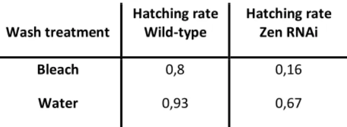
Table 1 – Hatching rate (hatched eggs/total eggs) for preparing the eggs treatments; De-corionized with bleach, Washed with water
Results
1. Wash in water vs. wash in bleach First we did an exploratory experiment, to see the effects of the washing treatments used to prepare the eggs. The results (Table 1) for the preparation of the eggs demonstrated a higher hatching rate for the Wt eggs in comparison with the Zen RNAi eggs for both treatments, as expected. The Wt eggs had a hatching rate of 80% with the wash in bleach and a
hatching rate of 93% with the wash in water. The Zen RNAi eggs had a hatching rate of 16% with the wash in bleach and 67% with the wash in water. When we compared the hatching rates between eggtypes, we could verify that the wt eggs were much more resilient than the Zen RNAi eggs, for both treatments. The difference with the wash with bleach was quite high between populations, whereas the difference with the wash in water was not as significant. This might be due to the lack of serosa of the Zen RNAi and the bleach treatment being much harsher then the water treatment.
2. Submergence protocol
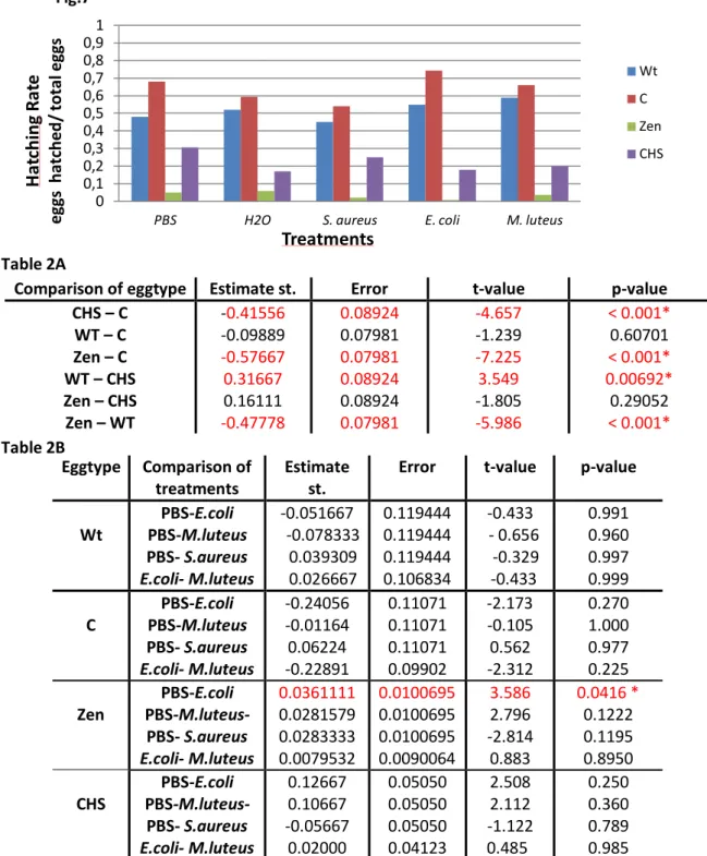
In the submergence protocol we submerged the eggs from the different populations in solutions with or without bacteria and checked how many eggs hatched. The hatching rate was measure by the ratio between the number of eggs hatched and and the number of eggs treated (Fig. 7). For the statistic analysis of the submergence protocol, we did a simple ANOVA with a multi-comparison of means for the eggtype and within eggtype for the different treatments used, known as the Tukey rage test (Table 2).
When we compared the eggtypes we could see statistically significant differences between hatching rates for the Zen RNAi eggtype and the C RNAi eggtype (p-values <0.001), and also between the CHS RNAi eggtype and the C RNAi eggtype (p-values <0.001).
The results demonstrated a relatively high hatching rate for the Wt population, always higher than 45 %. We could not see statistically significant differences between treatments with or without bacteria for the Wt population, but the S. aureus seemed to be slightly more aggressive than the rest of the tested bacteria.
The hatching rates for the C RNAi eggs were also relatively high for all treatments, always higher than 50%. The hatching rate of the C RNAi population was always higher when we compared with the Wt population; this eggtype was more resilient than the Wt to the submergence protocol. There were no statistically significant differences between the PBS treatment and the treatments with the submergence in bacterial solution- E. coli, M. luteus and S. aureus (1a/1b). The wild-type egg (Control RNAi) had no reaction to the bacterial challenge. The S. aureus seemed again to be more aggressive than the other bacteria.
When we observed the hatching rates for the Zen RNAi eggs we could see they were considerably lower for all treatments, did not reach the 10%. When we compared the hatching rates of Zen RNAi population there was no statistically significant differences for the hatching between treatment with PBS and treatments with M. luteus and S. aureus. There was a significant difference between the treatment with submergence in E.coli and the PBS treatment (p-value of 0.0416) (2a). This indicated a reaction to the treatment with the gram negative bacteria. When we compared the results of the Zen RNAi, for the washing treatment tryouts with the submergence protocol, we could see that the hatching rate of the Zen RNAi
Wash treatment Hatching rate Wild-type Hatching rate Zen RNAi Bleach Water 0,8 0,93 0,16 0,67
Fig.7: A-Graphic for Hatching Rate of the eggs for the submergence protocol - for different populations Wt (blue), C(red), Zen(green), CHS(purple) and different treatments of submergence:H2O, PBS, E. coli, M. luteus and S. aureus
Table 2: A) Tukey range test for eggtype; B) Tukey range test for treatments for C RNAi population, Zen RNAi population and CHS RNAi population;
and PBS) was around 5%. This demonstrated how the maintenance in relatively high humidity, decreased the hatching rate in serosa-less eggs.
In respect of the CHS RNAi eggs we could see a quite oscillating hatching rate, with an average of about 20%. For the CHS RNAi population there was no statistically significant differences between the treatment with PBS and the treatments with bacterial challenge - E.
coli, M. luteus and S. aureus (2b). So there seemed to be no reaction to the bacterial challenge
in this population. The submergence in water seem to be quite harsh for the CHS RNAi population (Fig. 7)
Comparison of eggtype Estimate st. Error t-value p-value
CHS – C WT – C Zen – C WT – CHS Zen – CHS Zen – WT -0.41556 -0.09889 -0.57667 0.31667 0.16111 -0.47778 0.08924 0.07981 0.07981 0.08924 0.08924 0.07981 -4.657 -1.239 -7.225 3.549 -1.805 -5.986 < 0.001* 0.60701 < 0.001* 0.00692* 0.29052 < 0.001* Eggtype Comparison of treatments Estimate st.
Error t-value p-value
Wt PBS-E.coli PBS-M.luteus PBS- S.aureus E.coli- M.luteus -0.051667 -0.078333 0.039309 0.026667 0.119444 0.119444 0.119444 0.106834 -0.433 - 0.656 -0.329 -0.433 0.991 0.960 0.997 0.999 C PBS-E.coli PBS-M.luteus PBS- S.aureus E.coli- M.luteus -0.24056 -0.01164 0.06224 -0.22891 0.11071 0.11071 0.11071 0.09902 -2.173 -0.105 0.562 -2.312 0.270 1.000 0.977 0.225 Zen PBS-E.coli PBS-M.luteus- PBS- S.aureus E.coli- M.luteus 0.0361111 0.0281579 0.0283333 0.0079532 0.0100695 0.0100695 0.0100695 0.0090064 3.586 2.796 -2.814 0.883 0.0416 * 0.1222 0.1195 0.8950 CHS PBS-E.coli PBS-M.luteus- PBS- S.aureus E.coli- M.luteus 0.12667 0.10667 -0.05667 0.02000 0.05050 0.05050 0.05050 0.04123 2.508 2.112 -1.122 0.485 0.250 0.360 0.789 0.985 0 0,1 0,2 0,3 0,4 0,5 0,6 0,7 0,8 0,9 1
PBS H2O S. aureus E. coli M. luteus
H
at
ch
in
g
R
at
e
eg gs h atc h ed / to ta l e gg sTreatments
Wt C Zen CHS 13 Fig.7 Table 2A Table 2BWhen we compared the bacteria treatments: E. coli and M. luteus, there was no statistically significant difference between them. The C RNAi and the Zen RNAi eggs did not have a different reaction to the Gram positive or Gram negative bacteria. We only tested the difference between these two bacteria, because S. aureus showed to be much more aggressive than the other bacteria.
Overall there were no results to accept or deny our hypothesis. There seemed to be a difference on the reaction to the bacterial challenge of Zen RNAi population, but not to all the bacteria treatments. In this protocol there might have been some problems with the relative humidity and the survival of the eggs with no serosa and no serosal cuticle.
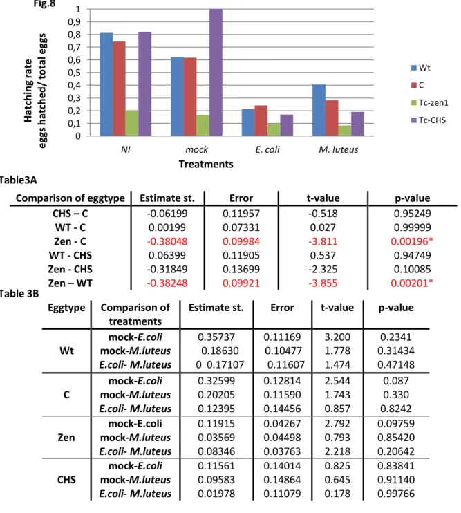
3. Pricking protocol
For the pricking protocol we submitted the different populations to a prick with a tungsten needle, the needle was dipped or not in bacteria solution. The hatching rate was determent for all treatments by the ratio: number of eggs hatched/ number of eggs treated (Fig.8). For the statistic of the pricking protocol (Table 3), we did a multi-comparison of means, known as the Tukey range test for the eggtype and between treatments of each eggtype. The Tukey rage test for the eggtype, had statistically significant differences between Zen RNAi and control RNAi hatching rates (p-value of 0.00196). We also had a statistically significant difference between the Zen RNAi hatching rate and the one from the wt population (p-value of 0.00201). The rest of the eggtypes did not appear to have any difference between the hatching rates.
When we observed the hatching rate of the wt eggs, we could see the higher hatching rate for the treatments without bacteria; 81% for no prick and 62% for mock prick. And a lower hatching rate for bacteria treatments, the difference was statically significant; the pricking with
E.coli had 20% hatching rate and the pricking with M. luteus about 40%.
For the C RNAi eggs we could see similar results. For the control RNAi population we could not see statistically significant differences between the treatments with bacterial challenge - E. coli and M. luteus- and the treatments without bacterial challenge -mock prick (1a/1b). Even though the in the graphic there seem to be a difference between them. The results indicate that this type of egg might do not have a reaction to the prick with bacteria.
For the Zen RNAi eggs we found again quite low hatching rates for all treatments. There were no statistically significant differences between the bacterial challenge and without bacterial challenge, in the Zen RNAi population (2a). This population showed no differences between the treatments with bacteria- E.coli and M. luteus. The absence of serosa did not affect the response of the prick with bacteria.
Finally by observed the CHS RNAi hatching rates we could see a quite high hatching rate for the treatments without bacteria than for treatments with bacteria. For the treatments mock for the Zen RNAi and CHS RNAi population there was not enough data to be correctly analyze and compared the hatching rates. There were also no statistically significant differences between the mock pricking treatments and the bacterial challenge for the CHS RNAi eggtype (2b). The absence of serosal cuticle did not affect the survival of the eggs pricked with bacteria, so the serosal cuticle might not have a function in the defense against pathogens.
0 0,1 0,2 0,3 0,4 0,5 0,6 0,7 0,8 0,9 1
NI mock E. coli M. luteus
Ha tc h in g ra te eg gs h atc h ed / to ta l e gg s Treatments Wt C Tc-zen1 Tc-CHS
Fig.8: A-Graphic for Hatching Rate of the eggs for the pricking protocol - for different populations Wt (blue), C(red), Zen(green), CHS(purple) and different treatments of pricking: no pricking (NI), mock pricking, E. coli and M. luteus
Table 3: A) Tukey range test for eggtype; B) Tukey range test for treatments for C RNAi population, Zen RNAi population and CHS RNAi population;
No statistically significant difference resulted from the comparison between bacteria treatments: E. coli and M. luteus. The C RNAi and the Zen RNAi eggs did not have a different reaction to the Gram positive or Gram negative bacteria.
Summarizing there were no data to accept our hypothesis. The Zen RNAi and CHS RNAi population did not have enough data to analyze the hypothesis but there seemed to be a trend of lower hatching rates for the treatments with bacteria in comparison with the treatments
Comparison of eggtype Estimate st. Error t-value p-value
CHS – C WT - C Zen - C WT - CHS Zen - CHS Zen – WT -0.06199 0.00199 -0.38048 0.06399 -0.31849 -0.38248 0.11957 0.07331 0.09984 0.11905 0.13699 0.09921 -0.518 0.027 -3.811 0.537 -2.325 -3.855 0.95249 0.99999 0.00196* 0.94749 0.10085 0.00201* Eggtype Comparison of treatments
Estimate st. Error t-value p-value
Wt mock-E.coli mock-M.luteus E.coli- M.luteus 0.35737 0.18630 0 0.17107 0.11169 0.10477 0.11607 3.200 1.778 1.474 0.2341 0.31434 0.47148 C mock-E.coli mock-M.luteus E.coli- M.luteus 0.32599 0.20205 0.12395 0.12814 0.11590 0.14456 2.544 1.743 0.857 0.087 0.330 0.8242 Zen mock-E.coli mock-M.luteus E.coli- M.luteus 0.11915 0.03569 0.08346 0.04267 0.04498 0.03763 2.792 0.793 2.218 0.09759 0.85420 0.20642 CHS mock-E.coli mock-M.luteus E.coli- M.luteus 0.11561 0.09583 0.01978 0.14014 0.14864 0.11079 0.825 0.645 0.178 0.83841 0.91140 0.99766 Fig.8 Table3A Table 3B 15
without bacteria. This indicates that perhaps the serosa or the serosal cuticle might be related to the immune defense of T. castaneum.
4. Microscopy
We analyze the images we retrieved from the microscopy, with infection by S. aureus of eggs from the wt and Zen RNAi population. Overall we saw the eggs infected with bacteria, but we could not differentiate if the bacteria were inside or at the surface of the egg. We also used the DIC microscopy, because the Wt infection had a pattern, when we did the DIC microscopy we could see the pattern was the granules inside the vitelin membrane. These results were not as good as we wanted because we could not discard from being auto-fluorescence.
Discussion
Fig.9: A- Wt eggs infected with S. aureus; B- Zen eggs infected with S. aureus; C- Wt eggs infected with S. aureus, granules; D- Wt eggs infected with S. aureus DIC microscopy
C D
Discussion
The overall results could not give a concrete answer to our hypothesis. But there were still interesting results for us to speculate on what might be the relationship of the serosa and serosal cuticle with innate immunity of the T. castaneum egg. In addition we summarized several protocol problems that could be overcome in the future.
1. Wash in water vs wash in bleach
When we analyzed the exploratory results for the wash, we found the wt eggs were more resilient than the Zen RNAi eggs. The wt population seemed to survive the wash in water and in bleach better (93% for water and 80% for bleach). Zen RNAi on the other hand, appeared to be more susceptible to the washing in bleach and in water (62% for water and 16% for bleach). The reason why the hatching rate of the Zen RNAi eggs was low, might be that the eggs with no serosa are more sensitive to high humidity. This conclusion as been demonstrated in Chris Jacob’s work (Jacobs et all, in prep). The reason why the bleach wash had lower hatching rates, especially for the Zen RNAi eggs, was because it removed the chorion of the eggs. If the egg did not have a serosa it would be more susceptible to mechanical shocks than an egg with serosa.
In conclusion, the bleach treatment is quite aggressive to the eggs and almost lethal to the eggs that do not possess a serosa. So we can say, by removing the chorion we made the eggs more susceptible to mechanical shocks and to high humidity, making the chorion a fundamental layer, used to protect the integrity of the T. castaneum egg.
The reason why we used the wash in bleach in the submergence protocol was because it was essential to remove the chorion, to verify if it was the serosa and not the chorion that protected against pathogens. For the pricking protocol we did not use the wash with bleach because there was no need to do so, the needle could make a hole through the chorion and the serosa.
2. Submergence protocol
When we reviewed the results of the submergence protocol, we could see that our hypothesis could not be tested due to the low hatching rate of the Zen RNAi eggs, for all treatments. The submergence protocol required some resilience to: 1) wash of the chorion with bleach, 2) mechanical shocks by sieving and 3) high humidity.
There were significant differences between eggtypes, revealing that the different eggs had hatching rates quite different from each other. A curious result was the high survival of the C RNAi eggs, comparative to the Wt population, even though the difference was not statistically significant. Both population have a Wild-type egg, the C RNAi population has an insert of dsRNA for a non-codifying sequence in T. castaneum, so there would be no reason for the higher survival rate. Only if the technique produced some artifacts not known that would improve the resilience of these eggs to harsh treatments, like the removal of the chorion5.
When we analyzed the effect of the treatments, we did two control experiments: the submergence in water and the submergence in PBS. We did both of this treatments because we wanted to control the osmotic potential our eggs were exposed to. With this experiment we could see some populations survived better in water- wt population- and others in PBS- C RNAi population and CHS RNAi population. The Zen RNAi population has a low hatching rate for both treatments, so it was affected for the solution itself, not for the osmotic potential of the solutions. We expected this response of the CHS RNAi population due to the lack of serosal cuticle, which exposes the serosa, this extra-embryonic membrane can be quite sensitive to certain changes in the osmotic potential, leading most of the times to a apoptosis of the cells (Jacobs et all, in prep).
The effect of the treatments also show that the submergence with S. aureus had the lowest hatching rate, this is expectable because S. aureus is a quite aggressive bacteria, that most insects are not able to defend.
1a and 2a) The control population did not have statistically significant differences between the treatments with or without submergence in bacteria, which lead us to believe the wild-type egg can protect itself to the submergence in bacteria. The protection could be passive, the serosa or the serosal cuticle could be functioning as a barrier, or active, when the serosa cells can active the immune defense. For answering these questions we had to analyze the hatching rate of the serosa-less eggs and of the eggs with no serosal cuticle:
The Zen RNAi population had in general the lowest survival for all treatments, the reasons could be, as seen before, the aggressiveness of the bleach treatment but mainly we thought it was due to the sensitivity the Zen RNAi eggs show to high and low humidity (Jacobs et all, in prep). The submergence protocol has of course high levels of humidity in the steps but also the oscillations of humidity in the chamber where we matured the eggs could lead to the low survival of the eggs.
1b) The Zen RNAi population showed a statistically significant difference for the hatching rate of the submergence in E.coli and the PBS treatment, but it was only lower for E.
coli. The other bacteria did not affect the hatching rate of the eggs with no serosa. This lead us
to think that the serosa might have an immune defense mechanism for the gram negative bacteria. If true, we are between two case scenarios: First, the immune response of T.
castaneum egg is like the immune response in D. melanogaster and upon entrance of gram
negative bacteria the IMD pathway is activated28,29 . Second the immune response of the T.
castaneum egg is like the larvae of T. castaneum; upon entrance of gram negative bacteria
both pathways- Toll and IMD- are activated, this is based on the recent studies of T. castaneum immunity30 and it is in agreement with the preliminary results 19. The experiments made cannot distinguish which scenario is correct, our suggestion is to make a quantitive PCR for the elements of both pathways and with and without E. coli challenged.
2b) The survival of the CHS RNAi population when we compared the treatments with or without bacterial challenge, showed no statistically significant differences between them. The serosal cuticle seems to have no critical role in the defense against pathogens, in this case pathogens in solution. We could only speculate that the main reason for the lower survival of the CHS RNAi is the absence serosal cuticle which could make the eggs more sensitive to the mechanical shocks of this protocol or even to the humidity. Even though there is no proof that the serosal cuticle has any function in the survival of the eggs to high humidity.
In conclusion we could not say that the absence of serosa could affect the entrance of pathogens by acting as a barrier or has an active immune defense; because we were not able to find statistically significant differences for all treatments with bacteria, in the eggs without serosa. At least for the gram negative bacteria, E.coli, there seemed to be a significant reaction upon the absence of the serosa. On the other hand, there seemed to be no influence in the presence of the serosal cuticle, there were no major differences between the submergence with or without bacteria, so the serosal cuticle might not have a function in the defense against pathogens via solution.
3. Pricking protocol
For the pricking protocol the results were similar to what we expected. In general, the pricking without bacteria had a higher survival than the prick with the needle dipped in bacteria. This shows that there was an effect of the bacteria on the populations. This protocol had less shocks to the integrity of the eggs; the main problem was the thickness of the needle because the needles were too thin, they broke between treatments, making the holes in the eggs too big. When the hole was too big could cause leaking of the egg. The leaking of the egg could lead to the dry out and sequential death of the egg.
1a and 2a) For the pricking protocol the Control RNAi population, performed with no differences between the treatments with or without bacterial challenge, leading us to believe the wild-type eggs, are not affected by the pricking with bacteria.
The Zen RNAi population had survival problems even in the treatments without bacteria. This can be explain by the protocol, where the eggs are align in water on the slide, so the eggs spent at least 15 minutes at high humidity levels. Another problem with the survival of the Zen RNAi eggs, is the absence of serosa and serosal cuticle that makes them quite sensitive to the mechanical shocks, like pricking. The pricking with the needle ruptures the integrity of the egg because the membranes that protect the wild-type egg are normally 3: chorion, serosal cuticle and serosa. While in the case of serosa-less eggs, only the chorion was present, so when we pricked the egg, there was more leaking than in any other population. The leaking caused the desiccation of the egg and high mortality rate. Even thought the hatching rate of the eggs with no serosa was low, it was higher than for the submergence protocol, the reason might be the less time spent on relative high humidity (Jacobs et. all, in prep).
2a) The results were not in agreement with our hypothesis, there were no statistically significantly differences between mock pricking and the pricking with E.coli or M. luteus. So we could not say that the serosa had any immune defense activity. But we did not have enough data to analyse correctly the mock pricking treatment for this population, leading us to believe that we did not have the correct result. Our suggestion is to repeat this protocol with enough data (at least 300 eggs) to correctly test our hypothesis.
2b) In this Protocol, the CHS RNAi population performed much better than in the submerged protocol. There was no statistically difference in the survival between the treatments without bacteria and with bacteria. We could not speculate any function of the serosal cuticle because there was not enough data to analyze correctly the hatching rate numbers for the mock prick treatment, a fundamental treatment to compare the influence of the serosal cuticle in the immune response. But taking in account the results we had, they demonstrate that the serosal cuticle might not act in the innate immune defense. In the pricking protocol we were testing an active defense against the entrance of pathogens, but the serosal cuticle could still act as a barrier.
Overall there were no statistically significant differences between the mock pricking and the pricking with bacteria for the eggs without serosa and without serosal cuticle, so we concluded there was no active immune defense on the serosa or the serosal cuticle. Nevertheless there were significant differences for the Control and Zen RNAi populations, but we could not see if the reason for the differences was the absence of serosa or not. Also there were no significant differences between the Control RNAi population and the CHS RNAi population, leading us to think that the presence of serosal cuticle is probably not affecting the survival of the eggs that are pricked with bacteria.
4. Comparison of bacteria treatments
For both protocols there was no statistically significant difference between the bacteria treatments- when E.coli and M. luteus treatments were compared. But analyzing the data we had we could see a trend where E. coli was more aggressive than the gram positive bacteria M. luteus. This could be due to the virulence of the bacteria or with the mechanism of innate immune defense. With this experiment we could not distinguish if there was a differentially response for the two bacteria used. On the other hand, we know now that in T.
castaneum larvae there is normally a common response of the two pathways against a single
bacterium infection, gram positive or gram negative30. This hypothesis was in agreement with our data.
5. Conclusions and suggestions
Concluding about the importance of the serosa in the defense against pathogens, we where the firsts to develop a protocol where you can test issue. Even thought we cannot say much about it, because there were no significant differences for all the treatments and not enough data for some. There was still a trend of lower survival for serosal-less eggs and even lower for eggs injected with bacteria. Our suggestion is that more data should be collected to test our hypothesis, especially for the pricking protocol; for the mock pricking treatment with the CHS RNAi population and Zen RNAi population. In our opinion is better to use the pricking protocol, instead of the submergence protocol, because it is less aggressive to the integrity of the eggs: humidity-wise and in terms of physicals shocks. The thing to care in this protocol is the size and the cut of the needle, these characteristics can be control by the use of tungsten needles. Another advantage of this protocol was we could check if the serosa had an active function in the immune defense, distinguished the barrier function of the serosa.
Finally our suggestion is to collect more information about the innate immune system in Tribolium castaneum, by the means of making some hybridization in situs for the genes we already know are involved in the innate immune response of this specie- for both pathways. As well as perform some quantitative PCR for the same genes using embryos submitted or not to bacterial challenge. In this case we suggest the pricking with E. coli, so we can see if there is activation or inhibition of genes.
Bibliography
1. Evolution of the Insects , Grimaldi, D., Engel, M. S., 1st Edition, 2005
2. The genome of the model beetle and pest Tribolium castaneum. Consortium, Tribolium Genome Sequencing. Abril 2008, Nature, pp. 949-955.
3. The Biology of Tribolium with special emphasis on Genetic Aspects. Sokoloff, A. Oxford : Clarendon Press and Oxford University Press, 1977. 2.
4. Knockdown stands up; Voorhoeve, P.M.; Agami, R.; Thends Biotechnology 21 2-4;2003 5. Parental RNAi in Tribolium (Coleoptera). Gregor Bucher, Johannes Scholten and Martin
Klingler. s.l. : Current Biology, 2002, Vol. 12. 3.
6. Tribolium castaneum beetle book 1; Gregor Bucher; version 1 and 2; 2008 7. Segmentation. Tautz, D. s.l. : Development Cell, 2004, Vol. 7.
8. Ovipositors, amnions and eggshell architecture in the diversification of terrestrial
arthropods; Zeh, D.W.; Zeh, J.A. & Smith, R.; Q. Rev. Biol. 64, 147-168 ; 1989harbor
9. Gastrulation: from cells to embryos; Roth, S.; Coldspring laboratory press New York; 2004 10. Evolutionary Changes in Developmental Pontentials of the Embryo proper and Embryonic
Membranes along with Derivative structures in Atelocerata with Special Reference to hexapoda (Arthropoda). Machida, R., Ando, H. Proc. Arthropod. Embryology Society of
Japan, 1998.
11. The Amnioserosa is an apomorphic character of cyclorrhaphan flies. Schmidt-Ott, U., Development Genes Evolution 210, 373-6, 2000
12. Distinct Functions of the Tribolium zerknullt Genes in Serosa Specification and Dorsal
Closure. Maurijn van der Zee, Nicola Berns and Siegfried Roth. s.l. : Current Biology, 2005,
Vol. 15.
13. Tribolium Embryogenesis A SEM study of cells shapes and moements from blastoderm to
serosal closure, Handel, K. ,Grunfelder, C.G., Roth, S., and Sander, K., Development Genes
Evolution 210, 167-179, 2000
14. Extra-embryonic development in insects and the acrobatics of blastokinesis. Panfilio, K.A.; Akam, M.; et all; Dev Biol 313:471-91; 2008.
15. Schimidt-Ott, Urs. Hox3/zen and the Evolution of Extraembryonic Epithelia in Insects. s.l. : Landes Bioscience, 2010. Chapter10.
16. Case studies on the gene zerknüllt, decapenthaplegic and short-gastrulation in the beetle Tribolium castaneum illustrates concepts in evolutionary development biology; Zee, M.; Dissertation; Köln University; 2006
17. Chitin metabolism in insects: structure, function and regulation of chtin synthases and
chitinases; Merzendorfer, H.; Zimoch,L.; Experimental Biology 206; 4393-3312; 2003
18. Recent advances in understanding mechanisms molecular of insect cuticle differentiation; Moussaian, B.; Review Insect Biochemistry and Biology 40;363-375; 2010
19. The maternal NF-kb/Dorsal gradient of Tribolium castaneum: Dynamics of early
dorsoventral patterning in a short-germ beetle; Chen, G.; Handel, K.; and Roth, S.;
Development 127, 5145-5156, 2000
20. Comparative genomic analysis of Tribolium immune system; Zou,Z.; Evans, J.D.; Lu, Z; Zhao,P.; Williams, M.; Sumathipala, N.; Hetru, C.;Hultmark, D. and Jang, H.; Genome Biology, 2007
21. Expression and nuclear translocation of the rel/NF-kB-related morphogen dorsal during the
immne response of Drosophila; Reichhart, J.M.; Georgle,P.; Meister,M.; Lemaitre, B.; Kappler,C.; Hoffmann, J.A.; . C.R. Acad. Sci. III 316:1218–24, 1993
22. Bacterial challenge stimulates innate immune responses in the extra-embryonic tissues of
tobacco hornworm eggs, Gorman, M. J., Kankanala, P. & Kanost, M. R., Insect Molecular
Biology 13, 19-24, 2004