1. Radiology and Research Division, Hospital Regional Monterrey, ISSSTE
2. Human Anatomy Department, Universidad Autonoma de Nuevo Leon, Facultad de Medicina
3. Rheumatology Department, Internal Medicine Division, Universidad Autonoma de Nuevo Leon, Hospital Universitario “Dr. José Eleuterio González”
4. Traumatology & Orthopedic Division, Universidad Autonoma de Nuevo Leon, Hospital Universitario “Dr. José Eleuterio González”
Conclusion: Synovitis and triquetral synovitis were the most prevalent lesion in three-studied phases. This could suggest the triquetrum as the first morphologi-cal site to be affected by RA; so it’s assessment should be considered in the RA evaluation when it’s clinically suspected.
Keywords: OMERACT-RAMRIS Score; Triquetrum bone; Magnetic Resonance Imaging; EULAR-OMER-ACT Atlas; Rheumatoid Arthritis.
IntroductIon
Rheumatoid arthritis (RA) is the most common chronic autoimmune disease, characterized by joint destruc-tion, disability, and life expectancy reduction due to progressive inflammatory complications1. Early dia -gnosis allows prompt treatment and reduces joint dama ge2,3. Magnetic resonance imaging (MRI) is the primary imaging study to visua lize anatomical struc-tures, abnormalities, variants, cortical bone shape ir-regularities, and morphological changes in RA accord-ing the American College of Radiology (ACR) Appropriateness Criteria4; it is the most sensitive (96%) diagnostic test and disease follow-up imaging method with a high specificity (94%) that can evidence patho-logic hand-wrist lesions in initial RA phases. The Eu-ropean League Against Rheumatism-Outcome Mea-sures in Rheumatology Clinical Trials (EULAR--OMERACT) Atlas (2005) and the OMERACT-RAM-RIS Rheumatoid Arthritis Magnetic Resonance Ima ging Score (2003) have established a Score Sheet for assess-ment with MRI5-7.
Resnick (1976) wrote about ERA phase describing frequents erosions in the pisiform and triquetrum bones after synovial proliferation. Other studies have reported a relation between age and erosion lesions in
The very early inflammatory triquetral
lesion by magnetic resonance imaging –
is this the first sign in rheumatoid arthritis?
Larios-Forte MC1, García-Coronado JM2, Skinner-Taylor CM3, Esquivel-Valerio JA3,
Vega-Morales D3, Vilchez-Cavazos F4, Quiroga-Garza A2, Elizondo-Omaña RE2
ACTA REUMATOL PORT. 2019;44:218-224
AbstrAct
Introduction: Rheumatoid Arthritis (RA) an autoim-mune, chronic, and disabling disease if untreated, af-fects wrist joints, with a diagnostic delay of up to 2 years. Triquetral bone allows rotational movement that pivots over the rest wrist bones, and maintains physi-ological loads during mobility. Magnetic resonance imaging (MRI) is the most sensitive (96%) method for diagnosis, shows lesions as early as in the initial RA stages. Our aim was to determine the most frequently affected structures in the hand-wrist joint by MRI us-ing the OMERACT-RAMRIS Score (2003) in three dif-ferent RA stages, including clinically suspicious arthral-gia (CSA) that haven’t reported before
Methods: We performed an exploratory, transverse, observational, descriptive study in 60 patients enrolled and classified by rheumatologists as: CSA, early rheumatoid arthritis (ERA), and established RA, prior to performing a dominant hand-wrist MRI for evalua-tion and descriptive analysis by an expert radiologist Results: Female predominance 83% (50), with a mean age 42.0+/-13.5 years; A total of 1,731 hand-wrist bone and joint sites were evaluated using EULAR-OMER-ACT Atlas (2005), identifying 56% (964 sites) with typ-ical RA lesions: synovitis, erosions, and bone marrow edema (BME or osteitis); synovitis was the most frequent with 46% (445 sitelesion), and triquetral syno -vitis the most frequent each clinical group: CSA 87% (20/23), ERA 91% (20/22), and RA 93% (14/15)
the carpal joint8. The hand-wrist complex is an anatomi cal joint structure that connects the radiusulna distal joint to the proximal metacarpals. It posses -ses great ligament variability, capsules, and bone struc-tures, each one with different functions. Carpal bones are joined by strong intrinsic ligaments (interosseous dorsal and radial ligaments with low flexibility) and
ex-trinsic ligaments (palmar and dorsal), that maintains the
biomechanics of the joint (physiological loads and the mobility arcs resistance), and provides wrist stability. These ligaments also have mechanoreceptors and pro-vide proprioception and neuromuscular control, which predominate at the triquetrum bone, where most liga-ments from the two carpals rows are inserted9-11.
At the first carpal row, which has the most mobili-ty, the scaphoid and triquetrum contribute to stabilize the central carpal column joint, allowing ample move-ments of flexion-extension and abduction-adduction (80% radial distribution and 20% ulnar)4,11-13. The tri-quetrum bone has a pyramid-shaped with 4 articular facets: the proximal, distal, lateral, and medial, articu -lating with the triangular fibrocartilage (first joint cap-sule), the hamate bone, the lunate bone, and the pisi-form (second joint capsule) respectively. This articulated triangular fibrocartilage absorbs and trans-mits the forces and pressures exerted on carpal bones12. Its stability depends on nine palmar and dorsal interosseous ligaments with mixed fiber directions, joi -ning all the carpal bones. The most morphologically controversial ligament according to Nozaki14is the ul-nar collateral ligament (as Hogikyan and Louis de-scribed as well as Bogumill) which it’s morphology in-cludes to the pisiform bone4,13,14 (Table I).
Some studies report in beginning early stages of RA, the triquetrum, capitate, and lunate bones are fre-quently affected with erosions15-17. Tamai et al.18, (2007) reported by MRI in ERA, the relationship between bone marrow edema (BME or osteitis) and serological markers (C-Reactive Protein, Antibody-IL-6, anti-CCP [anti-cyclic citrullinated peptide], HLA-DRB1 and DAS-28 [Disease Activity 28 joints Score]) with their autoimmune, anatomical, and traumatic factors, that play an important role with synovial and erosions pathogenesis. These appear predominantly in the metacarpophalangeal areas (MCP), influenced by the location of the ligaments18,19.
The aim of our study was to evaluate by MRI, pa-tients from three different RA clinical stages, including the clinically suspicious arthralgia (CSA), to assess the starting structural changes and determine the most
fre-quently affected site within the complex hand-wrist joint bones and topography.
MAterIAls And Methods
An exploratory, transverse, descriptive, non-blinded observational study, was performed in a northeastern Mexican cohort population (Monterrey, Nuevo Leon) from November 2016 to February 2017. Overall, 60 adult patients were enrolled from the rheumatology clinic (UANL University, School of Medicine), who cepted and signed informed consent; all assessed ac-cording to the American College of Rheumatology: ACR-EULAR (2010) criteria and were classified into three clinical groups, according to their RA phase (CSA, ERA, or RA). The CSA group included patients (RA direct relatives) with only arthralgia symptoms without clinically detectable inflammation, considered by the rheumatologists as being suspect to progress to arthritis over time; ERA group, consisted in patients with less than 2 years from the onset of arthralgia and joint inflammation, and the established RA patients group with more than 2 years with disease activity score (DAS28 greater than 2.6)
Simple T1 and STIR (Fat Sat - General Electric Signa Twin HDx 1.5 Tesla) MRI sequences were obtained from the dominant hand (Table II). One day prior to the study patients stopped anti-inflammatory medica-tion. Images were evaluated by a musculoskeletal ra-diologist, according the EULAR-OMERACT (Outcome Measures in Rheumatoid Arthritis Clinical Trials) At-las 2005 in order to identify the RA known lesions: synovitis, erosions, and BME/osteitis.
Descriptive statistics was performed with chi square test to differentiate between groups in SPSS (IBM
Sta-tistical Package for the Social Science SPSS, 22 version)
This study was reviewed and approved by the Ethics and Research Committees of the UANL University, School of Medicine: Registration number RE--17-00010, complying with the Helsinki Declaration.
results
A total of 60 MRIs were performed in patients with a mean age of 42.0+/13.5 (19–70) years. Most were wo -men 83% (50 wo-men/10 -men), and were distributed in 3 different RA stages as: CSA with 38% (23), ERA 37% (22), and RA 25% (15).
vitis more frequently than the MCP (metacarpopha-langes) joints. (Table III). Wrist synovitis was present in 46% (445 site-lesions) of the patients in all three phases, erosions in 38% (362), and BME/osteitis in We evaluated their hand-wrist bones and joints sites
including distal radio-ulnar joint (1,731), identifying 964 RA known lesions (synovitis, erosions and BME/osteitis). The carpal joints were affected by syno
-tAble I. proxIMAl cArpAl bone lIgAMent dIstrIbutIon
Bone Lig Location Specification
Triquetrum 9 1 I Lunotriquetral
4 P Long radiolunate (radiolunotriquetral), ulnotriquetral, capitotriquetral (triquetrohamocapitate), ulnar collateral
4 D Dorsal radiocarpal (dorsal radiotriquetral), dorsal intercarpal (dorsal scaphotriquetral), triquetrohamate (dorsal), scaphotriquetral Scaphoid 7 2 I Scapholunate, scaphotrapeziotrapezoid (palmar)
4 P Radioscaphoid (radial collateral), scaphocapitate, scaphotriquetral, radioscaphocapitate (palmar)
1 D Dorsal Intercarpal (dorsal scaphotriquetral)
Lunate 6 2 I Scapholunate, lunotriquetral
3 P Long radiolunate (radiolunotriquetral), short radiolunate, ulnolunate 1 D Dorsal radiocarpal (dorsal radiotriquetral)
Pisiform 1 0 I –
0 P –
1 D Ulnar collateral Lig: Ligaments involving the carpal bone; I: Interosseous; P: Palmar; D: Dorsal.
tAble I. MAgnetIc resonAnce hAnd protocol
Slice Interslice Time Angle
TR/TE Thickness Gap # Scan Field
Sequences (msec) (mm) (mm) Slices Matrix (Min) Nex Flip of View
Locator (3 levels) 6 / 2 3 5 27 256 x 256 3-4 1 30 300 1.Axial T1 FSE MCP 716 / 12 2 2 19 512 x 512 5 2 90 120 2.Coronal T1 FSE 550 / 11 2 0 17 512 x 512 4 2 90 170 Wrist and MCP 3. Coronal STIR 5116 / 29 2 0 17 512 x 512 4 2 90 170 Wrist and MCP 4. Axial T1 FSE Wrist
Right or Left 733 / 11 2 0 19 512 x 512 5 2 90 120
5. Coronal T1 FSE w/
Gad IV Wrist and MCP 550 / 11 2 0 17 512 x 512 4 2 90 170
6. Axial T1 FSE 733 / 11 2 0 19 512 x 512 5 2 90 120
w/Gad IV MCP
7. Axial T1 FSE 750 / 11 2 0 19 512 x 512 5 2 90 120
w/Gad IV Wrist
Sequences parameters for visualization from the first-through-to-fifth MCP joints and wrist of the most painful hand.
msec: milliseconds; mm: millimeters; min: minutes; MCP: metacarpal-phalanges; FSE: Fast spin echo; STIR: Short tau inversion recovery; w/Gad IV: with gadolinium intravenously (MRI Scanning Signa Twin HDx 1.5 Teslas of General Electric (GE))
synovitis in 76.7% (46/60), erosions in 71.7% (43/60), and osteitis 35% (21/60) of patients. (Table III & Table IV), (Figure 1, Figure 2, Figure 3).
In the inferential analysis we only found significant difference at the Pearson chi-square test (p< 0.034), 16% (157). The triquetral bone was the most
fre-quently affected 76.4% (135 site-lesions) with synovi-tis in 90% (54/60) of patients, erosions in 93.3% (56/60), and osteitis 41.7% (25/60). The scaphoid was the second most affected 66.7% (117 site-lesions) with
tAble III. totAl sIte-lesIons In the 60 pAtIents, froM the three dIfferent rheuMAtoId ArthrItIs stAges
Synovitis Erosion BME
n (%) n (%) n (%) Triquetrum 54 (90.0) 56 (93.3) 25 (41.7) Scaphoid 46 (76.7) 50 (83.3) 21 (35.0) Lunate 40 (66.7) 43 (71.7) 24 (40.0) Capitate 33 (55.0) 41 (68.3) 20 (33.3) Hamate 40 (66.7) 34 (56.7) 16 (26.7) Trapezium 42 (70.0) 28 (46.7) 10 (16.7) Pisiform 36 (60.0) 36 (60.0) 5 (8.3) Trapezoid 30 (50.0) 34 (56.7) 10 (16.7)
Ulnar Carpal Joint 40 (66.7) 19 (31.7) 14 (23.3)
Radio Carpal Joint 41 (68.3) 21 (35.0) 10 (16.7)
Distal Radio-Ulnar Joint 43 (71.6) 0 (0) 2 (3.3)
Total Site-Lesions 964 445 (46%) 362 (38%) 157 (16%)
n: number of sites-lesions; %: percentage within all arthritis rheumatoid (RA) stages; BME: bone marrow edema
tAble IV. pAtIents sIte-lesIons In eAch rheuMAtoId ArthrItIs phAse (froM MAjor to MInor frequency)
Total Lesions
CSA ERA RA per Bone
n (%) Synovitis BME Erosions Synovitis BME Erosions Synovitis BME Erosions m% (n%) Triquetrum 20 (87) 21 (91) 2 (9) 20 (91) 21 (96) 12 (55) 14 (93) 14 (93) 11 (73) 76.4 135 (14.0) Scaphoid 14 (61) 16 (70) 4 (17) 18 (82) 20 (91) 10 (46) 14 (93) 14 (93) 7 (47) 66.7 117 (12.1) Lunate 11 (48) 22 (96) 2 (9) 16 (73) 21 (96) 12 (55) 13 (87) 0 (0) 10 (67) 59.0 107 (11.1) Capitate 10 (44) 21 (91) 4 (17) 12 (55) 20 (91) 7 (32) 11 (73) 0 (0) 9 (60) 51.4 94 (9.8) Hamate 11 (48) 13 (57) 4 (17) 16 (73) 12 (55) 6 (27) 13 (87) 9 (60) 6 (40) 51.6 90 (9.3) Trapezium 15 (65) 5 (22) 2 (9) 16 (73) 14 (64) 3 (14) 11 (73) 9 (60) 5 (33) 45.9 80 (8.3) Pisiform 14 (61) 14 (61) 0 (0) 14 (64) 14 (64) 5 (23) 8 (53) 8 (53) 0 (0) 42.1 77 (8.0) Trapezoid 10 (44) 9 (39) 0 (0) 11 (50) 14 (64) 5 (23) 9 (60) 11 (73) 5 (33) 42.9 74 (7.7) Ulnocarpal 0 (44) 3 (13) 1 (4) 19 (86) 10 (46) 8 (36) 11 (73) 6 (40) 5 (33) 41.6 73 (7.6) Joint Radiocarpal 11 (48) 6 (26) 0 (0) 21 (96) 8 (36) 5 (23) 9 (60) 7 (47) 5(33) 41.0 72 (7.5) Joint Distal 11 (48) 0 (0) 2 (9) 18 (82) 0 (0) 0 (0) 14 (93) 0 (0) 0(0) 25.7 45 (4.6) Radioulnar joint
n: 60 patients; (%): percentage in the RA stage of clinically suspicious arthralgia (CSA), early rheumatoid arthrtitis (ERA) and rheumatoid arthrtitis (RA); m%: total mean percentage of lesions in each bone; in overall 964 lesions there are synovitis n = 445 (46%), erosions n = 362 (38%), BME (osteitis) n = 157 (16%)
from presence of synovitis in all phases, dependent of the triquetral bone.
dIscussIon
The timely diagnosis in patients with arthralgia
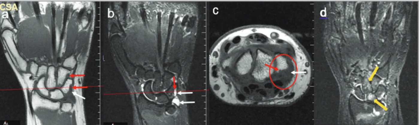
sus-pected of developing RA, is an essential objective to al-low the benefit of optimum treatment and improve prognosis2,20. Our study, based on a balanced Mexican population including three different RA stages accord-ing the ACR-EULAR criteria (2010) and EULAR-OMERACT RAMRI score reference image atlas, pro-vides an important strength, in which, the un-reported fIgure 1. Hand-wrist MRI Coronal T1 and STIR in a clinically suspicious arthralgia (CSA) patient. First Carpal line lesions in a 59 year-old woman diagnosed with CSA. White arrows show synovitis in triquetrum bone; red arrow shows erosion in trapezoid; yellow arrows show bone marrow edema (osteitis) in hamate and scaphoid bones. A) T1 sequence synovitis in triquetrum and prestyloid--triquetral recess is seen hypointense (dark gray signal) (white arrow); erosions in triquetral and trapezoid cortical bones edge that usually show “no signal” (black signal), are seen hypointense in T1 due to joint effusion sinovitis (red arrow). B) STIR sequence synovitis in triquetrum and prestyloid-triquetral joint recess is seen hyperintense (bright white signal) (white arrow); erosion in triquetrum due to effusion synovitis within it (red arrow). C) T1 axial slice (corresponding to the red line level in the coronal plane), synovitis in the triquetrum is seen hypointense (white arrow in red circle) and the erosion is seen hypointense (red arrow); D) BME/osteitis is seen hyperintense on STIR in lunate and capitate bones (yellow arrow).
fIgure 2. Hand-wrist MRI Coronal T1 and STIR in a early rheumatoid arthritis (ERA) patient. First Carpal line lesions in a 35 year-old man diagnosed with ERA. White arrows show synovitis; yellow arrows show bone marrow edema (osteitis). A) T1 sequence synovitis in triquetrum and prestyloid-triquetral recess is seen hypointense (dark gray signal) (white arrow). B) STIR sequence synovitis is seen hyperintense (bright white signal) in triquetrum and prestyloid-triquetral joint recess and radio-scaphoid-trapezoid joint (white arrows); BME/osteitis is seen as hyperintense signal in triquetrum, lunate and scaphoid bones (yellow arrow heads); C) T1 axial slice (corresponding to the red line level in the coronal plane), joint effusion synovitis triquetrum is seen hypointense (white arrow)
finding was obtained, regarding the predominance of triquetral bone lesion, present in 76.4% of all site-le-sions in the three stages; that in the CSA stage (not pre-viously reported in literature) reached 62.3%, followed by the lunate (51%) and scaphoid (49.3%); in ERA stage the triquetrum, lunate and scaphoid were the most prevalent sites affected with 80.6%, 74.6%, and 73% respectively, and similarly in establish RA with 86.3%, 77.6% and 51.3% for the triquetrum, scaphoid and lunate (Table IV).
This triquetral bone affectation may be due to the different ligamentous relationships as well as morpho-logical factors, that allows rotational mobility and that pivots over the rest of the carpal bones maintaining physiological loads in the medial column wrist arch mobility. It also has the largest number of ligamentous insertions, compared to the other carpal bones, which triggers initial damage with an inflammatory process in this area4,9(Table I).
Patients with triquetral synovitis and erosion were also high in CSA phase, 87% and 91% respectively. It could be suggested that this higher prevalence is in re-lation with the major amount of ligaments and os-teomyoarticular system inserted on triquetral bone, that allows it to be the pivot of the rotational mobility, loads and resistance over the rest of the carpal
struc-tures12,21. The scaphoid and lunate bones were the next most frequently affected bones overall, affected in 66.7% and 59% of patients respectively (Table IV).
This triquetral injury prevalence is probably in fluenced by its instability, since it does not directly arti -culate to the ulnar bone due to the presence of the trian-gular fibrocartilage of the wrist, which absorbs and transmits the pressure forces exerted on the carpal bones12,13. It has a higher tendency for traumatic injuries since it’s the second one in carpal fractures frequency (38%), and represents 3-4% of all the carpal bone le-sions22-25.
There are also other factors to consider like the ex-pected degenerative cortical bone changes by age. Miki8 (1978) reported the articular disc wrist presents major ulnar damage at triangular cartilage level and at the in-terosseous ligaments between scaphoid, lunate or tri-quetral bones when AR begins at an early age8. Similarly, we found lesions in all carpal bones, most predominan -tly in the triquetral and lunate. The triquetral bone is probably the first and most affected due to the mechani-cal forces, and possibly a lower blood vessel density4,26.
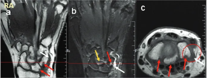
Our study included CSA, ERA, and RA stages in pa-tients at the time of evaluation for wrist morphological changes with MRI. We report different types of lesions (synovitis, erosions, and BME/osteitis) at different sites, fIgure 3.Hand-wrist MRI Coronal T1 and STIR in a rheumatoid arthritis (RA) patient. First Carpal line lesions in a 45 year-old woman. White arrows show synovitis; red arrow shows erosion; yellow arrows show bone marrow edema (osteitis). A) T1 sequence synovitis in triquetrum is seen hypointense (white arrow) and erosions are seen in the triquetrum, lunate and scaphoid bones edge with “no signal” (black signal), here seen hypointense in T1 due to joint effusion synovitis (red arrows). B) Synovitis on STIR sequence is seen hyperintense (bright white signal) in the triquetrum and prestyloid-triquetrum joint recess (white arrow); erosion in triquetrum is seen hyperintense with synovitis (red arrow); BME/osteitis is seen hyperintense in the capitate bone (yellow arrow). C) T1 axial slice, hypointense synovitis in prestyloid-triquetral joint recess (white arrow in red circle) and erosions with synovitis inside them, are seen hypointense in the triquetral, lunate and scaphoid bones (red arrow).
making it more detailed than previously published pa-pers4,10,17,19,27. A total of 23 MRI cases of patients with CSA were included, which has not been reported be-fore. However, a limitation is the overall sample size of patients included.
conclusIons
Carpal synovitis was the most frequent lesion in all three RA phases, and the triquetrum bone the most af-fected. Triquetrum evaluation could be taken into account as a candidate and possible clinical biomar ker of RA from its initial stages or when an initial evalua -tion of the disease is performed. It could be assessed as a first joint site for lesions in RA’s natural history (this due to its anatomicaltopographic characteristics). Ne -vertheless, it is necessary to perform further studies in order to obtain greater evidence for this finding. Limi-tations of the present study are the relatively small number of patients evaluated in each phase and that the treatment variability was not controlled, therefore that might influence on the MRI findings.
correspondence to
Rodrigo Enrique Elizondo-Omaña Av Francisco I. Madero S/N E-mail: rod_omana@yahoo.com
references
1. Ruiz-Esquide V, Sanmartí R. Tobacco and other environmental risk factors in rheumatoid arthritis. Reumatol Clin 2012; 8:342–350. 2. Combe B, Landewe R, Daien CI, Hua C, Aletaha D, Álvaro-Gracia JM,
et al. 2016 update of the EULAR recommendations for the man-agement of early arthritis. Ann Rheum Dis 2017; 76:948–959. 3. Erol AM, Ceceli E, Ramadan US, Borman P. Effect of rheumatoid
arthritis on strength, dexterity, coordination and functional status of the hand: relationship with magnetic resonance imaging find-ings. Acta Reumatol Port 2016; 41:328–337.
4. Ringler MD, Murthy NS. MR imaging of wrist ligaments. Magn Re-son Imaging Clin N Am 2015; 23:367–391.
5. Conaghan P, Bird P, Ejbjerg B, O’connor P, Peterfy C, McQueen F, Østergaard M. The EULAR–OMERACT rheumatoid arthritis MRI reference image atlas: the metacarpophalangeal joints. Annals of the rheumatic diseases 2005; 64 (suppl 1): i11-i21.
6. Ejbjerg B, McQueen F, Lassere M, Haavardsholm E, Conaghan P, O’connor P, Genant, H. The EULAR–OMERACT rheumatoid arthri-tis MRI reference image atlas: the wrist joint. Annals of the rheumat-ic diseases 2005; 64 (suppl 1): i23-i47.
7. Bird P, Conaghan P, Ejbjerg B, McQueen F, Lassere M, Peterfy C, Emery P. The development of the EULAR–OMERACT rheumatoid arthritis MRI reference image atlas. Annals of the rheumatic dis-eases 2005; 64 (suppl 1): i8-i10.
8. Miki ZD. Age changes in the triangular fibrocartilage of the wrist joint. J Anat 1978; 126:367–384.
9. Resnick D. Early abnormalities of pisiform and triquetrum in rheumatoid arthritis. Ann Rheum Dis 1976; 35:46–50.
10. Garcia-Elias M, Puig de la Bellacasa I, Schouten C. Carpal ligaments: a functional classification. Hand Clin 2017; 33:511–520. 11. Spina JC, Dutruel S, Colombo O, Badano F, Aliaga L, Barreira JC.
Evaluación del daño estructural de manos mediante RM en pa-cientes con artritis reumatoidea temprana, sin evidencia de ero-siones radiográficas. Rev argent radiol 2009; 73:439–448. 12. Medina Gonzalez CE, Benet Rodríguez M, Martínez FM. El
com-plejo articular de la muñeca: aspectos anatofisiológicos y biomecáni-cos, características, clasificación y tratamiento de la fractura distal del radio. MediSur 2016; 14:430–446.
13. Totterman S, Miller R, Wasserman B, Blebea JS, Rubens DJ. Intrin-sic and extrinIntrin-sic carpal ligaments: evaluation by three-dimension-al fourier transform mr imaging. Am J Roentgenol 1993; 160:117–123.
14. Nozaki T, Wu W Der, Kaneko Y, Rafijah G, Yang L, Hitt D, et al. High-resolution mri of the ulnar and radial collateral ligaments of the wrist. Acta radiol 2017; 58:1493–1499.
15. Lisbona MP, Maymó J. Magnetic resonance of the hand in rheumatoid arthritis. review of methodology, and its use in diagno sis, monitoring and prognosis. Reum Clin 2007; 3:126-136.
16. Boutry N, Larde A, Lapegue F, Solau-Gervais E, Flipo R, Cotten A. Magnetic resonance imaging appearance of the hands and feet in pa-tients with early rheumatoid arthritis. J Rheumatol 2003; 30:671–679.
17. Lee KA, Min SH, Kim TH, Lee SH, Kim HR. Magnetic resonance imaging-assessed synovial and bone changes in hand and wrist joints of rheumatoid arthritis patients. The Korean journal of inter-nal medicine 2017.
18. Tamai M, Kawakami A, Uetani M, Takao S, Tanaka F, Fujikawa K, et al. Bone edema determined by magnetic resonance imaging re-flects severe disease status in patients with early-stage rheumatoid arthritis. J Rheumatol 2007; 34:2154–2157.
19. van Steenbergen HW, Mangnus L, Reijnierse M, Huizinga TWJ, van der Helm-van Mil AHM. Clinical factors, anticitrullinated peptide antibodies and mri-detected subclinical inflammation in relation to progression from clinically suspect arthralgia to arthritis. Ann Rheum Dis 2016; 75:1824–1830.
20. Arana-Guajardo A, Pérez-Barbosa L, Vega-Morales D, Riega-Torres J, Esquivel-Valerio J, Garza-Elizondo M. Aplicación de un modelo de predicción de progresión de artritis reumatoide en pacientes con artritis indiferenciada. Reumatol Clínica 2014; 10:360–363. 21. Østergaard M, Peterfy C, Conaghan P, McQueen F, Bird P, Ejbjerg B,
et al. OMERACT rheumatoid arthritis magnetic resonance imaging studies. core set of mri acquisitions, joint pathology definitions, and the OMERACT RA-MRI scoring system. J Rheumatol 2003; 30:1385–1386.
22. Paruchuri RK, Rajasekhar L, Reddy VS. Role of mri in evaluation of asymmetric undifferentiated hand arthritis [Letter]. Indian J Rheumatol 2016; 11:60–61.
23. van Steenbergen HW, Aletaha D, Beaart-van de Voorde LJJ, Brouw-er E, Codreanu C, Combe B, et al. EULAR definition of arthralgia suspicious for progression to rheumatoid arthritis. Ann Rheum Dis 2017; 76:491–496.
24. Díez Renovales F, Nates Uribe N, Korta I, Sarmiento de la Iglesia MM, Galíndez Agirregoikoa E, Grande Icaran D. Utilidad de la RM en la valoración de la AR. Estado actual. [Review]. Radiologia 2014; 56:871.
25. Narváez García JA. Valoración por imagen de la artritis reumatoide precoz. Reumatol Clin 2010; 6:111–114.
26. Daunt N. Magnetic resonance imaging of the wrist: anatomy and pathology of interosseous ligaments and the triangular fibrocartilage complex. Curr Probl Diagn Radiol 2002; 31:158–76.
27. Honing EW, Moeller JL. Wrist Injuries. Phys Sportsmed1998; 26:40–49.