Image
Mailing Address: Viviane Tiemi Hotta •
Av. Dr. Enéas de Carvalho Aguiar, 44 - Cerqueira César - 05403-000 - São Paulo, SP - Brasil
E-mail: [email protected]
Manuscript received March 22, 2011; revised manuscript received June 14, 2011; accepted June 22, 2011.
Keywords
Mass, right atrium, echocardiography, resonance.
Right Atrial Mass Assessment by Transthoracic Echocardiography
and Cardiac Magnetic Resonance
Viviane Tiemi Hotta e James Alberton
Instituto do Coração/Faculdade de Medicina da Universidade de São Paulo – SP - Brazil
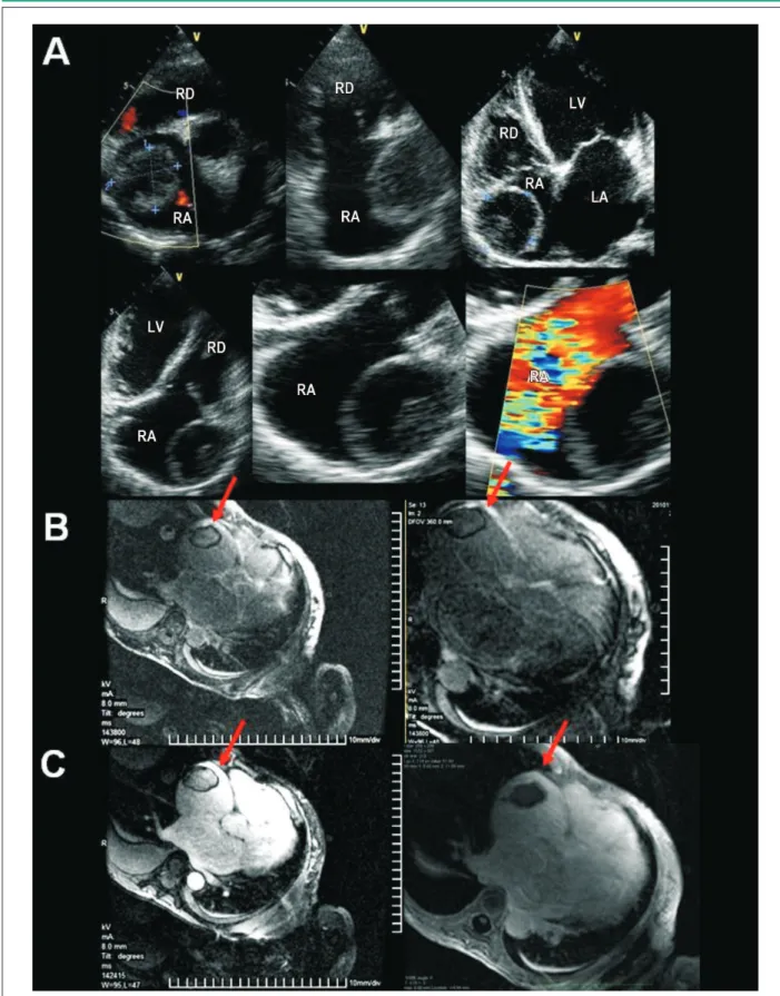
The patient underwent transthoracic echocardiography (TTE), which disclosed significant left ventricular dysfunction and cystic mass of heterogeneous content on the lateral wall of the right atrium (RA). Cardiac magnetic resonance (CMR) imaging confirmed the TTE findings. Due to the high probability of thrombus, oral anticoagulation and echocardiographic
control were chosen. The patient had refractory heart failure (HF) and died after twenty days of hospitalization. In this case, TTE showed good correlation with the MRI results. In addition to the unusual finding at the TTE, it shows the importance of integration between the imaging methods and the poor prognosis associated with masses found in the RA.
Image
(Arq Bras Cardiol 2011; 97(3) : e58-e59)
Hotta & Alberton RA Mass at TTE and CMR
Figure 1 - A) TTE disclosing image of cystic aspect on the lateral wall of the right atrium (RA). B) In the delayed enhancement CMR imaging (T1-weighted) and inversion time adjusted for the annulment of the myocardial signal, the lesion (red arrows) showed hypointense signal at the myocardium. C) CMR image with delayed enhancement and long inversion time (~ 600ms) also showed a hypointense signal, reinforcing the possibility of avascular structure, with the two main differential diagnoses being: cyst with high protein content and thrombus. (RA - right atrium, RV - right ventricle, LA - left atrium, LV - left ventricle).