Abstract
The Brazilian Cerrado is a highly diverse ecosystem, harboring a great variety of organisms; however, the mycodiversity
is still poorly documented. In this study, we record for the first time the bolete
Phlebopus beniensis
from the Cerrado
biome in the state of Goiás, located in Brazil’s Central-West Region. Description, macroscopic images, line drawings
and comments, as well as the geographic distribution of the genus for Brazil are presented herein.
Key words
: Basidiomycota, boletoid fungi, distribution, fungal diversity, Neotropical Region.
Resumo
O Cerrado brasileiro é considerado um ecossistema altamente diverso, incluindo uma grande diversidade de
organismos; no entanto, a diversidade fúngica ainda é pouco documentada. Este trabalho apresenta o primeiro
registro de
Phlebopus beniensis
para o bioma Cerrado no estado de Goiás, Região Centro-Oeste do Brasil. Descrição,
imagens macroscópicas, ilustrações microscópicas e comentários, assim como a distribuição geográfica do gênero
para o Brasil são aqui apresentados.
Palavras-chave
: Basidiomycota, fungos boletoides, distribuição, diversidade fúngica, Região Neotropical.
Short Communication / Nota Científica
Phlebopus beniensis (Boletinellaceae, Boletales)
in the Brazilian Cerrado biome
Francisco J. Simões Calaça
1,4,5,
Altielys Casale Magnago
2,
Renato Lúcio Mendes Alvarenga
3,4&
Solange Xavier-Santos
4http://rodriguesia.jbrj.gov.br
DOI: 10.1590/2175-786020186924x6
1 Universidade de Brasília, UnB, Inst. Ciências Biológicas, Depto. Ecologia, Lab. Ecologia de Ecossistemas, Prog. Pós-graduação em Ciências Ambientais
(Faculdade UnB de Planaltina), Campus Universitário Darcy Ribeiro, Asa Norte, 70919-900, Brasília, DF, Brazil.
2 Universidade Federal do Rio Grande do Sul, Campus do Vale, Depto. Botânica, Inst. Biociências, Prog. Pós-graduação em Botânica, Lab. Micologia, Av.
Bento Gonçalves 9500, Prédio 43433, Bl. 4, Sala 111, 91509-900, Porto Alegre, RS, Brazil.
3 Universidade Federal de Pernambuco, Centro de Ciências Biológicas (CCB), Depto. Micologia, Prog. Pós-graduação em Biologia de Fungos, Av. da
Engenharia s/n, 50740-600, Cidade Universitária, Recife, PE, Brazil.
4 Universidade Estadual de Goiás, Lab. Biodiversidade do Cerrado, Campus de Ciências Exatas e Tecnológicas, BR-153, nº 3105, Fazenda Barreiro do Meio,
75132-400, Mailbox 459, Anápolis, GO, Brazil.
5 Author for correspondence: calacafjs@gmail.com
Boletinellaceae P.M. Kirk, P.F. Cannon & J.C. David is a small family comprising only two genera, Boletinellus Murrill and Phlebopus (R. Heim.) Singer (Binder & Hibbett 2006; Kirk et al. 2008). Six species have been reported from Brazil: Phlebopus beniensis (Singer & Digilio) Heinem. & Rammeloo, P. brasiliensis Singer, P. braunii (Bres.) Heinem., P. harleyi Heinem. & Rammeloo, P. portentosus (Berk. & Broome) Boedijn and P. tropicus (Rick) Heinem. & Rammeloo, distributed in the Amazon and Atlantic forests biomes (Rick 1960; Singer & Digilo 1957, 1960; Singer et al. 1983; Putzke et al.1994; Oliveira & Sousa 1996; Watling & Meijer 1997). The Brazilian
Cerrado biome is considered one of the most diverse savannas in the world, as it is an important biodiversity hotspot (Myers et al. 2000; Klink & Machado 2005) and the second largest biome in Brazil. However, only 61% of the original Cerrado vegetation remains preserved, mostly represented by savanna vegetation of different densities and layers of herbaceous grasses (Rodrigues 2005; Sano et al. 2010).
940
Specimens were collected during the rainy season in the Brazilian Cerrado biome. Basidiomata were growing on grass in disturbed areas under Duranta repens L. (Verbenaceae) at Colégio Estadual Jad Salomão (Anápolis, Goiás, Brazil), on soil in a grazing area and on soil covered by litterfall in stational semi-deciduous forest both in municipality of Rio Quente, Goiás, Brazil. Photographs and macroscopic features were described taken from fresh basidiomata. Color codes are from the Online Auction Color Chart (Kramer 2004). Freehand sections of the basidiomata were made, rehydrated in KOH 3%, dyed in Congo Red and mounted for microscopic analyses. Melzer’s reagent was used for testing amyloidity of the microstructures. Qm refers to the quotient average of length/width ratio
range from the basidiospores. All microscopic features were drawn after digital micrographs from the specimens examined. Voucher materials were deposited at HUEG Herbarium (Thiers, continuously updated). The geographic distribution for Phlebopus presented here was compiled from recent fieldwork, herbaria and literature data (Maia et al. 2015).
Phlebopus beniensis (Singer & Digilio) Heinem. & Rammeloo, Mycotaxon 15: 390 (1982).
Figs. 1; 2 ≡ Phaeogyroporus beniensis Singer & Digilio, Lilloa 30: 150 (1960).
Type: R. Singer B1613, Bolivia, Guayaramerím (LIL) (as cited by Heinemenn & Rammeloo 1982).
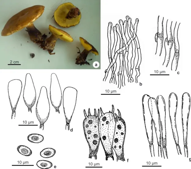
Figure 1
– a-g. Phlebopus beniensis – a. fresh basidiomata; b. pileipellis; c. clamp connections from pileipellis; d.
cheilocystidia; e. basidiospores; f. basidia; g. caulocystidia.
2 cm
10 µm
10 µm 10 µm 10 µm
10 µm
10 µm
a
b
c
e
f g
Pileus 80−100 mm diam., hemispheric at first to convex to plane convex when mature, slightly depressed in the center, smooth, slightly viscid in wet weather, light brown, caramel to sepia yellow colored (OAC 840, 838); straight margin, irregular, splitting when mature. Tubes 5−12 mm long centrally, yellow (OAC 895, 888) to yellow olive under pressure (OAC 887, 859), adnate to depressed; pores 2−3 per mm, rounded. Stipe 30−70 × 11−23 mm, central to slightly eccentric, subequal, tapering upwards, downwards to bulbous base, dark greysih brown (OAC 866, 868) yellowish near the apex, surface glabrous. Context pale yellow (OAC 899), unchanging when exposed. Spore print olive brown. Basidiospores 5.6–7.3 × 4.6–5 µm (Qm = 1.40), short ellipsoid,
yellow brown, inamyloid, guttulate, smooth, thick walled. Basidia 16.6–23 × 6–10 µm, clavate, thin walled, hyaline, guttulate, 4-sterigmate, 3−5 μm long. Pleurocystidia and cheilocystidia both with (15.5–)20.5−21.7 × (5–)7−10 μm, clavate to obclavate with obtuse tip, hyaline, inamyloid, smooth, thin walled. Pileipellis trichodermal, terminal elements 4.3−6.5 μm wide, round apex, hyaline, inamyloid, smooth, thin walled. Pileus trama parallel to subparallel, hyphae 2.5−10 μm wide, hyaline. Stipitipellis composed by clavate, fusiform to cylindrical caulocystidia, 3−13 μm, hyaline, inamyloid, thin walled. Stipe trama of vertically arranged hyphae, cylindrical, hyphae 4−9 μm wide, hyaline, inamyloid. Hymenophoral trama boletoid, gelatinized mediostratum with narrow
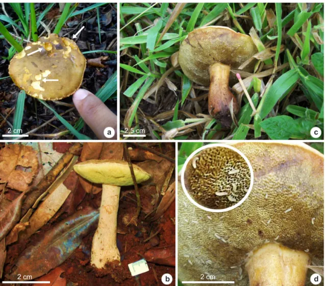
Figure 2
– a-d. fresh basidiomata of Phlebopus beniensis – a. growing on grass in disturbed areas (arrows show
signals of mycophagy); b. on soil covered by litterfall; c. on soil in a grazing area, with some insect larvae feeding
on the basidiome; d. detail of mycophagy by insect larvae.
2 cm
2 cm 2 cm
2,5 cm
a
b
c
942
hyphae 3−5(–5.3) μm wide, hyaline, inamyloid, oleiferous hyphae not observed; medium stratum with irregular to inflated hyphae 6−10 μm wide, divergent. Clamp connection present in all septa. (Figs. 1; 2).
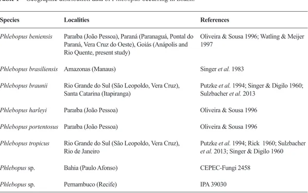
Distribution in Brazil: Paraíba (Oliveira & Sousa 1996), Paraná (Watling & Meijer 1997) and Goiás (present study).
Worldwide distribution: Once that Phlebopus is a genus of tropical boletes, P. beniensis occurs in Argentina, Bolivia, Brazil, Colombia, Costa Rica, Ecuador, Martinique, Puerto Rico, Venezuela in the Neotropics and from Liberia in Africa (Heinemenn & Rammeloo 1982; Ovrebo 1983; Singer et al. 1983; Miller et al. 2000; Guzmán et al. 2007; Palacio et al. 2014).
Specimens examined: BRAZIL. GOIÁS: municipality of Anápolis, Colégio Estadual Jad Salomão, 16o19’10.14’’S,
48o57’55.98’’W, growing gregariously on grass in
disturbed areas under Duranta repens L. (Verbenaceae), with some signals (Fig. 2a) of mycophagy (probably by insect, like shown in Fig. 2c,d), 12.XI.2014, F.J.S. Calaça FJSC48 (HUEG 10043). Municipality of Rio Quente, 17o46’S, 48°45’W, growing on soil covered by
litterfall (Fig. 2b) in a stational semi-deciduous forest, in a particular area, 17o47’35’’S, 48o47’36’’W, 22.I.2008,
S. Xavier-Santos SXS3549, SXS2215 and SXS2189. Growing on soil in a grazing area, with some insect larvae feeding on the basidiomes (Fig. 2c,d).
Additional specimens examined: BRAZIL. PARAÍBA:
João Pessoa, UFPB/CCEN/DSE, Mata do Biotério, 7o8’18.9”S, 34o50’40.1”W, 7.II.2009, M.A. Neves
MAN357 (ICN 184809); 7o8’18.5”S, 34o50’38.4”W,
11.III.2011, A.C. Magnago ACM 265, 267 (ICN 184911, 184912); 20.VI.2011, A.C. Magnago ACM 285 (ICN 184910).
Comments: These are the first records of Phlebopus beniensis for the Brazilian Cerrado, increasing the knowledge about fungi diversity in Brazil’s Central-West Region. Before this study, P. beniensis had been recorded only along the coastal Atlantic Forest (Oliveira & Sousa 1996; Watling & Meijer 1997). The current distribution in Brazil of P. beniensis and other Phlebopus species are shown on Table 1.
Phlebopus beniensis is morphologically very similar to P. brasiliensis, overlapping in many features, and differing by a few subjective characteristic: the bluing oxidation of pileus and stipe context when exposed, which are more visible when the material is quite fresh and humid, however, depending on dryer weather conditions this characteristic can be not so evident, like the ones observed in the examined specimens
as an unchanging reaction in the context. The size of the basidiomata is another differentiating characteristic: P. brasiliensis has pileus < 65 mm wide and P. beniensis usually larger when mature, reaching 130 mm wide. The cystidia in P. beniensis are more evident and numerous than in P. brasiliensis (Singer et al. 1983).
Singer et al. (1983) reported Phlebopus brasiliensis in the state of Paraíba, however, Oliveira & Sousa (1996) examined many collections of Phlebopus from the same area of Singer’s collections, and concluded that all these were misidentifications of P. beniensis. The species also differs from other Phlebopus found in Brazil for its wide, subgyrose to boletinoid pores, and larger basidiospores (8–9 × 6–7 µm) in P. tropicus; the darker colors of the basidiomes and most of all for its basidiospores larger than 7.5 µm in P. portentosus; stipe finely ornamented and globose to subglobose badiospores in P. harleyi; and pileus surface with olive tones and larger basidiospores [6.5–9.5(–11) × 5–7 µm] in P. braunii (Singer et al. 1983; Singer & Digilo 1960; Oliveira & Sousa 1996; Watling & Meijer 1997; Baroni et al. 2015). Phlebopus beniensis is regarded as a species that develops in moister tropical forests (Singer et al. 1983), but some works have recorded this species in many locations in Neotropical Regions and in Africa (Singer & Digilio 1960; Heinemenn & Rammeloo 1982; Guzmán et al. 2007; Palacio et al. 2014), like dry, semi-deciduous, seasonal subtropical forests in montane or sub-montane areas. Recently, Palacio et al. (2014) recorded P. beniensis in a tropical dry forest, in the northeast region of Colombia. As shown in Table 1, species of this genus seem to be more generalists with regard to its habitat since it has already occurred in Brazil, in tropical Atlantic and Amazonian Forests and seasonal savanna (Cerrado), as indicated in the present study. Moreover, as shown by our data, P. beniensis can be found in urban areas, associated or not to distinct plants species.
by Azara’s agouti (Dasyprocta azarae Lichtenstein 1823)] and Sulzbacher et al. (2015) reported mycophagy of Descomyces albus (Berk.) Bougher & Castellano by the invertebrate Balloniscus sellowii Brandt 1833 (Isopoda).
Once fungi are a rich source of nutrients (Fogel & Trappe 1978) for these animals, fungi can contribute to structure food webs, mainly to insect community (Yamashita et al. 2015), being a key components to maintenance of ecosystems. Our results highlight the importance of studies involving these ecological interactions between mycophagous invertebrates and fungi.
Although molecular data has not been addressed in this work, it should be investigated to better understand the relationship among species within Phlebopus, especially with the inclusion of Neotropic specimens, since morphological features which distinguish the species are generally subjective and confusing. As pointed out by Maia et al. (2015), it is still necessary to encourage new collection expeditions in Brazilian forests, especially in savanna areas in the Central-West Region in order to increase our knowledge about the mycodiversity for Brazil, as for the Neotropics, as well as including molecular data of Brazilian specimens in phylogenies.
Acknowledgements
The authors wish to thank Fundação de Amparo à Pesquisa do Estado de Goiás (FAPEG) for their financial support. ACM, FJSC and RLMA wish to thank the Conselho Nacional de Desenvolvimento Científico e Tecnológico (CNPq) for their fellowships. The authors also wish to thank the anonymous reviewers for their comments and suggestions, which greatly improved the quality of the manuscript.
References
Baroni TJ, Cifuentes J, Santana BO & Cappello S (2015) A new species of Phlebopus (Boletales, Basidiomycota) from Mexico. North American Fungi 10: 1-13.
Binder M & Hibbett DS (2006) Molecular systematics and biological diversification of Boletales. Mycologia 98: 971-981.
Fogel R & Trappe JM (1978) Fungus consumption (mycophagy) by small mammals. Northwest Science 52: 1-31.
Guzmán G, Castillo-Ayoui F, Gándara E & Vivero T (2007) First record of the genus Phlebopus (Basidiomycotina, Boletales) in Ecuador. Mycotaxon 99: 217-221.
Heinemann P & Rammeloo J (1982) Observations sur
Table 1
– Geographic distribution data of Phlebopus occurring in Brazil.
Species Localities References
Phlebopus beniensis Paraíba (João Pessoa), Paraná (Paranaguá, Pontal do Paraná, Vera Cruz do Oeste), Goiás (Anápolis and Rio Quente, present study)
Oliveira & Sousa 1996; Watling & Meijer 1997
Phlebopus brasiliensis Amazonas (Manaus) Singer et al. 1983
Phlebopus braunii Rio Grande do Sul (São Leopoldo, Vera Cruz), Santa Catarina (Itapiranga)
Putzke et al. 1994; Singer & Digilo 1960; Sulzbacher et al. 2013
Phlebopus harleyi Paraíba (João Pessoa) Oliveira & Sousa 1996
Phlebopus portentosus Paraíba (João Pessoa) Oliveira & Sousa 1996
Phlebopus tropicus Rio Grande do Sul (São Leopoldo, Vera Cruz), Rio de Janeiro
Putzke et al. 1994; Rick 1960; Sulzbacher et al. 2013; Singer & Digilo 1960
Phlebopus sp. Bahia (Paulo Afonso) CEPEC-Fungi 2458
944
le genre Phlebopus (Boletineae). Mycotaxon 15: 384-404.
Kirk PM, Cannon PF, Minter DW & Stalpers JA (2008) Ainsworth and Bisby’s dictionary of the fungi. 10th
ed. CAB International University Press, Cambridge. 771p.
Klink CA & Machado RB (2005) Conservation of the Brazilian Cerrado. Conservation Biology 19: 707-713.
Kramer LA (2004) The online auction color chart. Online Auction Color Chart Company, Stanford, Palo Alto. 12p.
Maia LC, Carvalho-Júnior AA, Cavalcanti LH, Gugliotta AM, Drechsler-Santos ER, Santiago ALCMA, Cáceres MES, Gibertoni TB, Aptroot A, Giachini AJ, Soares AMS, Silva ACG, Magnago AC, Goto BT, Lira CRS, Montoya CAS, Pires-Zottarelli CLA, Silva DKA, Soares DJ, Rezende DHC, Luz EDMN, Gumboski EL, Wartchow F, Karstedt F, Freire FM, Coutinho FP, Melo GSN, Sotão HMP, Baseia IG, Pereira J, Oliveira JJS, Souza JF, Bezerra JL, Araujo-Neta LS, Pfenning LH, Gusmão LFP, Neves MA, Capelari M, Jaeger MCW, Pulgarín MP, Menolli-Junior N, Medeiros PS, Friedrich RCS, Chikowski RS, Pires RM, Melo RF, Silveira RMB, Urrea-Valencia S, Cortez VG & Silva VF (2015) Diversity of Brazilian fungi. Rodriguésia 66: 1033-1045
Miller OKM, Lodge DJ & Baroni TJ (2000) New and interesting ectomycorrhizal fungi from Puerto Rico, Mona, and Guana Islands. Mycologia 92: 558-570. Myers N, Mittermeier RA, Mittermeier CG, Fonseca
GAB & Kent J (2000) Biodiversity hotspots for conservation priorities. Nature 403: 853-858. O l i v e i r a I C & S o u s a M A ( 1 9 9 6 ) B o l e t a l e s
(Hymenomycetes) no Campus I da Universidade Federal da Paraíba, João Pessoa: II - Gyrodontaceae. Revista Nordestina de Biologia 11: 97-117.
Ovrebo CL (1983) New records of fleshy fungi from
Venezuela. Mycotaxon 18: 355-356.
Palacio M, Gutiérrez Y, Franco-Molano AE & Callejas-Posada R (2014) Nuevos registros de macrohongos (Basidiomycota) para Colombia procedentes de un bosque seco tropical. Actualidades Biológicas 37: 319-339.
Editor de área: Dr. Anibal de Carvalho Junior Artigo recebido em 11/02/2016. Aceito para publicação em 23/05/2017.
Putzke J, Pereira AB & Maria L (1994) Os fungos da família Boletaceae conhecidos do Rio Grande do Sul (Fungi, Basidiomycota). Cadernos de Pesquisa Série Botânica 6: 75-100.
Rick J (1960) Basidiomycetes eubasidii in Rio Grande do Sul - Brasilia. 4. Meruliaceae, Polyporaceae, Boletaceae. Iheringia série Botânica 7: 193-295. Rodrigues MT (2005) A biodiversidade dos cerrados:
conhecimento atual e perspectivas, com uma hipótese sobre o papel das matas galerias na troca faunística durante ciclos climáticos. In: Scariot
A, Sousa-Silva JC & Felfili JM (orgs.) Cerrado:
ecologia, biodiversidade e conservação. Ministério do Meio Ambiente, Brasília. Pp. 235-246.
Sano EE, Rosa R, Brito JLS & Ferreira-Junior LG (2010) Land cover mapping of the tropical savanna region in Brazil. Environmental Monitoring and Assessment 166: 113-124.
Singer R & Digilio L (1957) Las boletáceas austro-sudamericanas. Lilloa 28: 247-268.
Singer R & Digilio L (1960) Las boletáceas de sudamerica tropical. Lilloa 30: 141-164.
Singer R, Araujo I & Ivory MH (1983) The ectotrophically mycorrhizal fungi of the Neotropical Lowlands, especially Central Amazonia. Beihefte zur Nova Hedwigia 77: 1-351.
Sulzbacher MA, Grebenc T, Jacques RJS & Antoniolli ZI (2013) Ectomycorrhizal fungi from southern Brazil - a literature-based review, their origin and potential hosts. Mycosphere 4: 61-95.
Thiers B [continuously updated] Index Herbariorum: a
global directory of public herbaria and associated staff. New York Botanical Garden’s Virtual Herbarium. Available at <http://sweetgum.nybg. org/science/ih/>. Access on January 2016. Trierveiler-Pereira L, Silva HCS, Funez LA & Baltazar
JL (2016) Mycophagy by small mammals: new and interesting observations from Brazil. Mycosphere 7: 297-304.
Watling R & Meijer AAR (1997) Macromycetes from the state of Paraná, Brazil. Edinburgh Journal of Botany 54: 231-251.
Yamashito S, Ando K, Hoshina H, Ito N, Katayama Y, Kawanabe M, Maruyama M & Itioka T (2015) Ecological Entomology 40: 390-400.