Rat Model of Myelomeningocele
By Jorge Correia-Pinto, Joaquim L. Reis, Grover M. Hutchins, Maria J. Baptista, Jose´ Esteva˜o-Costa, Alan W. Flake, and Adelino F. Leite-Moreira
Porto, Portugal; Baltimore, Maryland; and Philadelphia, Pennsylvania
Background/Purpose: The rationale for in utero repair of myelomeningocele has been supported experimentally by the observation of preserved neural function after prenatal closure of surgically created defects compared with nonre-paired controls. The mechanism of injury to the exposed neural elements is unknown. Postulated mechanisms in-clude trauma to the herniated neural elements or progres-sive injury from amniotic fluid exposure as gestation pro-ceeds. A component of amniotic fluid that may contribute to neural injury is meconium. In the current study the effect of human meconium on the exposed spinal cord in a fetal rat model of myelomeningocele was examined.
Methods: Twenty time-dated pregnant rats underwent lapa-rotomy at 181⁄2days of gestation. The exposed uterus was bathed in ritrodrine for tocolysis. The amniotic cavity was opened over the dorsal midline of the fetal rat, and, under a dissecting microscope (⫻25), a 2- to 3-level laminectomy was performed. Under magnification (⫻40), the translucent dura was opened using a 25-gauge needle as a knife. Two fetuses per dam were operated on. In the control group, the amniotic fluid was restored with saline solution, whereas in the exper-imental group a solution of Human meconium diluted (10%) in saline was used to restore the amniotic fluid. Fetuses were harvested by cesarean section at 211⁄2days’ gestational age. The liveborn pups were then killed and fixed in 10%
forma-line. Sections 10m thick were stained with H&E and studied by light microscopy for evidence of spinal cord injury. Results: Seven of 20 (35%) experimental rat pups and 6 of 20 (30%) control rat pups were liveborn. All liveborn pups had severe paralysis of the hindlimbs and tail, so that functional differences between the 2 groups could not be detected. Histologic examination of 13 spinal cords at the site of sur-gical exposure showed that necrosis of neural tissue in 5 of 7 meconium-exposed rat pups was increased when com-pared with that observed in the 6 fetuses exposed to amni-otic fluid without meconium. In general, inflammation was greater and repair processes appeared delayed in meco-nium-exposed rat pups.
Conclusions: Exposure of the spinal cord of fetal rats to amniotic fluid by surgically created myelomeningocele leads to severe functional impairment. Histologically recognizable necrosis of neural elements was increased in those animals that were exposed to diluted human meconium in the amni-otic fluid. The results support the hypothesis that meconium may contribute to the pathophysiology of spinal cord injury observed in myelomeningocele.
J Pediatr Surg 37:488-492. Copyright © 2002 by W.B. Saunders Company.
INDEX WORDS: Myelomeningocele, rat, fetal surgery, meco-nium.
M
YELOMENINGOCELE is a congenital anomaly associated with significant lifelong disabilities that include paraplegia, hydrocephalus, sexual dysfunc-tion, skeletal deformadysfunc-tion, and bowel and bladder incon-tinence. These disabilities limit the average longevity of patients born with myelomeningocele to less than 40years1 and are, in part, secondary to the severe spinal cord damage sustained before birth.2
According to the “two-hit hypothesis” the exposed neural elements in myelomeningocele primarily are the result of defective spinal cord development (myelodys-plasia), whereas a secondary event resulting in erosion and necrosis of the exposed spinal cord results in pro-gressive damage with gestational age.2-4This hypothesis is supported by several observations suggesting that neural damage acquired during fetal life is increased by mechanical trauma or chemical toxicity of the amniotic fluid.2-8
These findings have led some investigators to propose repair of myelomeningocele in utero as an alternative approach to minimize neural damage before birth.9,10 However, the specific mechanism of neural injury re-mains unknown. There is now evidence that in utero defecation may normally occur11and that physiological passage of meconium increases late in gestation. This led
From the Departments of Physiology and Pediatric Surgery, Faculty of Medicine, Porto, Portugal; Department of Anatomy, ICBAS, Porto, Portugal; Department of Pathology, The Johns Hopkins University School of Medicine, Baltimore, MD; and The Children’s Institute for Surgical Science, The Children’s Hospital of Philadelphia, Philadel-phia, PA.
Presented at the 32nd Annual Meeting of the American Pediatric Surgical Association, Naples, Florida, May 20-23, 2001.
Address reprint requests to Jorge Correia-Pinto, MD, Servic¸o de Fisiologia, Faculdade de Medicina da Universidade do Porto, A1 Prof Hernaˆni Monteiro, 4200-319 Porto, Portugal.
Copyright © 2002 by W.B. Saunders Company 0022-3468/02/3703-0049$35.00/0
doi:10.1053/jpsu.2002.30872
us to hypothesize that spinal cord damage in myelome-ningocele may be related to contamination of the amni-otic fluid by meconium. To test this hypothesis, we carried out an experimental study in the fetal rat model of myelomeningocele. Our results support the conclusion that prenatal exposure to meconium may contribute to damage of the neural elements in myelomeningocele.
MATERIALS AND METHODS
All experimental protocols were reviewed and approved by the Institutional Animal Care and Use Committee and followed guidelines set forth in the National Institutes of Health Guide for the Care and Use of Laboratory Animals.
Experimental Model
Female rats were obtained from a commercial breeder (225 g, Criffa, SA, Barcelona, Spain). Animals were mated, and females were checked daily for introital plugging. The day of plugging was defined as gestational day 0 for time dating. The pregnant rats were kept in individual cages and maintained under 12 hours light/12 hours dark cycling conditions. On day 181⁄2(term, 22 days) anesthesia was induced by inhalation of sevoflorane (Sevorane, Abbot Laboratories) supple-mented by intraperitoneal sodium pentobarbital (15 mg/kg intraperito-neal).
Open spinal dysraphism was created as described by Heffez et al.3 Briefly, a hysterotomy was performed by placement of a 6-0 nylon purse-string suture incorporating the amniotic membranes followed by a 3-mm incision placed within the pursestring overlying the back of the fetal rat. For fetal immobilization, the dorsal skin of the fetus was sutured to the border of the opened uterus with a 6-0 nylon. Under 25⫻ magnification (Leica, Wild M651.MS-D, Herbrugg, Switzerland) the paraspinal muscles were dissected, and then a 2- to 3-level laminec-tomy was performed. The translucent dura was opened carefully under 40⫻ magnification using a 26-gauge needle. The fetus then was returned to the uterus, the amniotic fluid volume was restored, and the exposed uterus, after closure, was bathed with ritrodrine for tocolysis (10 mg/mL; Pre-par, Solvay). The maternal laparotomy was closed in 2 layers with a continuous 4-0 silk suture. Fetuses were harvested by cesarean section at 21.5 days’ gestational age.
Experimental Groups
Twenty dams were operated on with 2 fetuses per dam manipulated. In each dam, the amniotic fluid volume was restored with saline (37°C) in one operated fetus (control group), whereas the other received a sterile 10% (by weight) meconium in saline suspension (experimental group). Meconium was obtained from human newborns less than 24 hours of postgestational age. Sterility of meconium was confirmed by parallel aerobic and anaerobic cultures.
Data Analysis
Liveborn pups were examined for neurologic deficits assessing postural reactions and pain perception by pricking with a needle (superficial pain) and by pinching with a hemostat (deep pain). The pups were photographed and then killed and fixed in 10% formalin. Cross sections of spine from both groups were taken at the level of the surgical site and at the levels proximal and distal to the operative site. Sections (10-m thick) were stained with H&E and studied by light microscopy for evidence of spinal cord injury.
RESULTS
Operative Results
Seven of 20 (35%) experimental rat pups and 6 of 20 (30%) control rat pups were liveborn. All liveborn pups showed severe paralysis of the hindlimbs and tail, so that functional differences between the 2 groups could not be detected.
Histologic Study
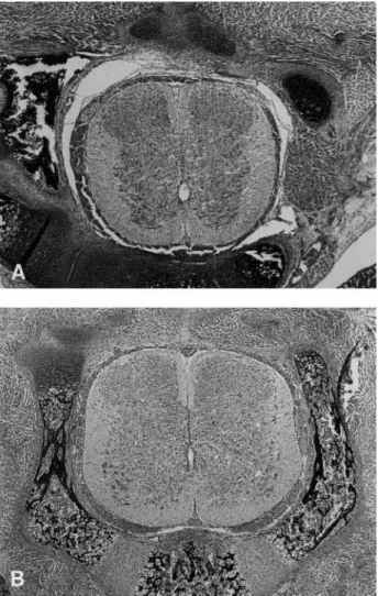
In contrast to nonexposed segments of spinal cord (Fig 1), histologic examination of the 13 spinal cords at the site of the defect showed necrosis of neural tissue that was increased in the meconium-exposed rat pups (Fig 2) relative to the rats exposed to amniotic fluid without meconium (Fig 3). In general, inflammation was greater and repair processes appeared delayed in the meconium-exposed rat pups.
Fig 1. Normal spinal cord. Sections of the spinal cord above (A) and below (B) the level at which the operative intervention was performed. A is at the thoracic level and B at the lumbar level. The dorsal aspect is above and the ventral below. Note the cellularity of the dorsal horns. (Both H&E, original magnificationⴛ50.)
489 MECONIUM INCREASES SPINAL CORD NECROSIS
DISCUSSION
Spinal cord damage is the primary cause of morbidity associated with myelomeningocele. The goal of perinatal management is to avoid or prevent the neural damage commonly observed in myelomeningocele. Improved understanding of the pathogenesis of neural injury is, therefore, required for rational perinatal strategies to evolve.
In this study, the observed fetal mortality rate was higher than reported in fetal rat models of other surgi-cally created anomalies.12 The mortality rate was the same as reported by Heffez et al3,5and is related to the duration (typically greater than 20 minutes) and extent of the surgical procedure required to expose the fetal rat spinal cord.
In our study, we found that spinal cord exposure induced severe functional impairment in both control and meconium-exposed groups. On histologic study,
how-ever, necrosis was increased in the meconium-exposed group suggesting that exposure to human meconium can increase the neurotoxicity of the amniotic fluid. Tradi-tionally, prenatal meconium passage was thought to occur only during fetal distress. Recently, Ciftci et al11 challenged this idea showing that meconium-stained am-niotic fluid reflects impaired clearance of amam-niotic fluid, which normally contains meconium from physiologic defecation.11The presence of intestinal enzymes in the amniotic fluid of healthy fetuses and its absence in fetuses with intestinal obstruction also supports the idea that meconium passage during fetal life is a normal physiological event.13-15Proximal intestinal dilatation in fetuses with colonic atresia or anorectal malformation also favors this theory.
Previous studies have shown that meconium increases amniotic fluid toxicity for other fetal tissues, such as intestine in experimental models of gastroschisis.16
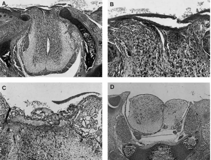
In-Fig 2. Meconium group. (A) The spinal cord protrudes into the surgically created defect, and the exposed surface is covered with proteinaceous material and admixed meconium. (B) The exposed dorsal portions of the cord shown in A show destruction of part of the dorsal horns with loss of the tissue and acute inflammation within the neural tissue. (C) Another subject animal with similar changes to those in B in the exposed portions of the spinal cord and a more brisk acute inflammatory cell reaction. (D) In this subject, there has been extensive loss of exposed dorsal spinal cord tissue. (All H&E; original magnifications: A,ⴛ50; B, ⴛ160; C, ⴛ160; D, ⴛ50).
flammation and induction of necrosis by meconium also has been shown in lung with meconium aspiration syn-drome. It therefore is not surprising that meconium increases damage of the exposed neural elements in this model.
According to our results, fetal spinal cord exposure leads to severe neural damage, which appears increased when human meconium is added to amniotic fluid. Thus, in contrast to other studies,17we believe that chemical toxicity secondary to amniotic fluid exposure is relevant to the pathophysiology of neural damage observed in myelomeningocele. Our hypothesis that meconium might be involved on the pathophysiology of spinal cord damage in myelomeningocele is in agreement with those of previous studies suggesting that most of neural dam-age occurs late in gestation.2,7 As mentioned above, in utero defecation is gestational age dependent and be-comes more prevalent the last weeks of gestation.11 Therefore, gastrointestinal waste products should be added to urea, ammonia and osmotic pressure gradients as potential causes of damage to the exposed spinal cord. However, our experimental protocol cannot exclude trauma as a contributing factor to spinal cord damage.
Although the results of in utero repair of myelomen-ingocele remain to be determined,18 our findings rein-force the theory that injury of the spinal cord is, at least in part, acquired during fetal life. Thus, the rationale of prenatal treatment remains promising for fetuses with myelomeningocele. There also may be a role for other prenatal strategies directed toward prevention of spinal cord damage and disability associated with myelomen-ingocele. For instance, the benefit of amnioinfusion, recently documented in experimental settings, may be attributable to withdrawal and dilution of the harmful chemical substances in amniotic fluid.19The identifica-tion of meconium as a source of those substances pro-vides new evidence supporting the potential benefit of prenatal treatment.
Exposure of the spinal cord of fetal rats to amniotic fluid by surgically created myelomeningocele leads to severe functional impairment. Histologically recogniz-able necrosis of neural elements was increased in those animals that were exposed to human meconium diluted in the amniotic fluid. The results suggest that meconium may contribute to the pathophysiology of spinal cord injury observed in myelomeningocele.
REFERENCES
1. Dillon CM, Davis BE, Duguay S, et al: Longevity of patients born with myelomeningocele. Eur J Pediatr Surg 10:33-34, 2000 (Suppl 1) 2. Hutchins GM, Meuli M, Meuli-Simmen C, et al: Acquired spinal cord injury in humans fetuses with myelomeningocele. Pediatr Pathol Lab Med 16:701-712, 1996
3. Heffez DS, Aryanpur J, Hutchins GM, et al: The paralysis associated with myelomeningocele: Clinical and experimental data implicating a preventable spinal cord injury. Neurosurg 26:987-992, 1990
4. Meuli M, Meuli-Simmen C, Hutchins GM et al: In utero surgery rescues neurological function at birth in sheep with spina bifida. Nat Med 1:342-347, 1995
5. Heffez DS, Aryanpur J, Rotellini NA et al: Intrauterine repair of experimental surgically created dysraphism. Neurosurg 32:1005-1010, 1993
6. Korenromp MJ, Van Good JD, Bruinese HW, et al: Early fetal movements in myelomeningocele. Lancet 1:917-918, 1986
7. Meuli M, Meuli-Simmen C, Hutchins GM, et al: The spinal cord lesion in Human fetuses with myelomeningocele: Implications for fetal surgery. J Pediatr Surg 32:448-452, 1997
8. Drewek M, Bruner JP, Whetsell WO, et al: Quantitative analysis of the toxicity of human amniotic fluid to cultured rat spinal cord. Pediatr Neurosurg 27:190-193, 1997
9. Tulipan N, Bruner JP: Myelomeningocele repair in utero: A report of three cases. Pediatr Neurosurg 28:177-180, 1998
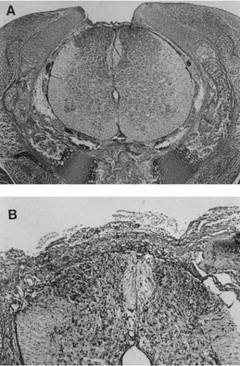
Fig 3. Control group. (A) Section of the spinal cord at the level of the operative exposure of the spinal cord shows a thin proteinaceous covering of the exposed cord within the defect. The spinal cord tissue appears intact. (B) Another control animal with a thicker layer of proteinaceous material covering the spinal cord. A small number of acute inflammatory cells also are present, but the neural tissue of the cord is well preserved. (Both H&E; original magnification: A,ⴛ50; B, ⴛ100.)
491 MECONIUM INCREASES SPINAL CORD NECROSIS
10. Adzick NS, Sutton LN, Crombleholme TM, et al: Successful fetal surgery for spina bifida. Lancet 352:1675-1676, 1998
11. Ciftci AO, Tanyel FC, Bingol-Kologlu M, et al: Fetal distress does not affect in utero defecation but does impair the clearence of amniotic fluid. J Pediatr Surg 34:246-250, 1999
12. Correia-Pinto J, Tavares M, Baptista MJ, et al: A new fetal rat model of gastroschisis: Development and early characterization. J Pe-diatr Surg 36:213-216, 2001
13. Pocknee RC, Abramovich DR: Origin and levels of trypsin in amniotic fluid throughout pregnancy. Br J Obstet Gynec 89:142-144, 1982
14. Kleijer WJ, Janse HC, Van Diggelen OP, et al: Amniotic fluid disaccharidases in the prenatal detection of cystic fibrosis. Prenat Diagn 5:135-143, 1985
15. Morin PR, Potier M, Dallaire L, et al: Prenatal detection of
intestinal obstruction: deficient amniotic fluid disaccharidases in af-fected fetuses. Clin Genet 18:217-222, 1980
16. Olguner M, Akgu¨r FM, Api A, et al: The effects of intraamniotic human neonatal urine and meconium on the intestines of the chick embryo with gastroschisis. J Pediatr Surg 35:458-461, 2000
17. McClone DG, Dias MS, Goossens W, et al: Pathological changes in exposed neural tissue of fetal delayed splotch (Spd) mice. Childs Nerv Syst 13:1-7, 1997
18. Holzbeierlein J, Pope JC IV, Adams MC, et al: The urodynamic profile of myelodysplasia in childhood with spinal closure during gestation. J Urol 164:1336-1339, 2000
19. Olguner M, Akgur FM, Ozdemir T, et al: Amniotic fluid ex-change for the prevention of neural tissue damage in myelomeningo-cele: An alternative minimally invasive method to open in utero surgery. Pediatr Neurosurg 33:252-256, 2000