GABRIEL ÁLVARES BORGES
EFFECTS OF CURCUMIN ON SIGNALING PATHWAYS AND CELL CYCLE IN HEAD AND NECK CARCINOMA CELLS
UNIVERSIDADE DE BRASÍLIA FACULDADE DE CIÊNCIAS DA SAÚDE
PROGRAMA DE PÓS-GRADUAÇÃO EM CIÊNCIAS DA SAÚDE
GABRIEL ÁLVARES BORGES
EFFECTS OF CURCUMIN ON SIGNALING PATHWAYS AND CELL CYCLE IN HEAD AND NECK CARCINOMA CELLS
Tese apresentada como requisito parcial para a obtenção do título de Doutor em Ciências da Saúde pelo Programa de Pós-Graduação em Ciências da Saúde da Universidade de Brasília.
Orientadora: Prof. Dr. Eliete Neves da Silva Guerra Coorientador: Prof. Dr. Rogério Castilho
Brasília 2019
GABRIEL ÁLVARES BORGES
EFFECTS OF CURCUMIN ON SIGNALING PATHWAYS AND CELL CYCLE IN HEAD AND NECK CARCINOMA CELLS
Tese apresentada como requisito parcial para a obtenção do título de Doutor em Ciências da Saúde pelo Programa de Pós-Graduação em Ciências da Saúde da Universidade de Brasília.
BANCA EXAMINADORA
Prof. Dr. Eliete Neves da Silva Guerra (presidente) Universidade de Brasília (UnB)
Prof. Dr. Francisco de Assis Rocha Neves (membro interno) Universidade de Brasília (UnB)
Dr. Cínthia Gabriel Meireles (membro externo) Ministério da Saúde
Prof. Dr. Cristiane Helena Squarize (membro externo) University of Michigan
„Die Wissenschaft fängt eigentlich erst da an, interessant zu werden, wo sie aufhört“
AGRADECIMENTOS
Agradeço à Universidade de Brasília, à Faculdade de Ciências da Saúde e ao seu Programa de Pós-Graduação em Ciências da Saúde, que de maneira tão importante contribuíram para a minha formação pessoal e acadêmica.
Agradeço ao Laboratório de Histopatologia Bucal da FS-UnB, ao Laboratório de Farmacologia Molecular da FS-UnB e a todos os que fazem destes marcos do esforço para a produção científica e para a pesquisa brasileira.
Agradeço a University of Michigan, a sua School of Dentistry, ao Laboratory of Epithelial Biology e aos colegas e professores que lá trabalham, por terem me recebido durante o doutorado-sanduíche e compartilhado comigo conhecimento e momentos únicos.
Agradeço à Fundação de Apoio à Pesquisa do Distrito Federal (FAPDF), ao Conselho Nacional de Desenvolvimento Científico e Tecnológico (CNPq) e à Coordenação de Aperfeiçoamento de Pessoal de Nível Superior (CAPES), instituições brasileiras que financiaram a minha pesquisa, as bolsas e os custos para apresentações em congressos durante o doutorado.
Agradeço a Deus, que me conduziu até aqui. Agradeço a cada pessoa que Ele colocou com zelo no meu caminho, para me apoiar, orientar, amar e ajudar.
ABSTRACT
Due to antioxidant, anti-inflammatory, antibiotic, and antitumor effects, curcumin has been extensively studied for its potential as therapy for a variety of diseases, especially in the last decade. Curcumin interacts with several transcription factors, inflammatory mediators, and protein kinases to modulate diverse signaling pathways and biological events. The systematic review and the original research article that constitute this thesis indicate that, on head and neck carcinoma cells and animal models, curcumin was able to decrease cell viability, modify the organization of cytoskeleton, arrest cell cycle at G2/M, induce apoptosis, and reduce the tumor burden. Curcumin also downregulated the expression of genes and proteins that are related to the PI3K-AKT-mTOR signaling pathway, which is known to be highly dysregulated and overactive in head and neck cancers. Such effects might be interrelated, and they highlight the promising therapeutic potential of curcumin to inhibit head and neck cancer growth and progression and to modulate the PI3K-AKT-mTOR signaling pathway.
RESUMO
Por conta dos efeitos antioxidante, anti-inflamatório, antibiótico e antitumoral, a curcumina e o seu potencial terapêutico contra várias doenças vêm sendo amplamente estudados, especialmente na última década. A curcumina pode interagir com diversos fatores de transcrição, mediadores inflamatórios e proteínas quinase para modular várias vias de sinalização e eventos biológicos. A revisão sistemática e o artigo de pesquisa original apresentados nessa tese indicam que, em linhagens celulares e modelos animais de carcinoma espinocelular de cabeça e pescoço, a curcumina foi capaz de reduzir a viabilidade celular, alterar a organização do citoesqueleto, interromper o ciclo celular em G2/M, induzir a apoptose e reduzir o volume tumoral. A curcumina também controlou a expressão de genes e proteínas relacionadas à via de sinalização do PI3K-AKT-mTOR, sabidamente desregulada e hiperativa nos cânceres de cabeça e pescoço. Tais efeitos podem estar associados, e evidenciam o potencial terapêutico da curcumina para inibir o crescimento e a progressão do câncer de cabeça e pescoço e para modular a via de sinalização do PI3K-AKT-mTOR.
Palavras-chave: curcumina, neoplasias de cabeça e pescoço, revisão sistemática, PI3K, AKT, mTOR.
LISTA DE FIGURAS
Figure 1 Estimates of new cases of head and neck cancer and deaths due to the disease Figure 2 Molecular structure of curcumin
Figure 3 Number of studies on curcumin published between 1995 and 2019 Figure 4 Schematic representation of the PI3K/AKT/mTOR signaling pathway Figure 5 Schematic representation of the cell cycle
Figure 6 Schematic representation of the extrinsic and intrinsic pathways of apoptosis
Article 1
Figure 1 Flow Diagram of literature search and selection criteria adapted from PRISMA
Article 2
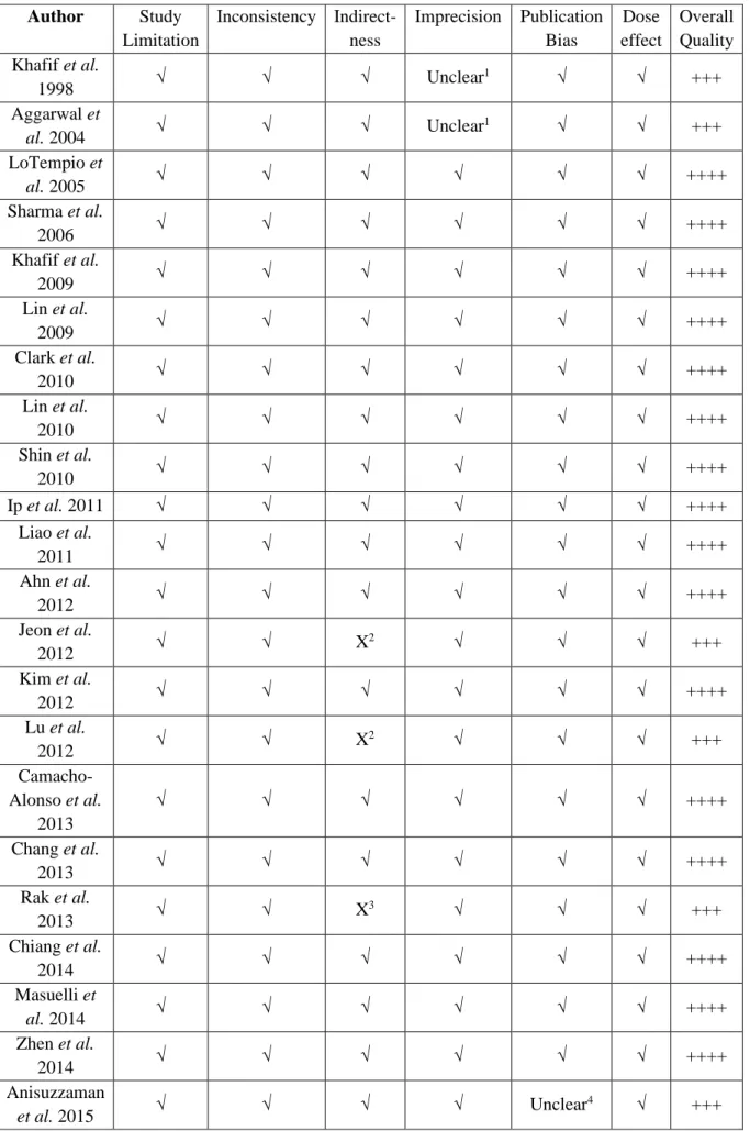
Figure 1 Effects of curcumin on HNC cells viability
Figure 2 Effects of curcumin on cell cycle and cell death for the SCC-9 cell line Figure 3 Effects of curcumin on cell cycle and cell death for the FaDu cell line Figure 4 Effects of curcumin on cytoskeleton organization and cell morphology
Figure 5 Effects of curcumin on the expression of genes that are related to the PI3K-AKT-mTOR signaling pathway
Figure 6 Effects of curcumin on the expression of proteins that are related to the PI3K-AKT-mTOR signaling pathway
LISTA DE TABELAS
Article 1
Table 1 Summary of descriptive characteristics of included studies Table 2 Description of PICOS for included studies
Table 3 Risk of bias in individual studies according to GRADE criteria.
Table 4 Risk of bias in individual studies according to SYRCLE’s RoB criteria Appendix 1 Database search
Appendix 2 Excluded articles and reasons for exclusion
Article 2
LISTA DE ABREVIATURAS E SIGLAS
4E-BP1 - 4E-binding protein 1 4NQO - 4-Nitroquinoline 1-oxide 5-FU - 5-Fluorouracil
AIF - Apoptosis-inducing factor AKT - Protein kinase B
Apaf-1 - apoptotic protease activating factor 1 ATCC - American Type Culture Collection Bax - Bcl-2-associated X protein
Bcl-2 - B-cell lymphoma 2 Bik - Bcl-2-interacting killer Bim - Bcl-2-like protein 11 BLI - Bioluminescence imaging
CDC25C - M-phase inducer phosphatase 3 CDC42 - Cell division control protein 42 CDK - Cyclin-dependent kinase
c-flip - Cellular FLICE (FADD-like IL-1β-converting enzyme)-inhibitory protein cIAP2 - Cellular inhibitor of apoptosis 2
COX2 - Cyclooxygenase-2
DAPI - 4′,6-diamidino-2-phenylindole DISC - Death-inducing signaling complex DMBA - 7,12-Dimethylbenz[a]anthracene DMEM - Dulbecco's Modified Eagle Medium DMSO - Dimethyl sulfoxide
DNA - Deoxyribonucleic acid E2F - E2 factor
EF-24 - a curcumin analog EGCG - Epigallocatechin gallate
EGFR - Epidermal growth factor receptor
eIF4E - Eukaryotic translation initiation factor 4E ELISA - Enzyme-Linked Immunosorbent Assay Endo G - Endonuclease G
FADD - Fas-associated protein with death domain FasL - Fas ligand
FBS - Fetal bovine serum
FITC - Fluorescein isothiocyanate FLLL12 - a curcumin analog GCO - Global Cancer Observatory
GLOBOCAN - Global Cancer Incidence, Mortality, and Prevalence GPCR - G-protein coupled receptor
GRADE - Grading of Recommendations, Assessment, Development, and Evaluations GTP - Guanosine triphosphate
HAT - Histone acetyltransferases HDAC - Histone deacetylase HNC - Head and neck cancer
HNSCC - Head and neck squamous cell carcinoma HPV - Human papillomavirus
IC50 - Half maximal inhibitory concentration IL - Interleukin
ISR - Insulin receptor mAb - Monoclonal antibody
MAPK - Mitogen-activated protein kinase mTOR - Mechanistic target of rapamycin mTORC - mTOR complex
MTS - 3-(4,5-dimethylthiazol-2-yl)-5-(3-carboxymethoxyphenyl)-2-(4-sulfophenyl)-2H-tetrazolium
MTT - 3-(4,5-dimethyl-2-thiazolyl)-2,5-diphenyl-2H-tetrazolium bromide NF-κB - Nuclear factor kappa B
p70 S6K - p70 ribosomal protein S6 kinase p85 S6K - p85 ribosomal protein S6 kinase pAb - Polyclonal antibody
PARP - Poly (ADP-ribose) polymerase PCNA - Proliferating cell nuclear antigen PCR - Polymerase chain reaction
PDK1 - Phosphatidylinositol-dependent kinase 1 PI - Propidium iodide
PI3K - Phosphoinositide-3-kinase
PICOS - Population, Intervention, Comparison, Outcome, and Study Design PIP2 - Phosphatidylinositol-4,5-biphosphate
PIP3 - Phosphatidylinositol-3,-4,-5-triphosphate PKC - Protein kinase C
pRB - Retinoblastoma protein
PRISMA - Preferred Reporting Items for Systematic Reviews and Meta-Analyses PTEN - Phosphatase and tensin homolog
PVDF - Polyvinylidene fluoride
RAC1 - Ras-related C3 botulinum toxin substrate 1 Rheb - Ras homolog enriched in brain
Rho GTPase - Ras homolog family of GTPases
RICTOR - Rapamycin-insensitive companion of mTOR ROS - Reactive oxygen species
RTKs - Receptor tyrosine kinases SGK - Glucocorticoid-induced kinase SRB - Sulforhodamine B
SYRCLE's RoB – Systematic Review Centre for Laboratory animal Experimentation Risk of Bias tool
TNF - Tumor necrosis factor
TRADD - TNF receptor type 1-associated DEATH domain protein TSC1 - Tuberous sclerosis complex 1
TSC2 - Tuberous sclerosis complex 2 TSI - Tumor Selectivity Index
TUNEL - Terminal deoxynucleotidyl transferase dUTP nick end labeling WHO - World Health Organization
XIAP - X-linked inhibitor of apoptosis protein
SUMÁRIO 1. INTRODUCTION 1 2. OBJECTIVES 9 3. HYPOTHESES 10 4. ARTICLE 1 11 5. ARTICLE 2 49 6. DISCUSSION 87 7. CONCLUSION 90 REFERENCES 91
1
1. INTRODUCTION
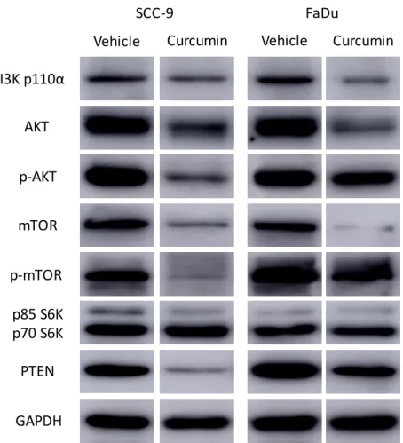
The Global Cancer Observatory (GCO) is an interactive web-based platform, maintained by the International Agency for Research on Cancer – World Health Organization (IARC-WHO), in which data on the epidemiological profile and world impact of cancer from different IARC’s cancer surveillance projects are gathered [1]. Considering all cancer types, IARC estimated for 2018 approximately 18 million new cases worldwide, and more than 9.5 million deaths related to the disease. The estimated numbers for Brazil in 2018 were 559,371 new cases and 243,588 deaths. Specifically concerning head and neck cancers (HNC), the data are presented in Figure 1.
Figure 1. Estimates of new cases HNC and deaths due to the disease in 2018, reported according to sex. Data source: GLOBOCAN 2018, IARC.
Lip and oral cav ity Lary nx Nas opha rynx Oro phar ynx Hyp opha rynx Saliv ary glan ds 0 100 200 300 400 New cases - HNC Worldwide N u m b e r o f c a s e s ( x 1 ,0 0 0 ) Male Female Lip and oral cav ity Lary nx Nas opha rynx Oro phar ynx Hyp opha rynx Saliv ary glan ds 0 2 4 6 8 10 12 New cases - HNC Brazil N u m b e r o f c a s e s ( x 1 ,0 0 0 ) Male Female Lip and oral cav ity Lary nx Nas opha rynx Oro phar ynx Hyp opha rynx Saliv ary glan ds 0 50 100 150 200 Deaths - HNC Worldwide N u m b e r o f d e a th s ( x 1 ,0 0 0 ) Male Female Lip and oral cav ity Lary nx Nas opha rynx Oro phar ynx Hyp opha rynx Saliv ary glan ds 0 1 2 3 4 5 6 Deaths - HNC Brazil N u m b e r o f D e a th s ( x 1 ,0 0 0 ) Male Female
2
As reported by GCO, the number of new cases of HNC totalized 887,659 worldwide in 2018, and the deaths 453,307. In Brazil, the new cases were 27,137 and the deaths were 14,574. That means that approximately 3% of the new HNC cases and 3.2% of deaths due to HNC were registered in Brazil.
The majority of HNCs are squamous cell carcinoma (HNSCC), to which tobacco and alcohol consumption is the most important etiological factor [2-4]. Betel-quid and areca-nut chewing also increase significantly the risk of HNSCC, which is especially prevalent in the Asia-Pacific region and among immigrants from this area [5, 6]. Infection with oncogenic subtypes of the human papillomavirus (HPV) is associated with the HNSCC carcinogenesis, particularly in the oropharynx, and the HPV+ status is related to an improved prognosis [7].
The treatment of HNSCC patients depends on the stage of the disease, and surgery is still the preferred approach, combined or not with radiotherapy and chemotherapy [4]. Aiming to reduce the morbidity that is inherent to HNSCC therapy and to improve the quality of life of patients, technological developments on the HNSCC therapy have been recently proposed, and evidence on their efficiency and efficacy has been produced. Regarding surgery, examples of such events are the microvascular reconstruction on management of advanced-stage primary HNSCC and the minimally invasive transoral surgical approaches, such as the transoral robotic surgery and the transoral laser microsurgery, on the treatment of early-stage cancers [4]. Radiotherapy benefits from the altered fractionation, intensity-modulated radiotherapy, adaptive radiotherapy, and proton beam therapy modalities, and an increasing number of studies report the advantages of distinct systemic therapy methods, such as different combinations of drugs or their association with other therapeutic modalities, targeted therapy (anti-EGFR therapy), and immunotherapy [4]. The advent of immunotherapy is regarded as the most remarkable development in the HNSCC therapy field [4], with a large body of evidence supporting its effectiveness on recurrent and/or metastatic HNSCC [8].
3
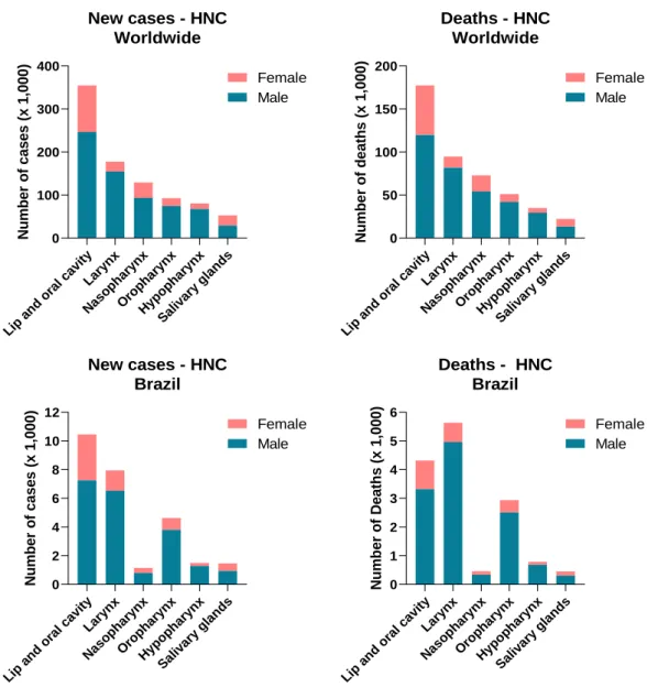
The research on nutraceuticals, natural diet-derived molecules that exert a potential effect on the control, treatment or prevention of a disease, is part of this effort to produce therapeutic options that are more specific and less detrimental to the patients who undergo cancer treatment. In fact, in 2017, the journal Seminars in Cancer Biology published an entire special issue with reviews on the role of different nutraceuticals in controlling oncogenesis [9]. Curcumin (Figure 2) is a natural polyphenol isolated from turmeric (Curcuma longa) rhizome, which has been used for thousands of years in the traditional Chinese and Ayurvedic medicine for wound healing, respiratory problems, and liver and dermatological disorders [10, 11]. It is also used as spice in the preparation of food, especially in Eastern Asia and Latin America [10]. Curcumin, desmethoxycurcumin, and bisdemethoxycurcumin are active molecules identified in turmeric and collectively referred to as curcuminoids [11]. The curcuminoids represent 3-5% of the constitution of turmeric, and among them, curcumin accounts for 77% [11]. Curcumin is practically insoluble in aqueous solutions at neutral and acidic pH, and solubility (and degradation) increases under alkaline conditions [11]. Due to its lipophilic nature, curcumin is soluble in organic solvents such as ethanol and dimethyl sulfoxide (DMSO) [11]. The therapeutic potential of curcumin is hindered by its objectionable pharmacological properties, such as poor bioavailability, solubility, and stability [12]. However, various types of chemical modifications of curcumin have been proposed as improvements to its pharmacokinetic profile and therapeutic properties [13].
Figure 2. Molecular structure of curcumin. Curcumin
4
Studies on curcumin have been increasingly published in the past decade (Figure 3), mostly due to its antioxidant, anti-inflammatory, and antitumor effects [14, 11, 15]. The presence of phenol groups in the molecular structure of curcumin (Figure 2) induces destabilization of reactive oxygen species (ROS) and justifies the antioxidant effects [10]. The anti-inflammatory effects derive from the inhibition of cyclooxygenase-2 (COX2) and the nuclear factor kappa B (NF-κB), proinflammatory proteins, and consequent reduction in synthesis of cytokines such as interleukin (IL)-1α, IL-1β, IL-2, IL-6, and IL-10 [16, 10]. The antitumor effects of curcumin will be further discussed in the studies presented in this thesis.
Figure 3. Number of studies on curcumin published between 1995 and 2019. The graph was prepared and published by Kotha and Luthria [11].
Phase I clinical trials with both healthy and cancer patients suggest treatment with curcumin, even in high daily doses, does not result in severe adverse events [17-19]. Additionally, several phase II and III clinical trials are currently under development [10]. A phase III clinical trial (NCT03769766) is being conducted to assess the potential of curcumin in preventing the progression of prostate cancer. Another (NCT02064673) is investigating its effect on recurrence-free survival in prostate cancer patients submitted to radical prostatectomy.
5
Ongoing phase II studies are assessing the association of curcumin with bevacizumab or the FOLFIRI regimen (folinic acid, fluorouracil, and irinotecan) on progression-free survival in colorectal cancer patients with unresectable metastasis (NCT02439385) and the association of curcumin to paclitaxel in advanced breast cancer (NCT03072992).
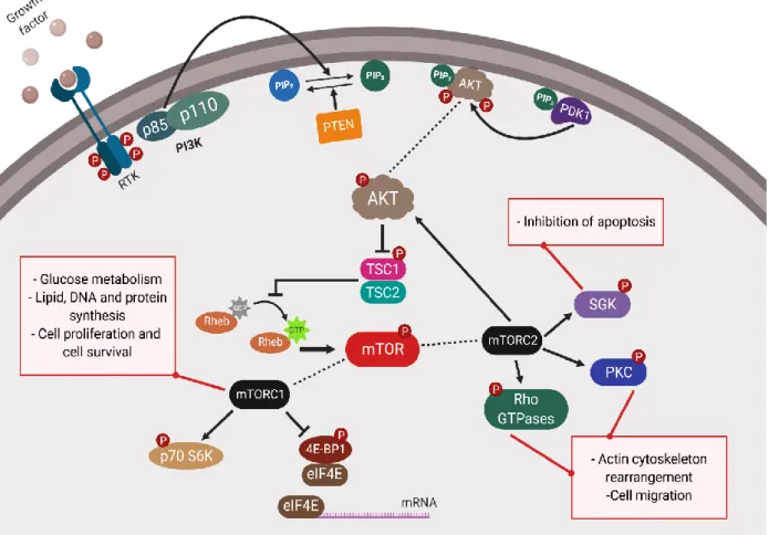
The phosphoinositide-3-kinase (PI3K) - protein kinase B (AKT) - mechanistic target of rapamycin (mTOR) signaling pathway controls cellular events such as cell growth and proliferation, metabolic homeostasis, protein and lipid synthesis, cell migration, and cytoskeleton organization [20]. This signaling pathway is described in the literature as markedly overactivated in cancer, which results in cell survival, cytoskeleton rearrangement, invasion, metastasis, and evasion of apoptosis [20].
The PI3K-AKT-mTOR signaling pathway is represented in Figure 4. The binding of growth factors to receptor tyrosine kinases (RTKs) such as the epidermal growth factor receptor (EGRF), the insulin receptor (ISR), and the G-protein coupled receptor (GPCR) triggers this pathway. The RTKs recruit to the membrane PI3K, which catalyzes the conversion of phosphatidylinositol-4,5-biphosphate (PIP2) into phosphatidylinositol-3,-4,-5-triphosphate (PIP3), which is negatively controlled by the phosphatase and tensin homolog (PTEN). PIP3 activates AKT and the phosphatidylinositol-dependent kinase 1 (PDK1). Both PDK1 and PIP3 phosphorylate AKT, which inhibits the tuberous sclerosis complex 1 and 2 (TSC1/2) by phosphorylating TSC2. The inhibition of TSC1/2 increases the activation of the Ras homolog enriched in brain (Rheb) by GTP-binding, which phosphorylates mTOR. mTOR forms different protein complexes (mTORC1 and mTORC2), each with their specific functions. mTORC1 enhances glucose metabolism and promotes lipid, DNA, and protein synthesis, as consequences of the phosphorylation of p70 ribosomal protein S6 kinase (p70 S6K) and 4E-binding protein 1 (4E-BP1), followed by the activation of the eukaryotic translation initiation factor 4E (eIF4E). mTORC2 results in actin cytoskeleton rearrangement and cell migration after phosphorylation of the protein kinase C (PKC) and the Ras homolog family of GTPases (Rho GTPases) and in
6
inhibition of apoptosis after phosphorylation of AKT and the serum and glucocorticoid-induced kinase (SGK) [20-22].
Figure 4. Schematic representation of the PI3K/AKT/mTOR signaling pathway.
Mutagenic events, mainly associated with tobacco and alcohol consumption, are considerably relevant to the HNSCC oncogenesis. In general, whenever the metabolic processing of carcinogens is inadequate, either due to genetic alterations that result in dysfunctional enzymes or because of a high concentration of carcinogens in the organism, the DNA is damaged [23]. That leads to an abnormal protein expression and mechanisms to evade apoptosis, prompt angiogenesis, maximize proliferation, and promote invasion and metastasis [23].
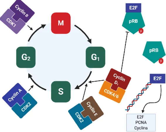
The cell cycle is a physiological process that results in cell proliferation, and that is essential for tissue development and repair. The cell cycle, represented in Figure 5, is characterized by two distinct phases: interphase (phases G1, S, and G2) and mitosis (phase M).
7
The transition from one phase to another is coordinated by complexes formed by a cyclin and a cyclin-dependent kinase (CDK), which are assembled and activated under diverse stimuli. The retinoblastoma protein (pRB) is another critical cell cycle regulator. It binds to transcription factors of the E2 factor (E2F) family, inactivates them, and keeps the cell cycle arrested at phase G1. The complexes cyclin-CDK then phosphorylate pRB, which releases E2F to take part in the transcription of genes that are vital for the cell cycle progression and cell proliferation [24, 25].
Many cell cycle and cell-cycle-related proteins are overexpressed or overactive in human cancers, particularly cyclin D, cyclin E, CDK4, CDK6, and CDK2 [26]. Research efforts focused on targeting such proteins have been made, with the objective of establishing the cell cycle inhibitors as a possible anticancer strategy [26].
Figure 5. Schematic representation of the cell cycle.
Apoptosis is a controlled and non-inflammatory cell death process that is observed on multicellular organisms from the earliest stage of development to adulthood. It is induced by a variety of intrinsic and extrinsic stimuli, such as irradiation, chemical agents, drugs, hormones,
8
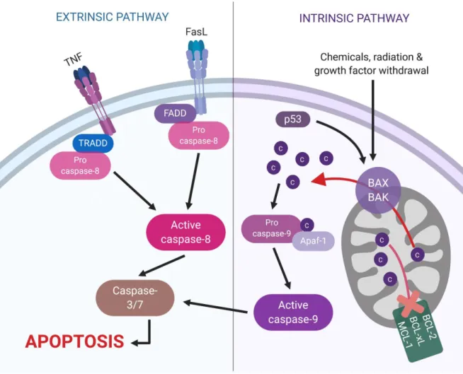
and inflammation-related proteins [27]. Apoptosis is characterized by biochemical and morphological changes in the cell, such as asymmetry, cell shrinkage, nuclear condensation, DNA fragmentation, and formation of apoptotic bodies [25]. Two main mechanisms trigger apoptosis, and they are represented in Figure 6: the extrinsic and the intrinsic pathways [28]. In both pathways, caspases are the main effector proteins. The initiator caspases, such as 8 and 9, cleave the inactive precursor forms of executioner caspases, such as caspase-3 and caspase-caspase-3, which, in their active forms, will lead the cell to an apoptotic cell death.
The extrinsic pathway is initiated by the binding of the Fas ligand (FasL) or the tumor necrosis factor (TNF) to their membrane receptors. The adaptor proteins Fas-associated protein with death domain (FADD) and TNF receptor type 1-associated DEATH domain protein (TRADD) are recruited by their respective receptors, and couple with procaspase-8, forming the death-inducing signaling complex (DISC). After DISC is formed, procaspase-8 is cleaved into its active form, which initiates apoptosis [27, 28]. The intrinsic or mitochondrial pathway is majorly controlled by members of the B-cell lymphoma 2 (Bcl-2) family, which might be either anti- or pro-apoptosis. These proteins coordinate the process by controlling the permeability of the mitochondrial membrane. When proapoptotic proteins are activated, the membrane is permeable, and the cytochrome C is translocated from the mitochondria to the cytosol. Once in the cytosol, cytochrome C forms with the apoptotic protease activating factor 1 (Apaf-1) and procaspase-9 a complex known as apoptosome. After the apoptosome is formed, procaspase-9 is cleaved into its active form [27, 28]. The protein p53, among other functions, activates apoptosis through the intrinsic pathway, especially in circumstances in which an irreversible DNA damage is observed [28].
Evasion of apoptosis as a means to keep the proliferative potential is a hallmark of cancer. The loss-of-function of proapoptotic genes, the reduced expression of proapoptotic proteins such as p53, the Bcl-2-associated X protein (Bax), and Fas, and the increased
9
expression of anti-apoptotic proteins such as Bcl-2 in HNSCC cell lines, animal models, and patient samples have been described [25, 29-31].
Figure 6. Schematic representation of the extrinsic and intrinsic pathways of apoptosis.
2. OBJECTIVES
In light of the evidence previously presented, the objectives of this thesis are:
1) Outline the state-of-the-art status of in vitro and in vivo research on curcumin and its effect on head and neck cancer;
2) Provide new insights into the biological effects of curcumin on head and neck carcinoma cell lines (SCC-9 and FaDu);
3) Demonstrate how curcumin modulates the PI3K-AKT-mTOR signaling pathway in head and neck carcinoma cell lines.
10
3. HYPOTHESES
1) The in vitro and in vivo studies published in the literature indicate that curcumin reduces cell viability, cell proliferation, and tumor growth on head and neck cancer cell lines and animal models;
2) When used in SCC-9 (tongue carcinoma) and FaDu (oropharynx carcinoma) cell lines, curcumin reduces cell viability, arrests cell cycle, induces apoptosis, and disorganizes the cytoskeleton, all effects that are expected of a drug against cancer and support curcumin as an antitumor agent;
3) Curcumin downregulates the PI3K-AKT-mTOR signaling pathway by reducing the expression of genes and proteins that are related directly or indirectly to it.
11
4. ARTICLE 1
Title: In vivo and in vitro effects of curcumin on head and neck carcinoma: A systematic review
Gabriel Álvares Borges1, Daniela Fortunato Rêgo1, Daniele Xavier Assad1,2, Ricardo D. Coletta3, Graziela De Luca Canto4,5, Eliete Neves Silva Guerra1
Affiliation:
1 Laboratory of Oral Histopathology, Health Sciences Faculty, University of Brasília, Brazil. 2 Hospital Sírio-Libanês, Brasília, Brazil.
3Department of Oral Diagnosis, School of Dentistry, University of Campinas, Piracicaba, São Paulo, Brazil.
4 Brazilian Centre for Evidence-Based Research, Department of Dentistry, Federal University of Santa Catarina, Florianópolis, Brazil.
5School of Dentistry, Faculty of Medicine and Dentistry, University of Alberta, Canada.
Article presented as published in the Journal of Oral Pathology Medicine, with minor grammar corrections and adaptations.
Full reference:
Borges GA, Rego DF, Assad DX, Coletta RD, De Luca Canto G, Guerra EN. In vivo and in vitro effects of curcumin on head and neck carcinoma: a systematic review. J Oral Path Med. 2017 Jan;46(1):3-20. doi: 10.1111/jop.12455. Epub 2016 May 24.
12 ABSTRACT
BACKGROUND: Head and Neck Squamous Cell Carcinoma (HNSCC) contributes globally to a great number of deaths and morbidity, in spite of new therapeutic strategies. There is a great need for new therapeutic drugs that are significantly effective and less deleterious to the patients’ general health. In this sense, the study of phytotherapy is a tendency, with results pointing to the use of phytochemicals as chemo-preventive and adjuvant agents. Therefore, the objective of this systematic review was to investigate the effects of curcumin on proliferation and survival of HNSCC.
MATERIALS AND METHODS: The search was conducted on six databases: Cochrane, LILACS, EMBASE, MEDLINE, PubMed, and Web of Science. In vitro and in vivo studies that evaluated the effects of curcumin on cell viability, tumor growth, cell cycle or cell death in HNSCC cell lines or animal models were selected.
RESULTS: Out of 525 initially gathered studies, 30 met the inclusion criteria. These studies demonstrated that curcumin induces cytotoxicity, apoptosis (via intrinsic pathway), and cell cycle arrest at G2/M phase in HSNCC cell lines. It also reduces tumor measurements in animal models. These events were mostly studied with MTT assay, flow cytometry, and expression of cell-cycle- and apoptosis-related proteins.
CONCLUSION: This systematic review demonstrated that curcumin is effective in HNSCC cell proliferation and survival, reinforcing the currently available evidence that curcumin could be an adjuvant drug in HNSCC treatment.
13 INTRODUCTION
Head and neck cancers, comprehended by oral cavity, pharynx and larynx cancers [1], are the sixth most common type of cancer worldwide, being especially incident in the south and southeast of Asia and parts of Europe and South America [2, 3]. Most of the head and neck cancers are squamous cell carcinomas (HNSCC) [4], to which the different forms of tobacco and alcohol consumption are considered the main causes [5, 3]. The human papillomavirus has also been recognized as an etiological factor, mainly for oropharyngeal cancers [6]. According to the National Cancer Institute in the United States, the occurrence of 45,780 new cases of HNSCC was estimated for 2015, as well as 8,650 deaths due to the disease [7].
Treatment of HNSCC is multimodal and involves surgery, radiotherapy, and chemotherapy. In spite of modern surgical techniques and therapeutic strategies such as cetuximab and taxanes, HNSCC mortality rates continue to be high in most countries, leading to an overall 5-year survival rate below 50% [5]. Therefore, new therapeutic strategies that are significantly effective and less deleterious to patients’ general health are required [8]. In this sense, targeted-therapy figures as an alternative, targeting proteins that have a considerable role in the oncogenic process [9, 10]. The study of plant- and diet-derived substances is also a tendency, with results pointing to their use as chemo-preventive and adjuvant agents, or even as a source of molecules that could be actively applied as HNSCC treatment [11-13]. One of these alternatives is curcumin, a yellow polyphenol derived from the rhizome of Curcuma
longa. Studies demonstrate its potential as an anti-inflammatory, antibiotic, and antioxidant
agent [14, 15]. Its anticancer activity, comprising the regulation of proliferation, survival, migration, invasion, angiogenesis, and metastasis, has also been reported for several types of cancer [16].
There is a relative number of in vitro and in vivo published studies that describe the activity of curcumin on HNSCC. However, a systematic review has never been written on this subject. Thus, the objective of this systematic review is to summarize the results of those
14
studies, especially concerning the effects of curcumin on proliferation and survival of HNSCC.
METHODS
Protocol and Registration
This systematic review was developed according to the Preferred Reporting Items for Systematic Reviews and Meta-Analyses PRISMA Checklist [17]. The protocol was not registered, given the nature of the investigated population.
Eligibility Criteria Inclusion Criteria
The inclusion criteria for this systematic review were based on PICOS (Population, Intervention, Comparison, Outcome, and Study Design). We considered in vitro and in vivo studies in which HNSCCs were evaluated. Cancers of the oral cavity, pharynx, and larynx were regarded as HNSCC [1]. The intervention was curcumin, and no treatment or treatment with other drugs and/or radiotherapy was the comparison. The expected outcomes were: (1) in vitro cell proliferation, viability or cytotoxicity or in vivo tumor volume or tumor incidence; and (2) apoptosis and/or cell cycle arrest, including analysis of protein expression.
Exclusion Criteria
Studies were excluded if: 1) Curcumin was not used, or used as treatment for conditions other than HNSCC; 2) Curcumin was not tested for both cell viability / cytotoxicity/ tumor growth and apoptosis / cell cycle arrest; 3) Only nasopharyngeal carcinoma cell lines were used; 4) Reviews, letters, personal opinions, book chapters, conference abstracts, and patents; 5) Clinical trials; 6) Written in alphabets other than Latin (Roman) alphabet; 7) Full copy was not available.
15 Information sources and search strategy
Individual search strategies were designed for each of the following bibliographic databases: Cochrane, LILACS, EMBASE, MEDLINE, PubMed, and Web of Science (Appendix 1). The search strategy used for PubMed was as following: (("oral cancer" or "oral carcinoma" or "oral cancers" or "oral carcinomas" or "OSCC" or "head and neck cancer" or "head and neck carcinoma" or "head and neck cancers" or "head and neck carcinomas" or "HNSCC" or (head and neck cancer[MeSH Terms]) or (head and neck neoplasms[MeSH Terms]) or (cancer of head and neck[MeSH Terms])) and (curcumin or curcuma)). The search included all articles published on or before January 26, 2016, with no time restrictions nor limits. In addition, the reference lists of selected articles were hand-screened for potentially relevant studies that could have been missed during the electronic database search.
Study Selection
Study selection was completed in two phases. In phase one, two authors (GAB and DFR) independently reviewed the titles and abstracts of all references. They selected articles that met the inclusion criteria based on their titles and abstracts. In cases of disagreement, a third author (DXA) intervened. Studies that clearly failed the inclusion criteria were discarded and those whose abstracts did not contain all needed information proceeded to phase two. Reference lists for all included articles were critically assessed by GAB for new articles. In phase two, full articles were read to determine the studies in which cell proliferation, cell viability, cytotoxicity, tumor volume or tumor incidence, and apoptosis or cell cycle arrest were studied. Two authors (GAB and DFR) independently participated in phase two. The third reviewer (DXA) resolved any disagreement. Final selection was always based on the publication full-text.
16 Data Collection Process and Data Items
One author (GAB) collected the required information from the selected articles. DFR and DXA crosschecked the collected information and confirmed its veracity. Any disagreement was resolved by discussion and mutual agreement. A fourth reviewer (ENSG) was involved as required.
Risk of Bias in Individual Studies
Quality of evidence was methodically assessed with the GRADE tool [18, 19] for in
vitro studies or the SYRCLE's RoB tool [20] for in vivo studies. The GRADE tool was adapted
to in vitro studies, according to Pavan et al. [21], given that no specific quality assessment method was developed for this type of study. For in vitro studies, two authors (DFR and DXA) categorized the articles as “high”, “moderate”, “low” or “very low” quality, according to their analysis of each study. For in vivo studies, the same two authors scored each item as “yes,” “no,” or “unclear” for each article. When they did not reach a consensus, a third author (GAB) intervened to make a final decision.
Summary Measures
Cell viability, tumor growth, cell cycle, and cell death in HNSCC cell lines and animal models after treatment with curcumin were the main evaluated outcomes.
RESULTS
In phase one, 525 articles were gathered from the databases. After duplicate removal, 236 articles remained. A comprehensive evaluation of titles and abstracts resulted in the exclusion of 152 articles. No additional article was identified from the reference lists of selected studies. A full-text review was conducted on 84 articles retrieved in phase one. This process led
17
to the exclusion of 54 studies (Appendix 2). In the end, 30 articles were maintained for final analyses [22-51]. A flow chart detailing this process is depicted in Figure 1.
Studies Characteristics
The studies were conducted in ten different countries: Brazil (n=1), China (n=5), Croatia (n=1), India (n=5), Israel (n=1), Italy (n=2), South Korea (n=4), Spain (n=1), Taiwan (n=5), and United States (n=8) [22-25, 29, 33, 46, 47]. All studies were published from 1998 to 2015, in English language.
Studies were divided into two subgroups: in vitro (n=28) and in vivo (n=10). All in vitro studies evaluated cell viability, cell proliferation, or cytotoxicity. For these evaluations, MTT assay was mostly applied, although XTT, MTS, and SRB (sulforhodamine B) assays, colony formation assay, and cell viability kits were also used. Cell cycle arrest was assessed by flow cytometry with propidium iodide (PI) staining. Apoptosis was assessed directly, mostly through flow cytometry with annexin V-FITC and PI staining or apoptosis detection kits, or indirectly, through the analysis of apoptosis-related proteins expression with western blot and immunofluorescence, caspase activity assays, DNA fragmentation, and nuclear morphology.
Most of the in vivo data were collected from studies that performed both in vivo and in
vitro experiments, and already fulfilled the inclusion criteria for the in vitro data. For this
reason, even though the in vivo data did not completely meet the criteria, they were still included. All in vivo studies evaluated tumor measurements, including tumor growth, volume, weight, multiplicity, and incidence. Studies also assessed apoptosis through western blot, TUNEL assay, and immunohistochemistry.
A summary of the descriptive characteristics of the included studies is presented in Table 1 and the PICOS information for each included study is described in Table 2.
18 Risk of Bias in Individual Studies
When assessed with GRADE, as seen in Table 3, six studies were graded as of moderate quality and one as of low quality. Three did not report the statistical analysis for considered results, even though the effects, especially concerning cell viability, were described in the text and figures. In one study, the curcumin concentrations used to establish IC50 values were not specified, and in another study the experimental design did not fully answer our question, considered that it aimed to evaluate the effects of curcumin and cisplatin on ototoxicity. In two other studies, experiments were conducted only with KB cell line, and in one HEp-2 and 7T, a HEp-2 subline, were the only cell lines used. According to the ATCC, KB and HEp-2 cell lines were originally thought to be derived from HNSCC, but found to be a HeLa (cervical cancer) cell contaminant [52, 53]. Considered their use as HNSCC cell lines by the authors, the aforementioned articles were still reviewed.
All studies that evaluated animals were considered of unclear risk of bias when assessed with SYRCLE’s RoB (Table 4). These studies did not fully describe information on allocation, randomization, and blinding, which are required for quality assessment.
Synthesis of Results In vitro studies
A great variety of HNSCC cell lines were used in the included studies, and CAL-27 (n=5), FaDu (n=4) and KB (n=3) were the most frequent.
Curcumin was used alone and compared to negative control (n=17), or used in association and compared to other treatments (n=11), which were irradiation (n=3), cisplatin (n=2), 5-fluorouracil (n=1), doxirubicin (n=1), EGCG (n=1), resveratrol (n=1), lycopene (n=1), photodynamic therapy (n=1), EF-24 (n=1), and FLLL12 (n=1) (the last two are curcumin analogs). Curcumin doses varied between 0.0001 and 400 µM, being the effective dosage rather variable among studies. IC50, when informed, ranged from 3 to 271.5 µM.
19
All in vitro studies (n=28) described an inhibition of proliferation and cell viability, reaching statistically significant levels in 14. A dose-dependent pattern was observed in most studies (n=19). When curcumin was compared to other treatments, it induced better effects, with the exception of photodynamic therapy and FLLL12, and when combined, the effects were better than with each treatment alone.
Cell cycle arrest was also observed (n=8), mostly at G2/M phase, as reported by seven studies. The sub-G1 cell population was monitored in six studies, and half of them described its increase after treatment with curcumin. In the other half, no significant difference was noticed when compared to control. Cell cycle-related proteins, such as cyclin D1 (n=6), cyclin B (n=1), cyclin A2 (n=1), cyclin E2 (n=1), CDK1 (n=1), CDK2 (n=1), and CDC25C (n=1), were evaluated in seven studies and found to have their expression decreased. Both cell cycle arrest and increased sub-G1 population were considered indicators of apoptosis by the authors.
All studies that evaluated apoptosis directly (n=10) confirmed its occurrence after treatment with curcumin. Apoptosis-related protein expression was thoroughly investigated (n=18). The pro-apoptotic proteins caspase-3 (n=12), caspase-7 (n=1), caspase-8 (n=3), caspase-9 (n=7), PARP (n=8), cytochrome c (n=3), Bax (n=4), AIF (n=2), Endo G (n=2), Apaf-1 (n=Apaf-1), Bik (n=Apaf-1) and Bim (n=Apaf-1) were increased in their active forms, whereas the anti-apoptotic proteins Bcl-2 (n=8), NF-κB (n=5), XIAP (n=2), cIAP2 (n=1) and c-flip (n=1) had their expression decreased. Three studies reported no alteration in Bcl-2 and caspase-3 expression. Nuclear morphology alterations (n=9) and DNA fragmentation (n=5) compatible with apoptosis were also observed.
In vivo studies
Out of ten in vivo studies, two adopted as animal model hamsters with DMBA-induced oral cancer and the other eight used mice inoculated with CCL23, CAL-27, SCC1, SCC40, SAS/luc, FaDu, Tu 686 or SALTO cells. One of these also used mice with 4NQO-induced oral
20 cancer [29].
Curcumin was tested alone and only compared to negative control (n=6), used in association with irradiation (n=2) or resveratrol (n=1), or compared to FLLL12 (n=1). Curcumin concentration varied between 0.6% to 1% when added to standard diet, 2 to 15 mg/100 µL when added to corn oil, and 35 to 200 mg/kg when delivered by gavage. Concerning experiment design, studies were heterogeneous regarding controls, period and regime of treatment, and delivery method.
Nine studies described an inhibition of tumor growth or volume as a consequence of treatment with curcumin, considered significant by some authors (n=4). In studies that associated curcumin with other treatments, the effects were better than each treatment alone. A cell-cycle-related protein, cyclin D1 (n=1), was down-regulated in curcumin-treated animals. The pro-apoptotic proteins caspase-3 (n=4), caspase-8 (n=1), caspase-9 (n=1), cytochrome c (n=2), and Bax (n=2) were increased in their active form and the anti-apoptotic proteins Bcl-2 (n=3), survivin (n=1) XIAP (n=1) and c-flip (n=1) had their expression decreased.
DISCUSSION
Summary of evidence
Curcuma has been used in culinary as a flavoring, coloring, and preservative agent, and in the traditional Ayurvedic and Chinese medicine for over 6,000 years [24]. Curcumin, one of its constituents, shows beneficial effects to human health, provoking interest in researchers. He
et al. [54] verified that curcumin might be used for the prevention and treatment of several
chronic conditions, such as diabetes, cardiovascular, neurological, and pulmonary diseases. The activity of curcumin against breast, colon and lung cancers, lymphoma, and leukemia has also been reported, and its chemo-preventive and therapeutic properties against oral cancer were experimentally and clinically demonstrated [55-57, 33, 58-60]. These data are supported by the
21
significant cytotoxicity and reduction of cell viability and tumor measurements observed in the reviewed studies.
Overexpression of EGFR, thus activating downstream signaling pathways such as PI3K/AKT/mTOR and Ras/MAPK, has been associated with the development and progression of HNSCC [5, 61]. These alterations result in induction of the proliferation, survival, angiogenesis, invasion, and metastasis behind the oncogenic process [61]. Overexpression and overactivation of proteins related to these pathways and others, such as NF-κB proteins, cyclins, and Bcl-2, all pro-oncogenic proteins, were observed in several included articles, and in all of them these proteins were found to be inhibited or suppressed by curcumin, pointing to an important effect of curcumin on HNSCC treatment.
Dysregulation of apoptosis is one of the most common molecular events known to be associated with the development of HNSCC, and the capacity of re-establishing its signaling, regulating and inducing it, is considered an essential characteristic of a possible anticancer agent [62]. The induction of cell cycle arrest is also desirable, given that an interruption in the cycle and apoptosis are closely related events [63]. Both apoptosis induction and cell cycle arrest were observed on HNSCC cells lines after curcumin treatment, which corroborates to the use of curcumin as an alternative anticancer agent, or at least it instigates further investigations.
Even though seven out of eight studies that evaluated the cell cycle showed a cell cycle arrest in G2/M phase, the most investigated cell cycle-related protein was cyclin-D1, associated with the G1/S transition [64]. Only one study assessed the expression of Cyclin-B, CDK1, and CDC25C, proteins related to the G2/M transition [64]. Such observation is important, as to direct future studies on which proteins should be further evaluated.
Apoptosis is triggered by two major mechanisms: the extrinsic and the intrinsic pathways [65]. In both pathways, the caspases play important roles as mediators of these processes. Initiator caspases such as Caspase-8 and Caspase-9, which cleave inactive pro-forms of the executor caspase-3 and caspase-7, were evaluated in eight included studies. Caspase-3
22
and caspase-7 were evaluated in 12 included studies [23, 28, 34-36, 40-43, 46, 49, 51]. The intrinsic or mitochondrial apoptotic pathway is mostly controlled by members of the Bcl-2 family, which may act as either inhibitors or promoters of apoptosis [65]. Bax, Bik, and Bim are pro-apoptotic proteins from the Bcl-2 family and had their increased expression found in seven included studies. Bcl-2 is an anti-apoptotic member of the Bcl-2 family, and its expression was found to be decreased by curcumin as well. The release of cytochrome c from the mitochondria to the cytosol, thus forming the apoptosome with Apaf-1 and caspase-9 and activating executor caspases, is an important process within the intrinsic pathway [66]. An increased expression of cytochrome c and Apaf-1 after treatment with curcumin was observed in four included studies. With these results it is possible to believe that the apoptosis observed in HNSCC cell lines and animal models might be triggered through the mitochondrial-dependent pathway.
The supra-additive effect of curcumin, when associated with currently prescribed chemotherapy agents, radiotherapy, novel therapeutic options or even other plant-derived substances, has been prompting a variety of studies with different types of cancer [67-69], all resulting in an additive effect when compared to treatments alone. Similar findings were observed when curcumin was associated with EGCG, resveratrol, and photodynamic therapy in HNSCC cell lines and animal models. The association of curcumin with irradiation or chemotherapeutic agents such as cisplatin and 5-Fluorouracil, currently prescribed as therapy for HNSCC, is of considerable importance, and was found to be significantly effective in five reviewed studies.
Although curcumin presents low toxicity to humans and animals [70, 55, 71], a number of limiting factors concerning its clinical use has been reported, especially when it comes to solubility, bioavailability and systemic absorption [72, 15]. Solutions for such limitations have been studied, aiming new drug delivery systems by oral administration, as proposed by three included studies. Lu et al. [38] demonstrated better solubility and stability of curcumin when it
23
was encapsulated in folate-linked liposomes, compared to its free form. Chang et al. [40] found that curcumin in nanoparticles induced apoptosis and a considerable cell growth inhibition in HNSCC cell lines, but little cytotoxicity in non-cancerous cell lines, evidencing the specificity of such delivery system. Mazzarino et al. [45], though, described better cytotoxic effects with free curcumin than with curcumin loaded in nanoparticles, with respective IC50 values of 93.40 µM and 271.5 µM after a 24h-treatment.
Limitations
Limitations regarding clinical application of this review should be considered. With our search strategy, we found eight clinical studies, but none of these evaluated the effects of curcumin on HNSCC directly. There is no standard quality assessment tool specific for in vitro studies. Therefore, we adapted the GRADE tool [18] to assess the quality of our selected in
vitro studies, classifying them through comparable baselines. Furthermore, four articles were
excluded in phase two because they were written in Chinese, according to our exclusion criteria, and the full copy of five articles could not be obtained. Nonetheless, if their data were included, our results might have been different, and therefore such exclusions were also considered a limitation.
Conclusion
This systematic review demonstrates that curcumin emerges as a potential promise in the treatment of HNSCC. The results showed that curcumin is effective on HNSCC cell lines, inhibiting cell proliferation and viability and inducing apoptosis (via intrinsic pathway) and cell cycle arrest at G2/M phase. In addition, curcumin reduced tumor burden in animal models. Altogether, this systematic review supports that curcumin might be potentially used in the HNSCC treatment, and that it should be further analyzed in clinical trials.
24
Figure 1. Flow Diagram of literature search and selection criteria adapted from PRISMA [17].
Records identified through database searching (n=525) Ident if ica ti on COCHRANE n=4 LILACS n=1 PUBMED n=162 MEDLINE n=101
Records after duplicates were removed (n=236) Scre en ing Records screened (n=84) Records excluded (n=152) E li g ibi li ty Incl u si on
Full text articles assessed for eligibility (n=84)
Additional articles identified from reference lists of selected articles
during initial search (n=0)
Full articles excluded with reasons (n=54) 1) Curcumin was not used, or used as
treatment for conditions other than HNSCC (n=5);
2) Curcumin was not tested for both cell viability / cytotoxicity / tumor growth and apoptosis / cell cycle arrest (n=31);
3) Only nasopharyngeal carcinoma cell lines were used (n=6);
4) Reviews, letters, personal opinions, book chapters, conference abstracts and patents (n=2);
5) Clinical trials (n=1);
6) Language restriction (n=4); 7) Full copy was not available (n=5). 8) Studies included (n=30) Web of Science n=150 EMBASE n=107
25 Table 1. Summary of descriptive characteristics of included studies (n=30).
Year Author Country Study Design
Assays Treatment Results Main Conclusions
1998 Khafif et
al.
USA In vitro Hoechst 33342 proliferation
assay; Cell cycle with ethidium bromide and flow cytometry
Curcumin and/or EGCG
ED50 was constant in both control and HNSCC cell lines
(3.45 and 5.18 µM). Combination index at ED50 was
0.7±0.08 for Curcumin+EGCG, indicating synergism. Cell cycle arrest at G2/M.
As single agent, curcumin was effective in suppressing growth of both
normal and malignant cells.
Combination treatment was more effective than each agent alone. 2004 Aggarwal
et al.
USA In vitro MTT assay; Cell cycle with
propidium iodide (PI) and flow cytometry; Apoptosis with annexin V-FITC and flow cytometry; Western blot
Curcumin Dose-dependent inhibition of proliferation in all cell lines. Cell cycle arrest at G1/S (54% in control, 87% at 25 µM)
in 24h. dependent increased apoptosis. Dose-dependent cleavage of caspase-3 and -9 and PARP. Decreased expression of NF-κB, cyclin-D1, and Bcl-2.
Curcumin suppressed proliferation and induced G1/S phase arrest in all
HNSCC cell lines studied, very likely through downregulation of cyclin-D1 and Bcl-2. 2005 LoTempio et al. USA In vitro and in vivo
In vitro: MTT assay; Cell
sorting and apoptosis with PI and annexin V-FITC and flow
cytometry;
Immuno-fluorescence; Western Blot
In vivo: Tumor measurement
Curcumin; Curcumin injections and topical paste (in vivo)
In vitro: Significant dose-dependent cytotoxicity in all cell
lines (P<0.0001). Increased cell death (27-33%, depending on the cell line) including early apoptosis (6-17%), at 50 µM. Decreased expression of NF-κB and cyclin-D1.
In vivo: Curcumin injections had little to no effect on
tumors. Saline/curcumin paste inhibited tumor growth.
Curcumin treatment resulted in growth inhibition, which was due to downregulation of the anti-apoptotic NF-κB. In vivo, topical applications of saline/curcumin paste were more efficient than injections.
2006 Sharma et
al.
India and USA
In vitro MTT assay; Cell cycle with PI
and flow cytometry
Curcumin Dose-dependent decrease in viability. IC50 was 25-50 µM,
depending on the cell line. Increase in sub-G1 population
(14.4% in control, 75.1% in treatment).
Curcumin induced cell death by apoptosis in HNSCC cells, as observed by a sub-G1 peak in cell cycle analysis.
2008 Garg et al. India In vivo Tumor measurements; Western blot; TUNEL assay
Curcumin Decrease in tumor incidence (by 45%), tumor burden (from 700 to 300 mm3 i.e. by 43%) and tumor multiplicity
(by 51%). Decreased latency period of DMBA-induced SCC (10 weeks in control, 12 weeks in treatment). Significant decrease in expression of PCNA and Bcl-2 and increase in Bax and Caspase-3 (P≤0.05).
Curcumin decreased the DMBA-induced tumor burden and multiplicity and enhanced the latency period of SCC tumors. Higher Bax/Bcl-2 ratio and apoptotic index suggested apoptotic cell death.
2009 Khafif et
al.
Israel In vitro
and in vivo
In vitro: XTT assay; Cell cycle
with PI and flow cytometry
In vivo: Tumor measurements
Curcumin and/or Irradiation
In vitro: Dose-dependent inhibition of viability (at 15 µM,
P<0.05). IC50 was 15-22 µM, depending on the cell line.
Better results when treatments were combined (P<0.05). Minor effects on sub-G1 population with treatments alone.
Additive effect on sub-G1 population with combination.
In vivo: No significant decrease in neither tumor volume
nor tumor weight with treatments alone. Combined treatment reduced tumor weight (25%, P=0.09) and tumor sizes (15%, P=0.23).
Curcumin inhibited HNSCC cell growth and enhanced effect of radiation in vitro. Combined treatment showed a trend towards improvement in tumor weight compared to curcumin or irradiation alone.
26
2009 Lin et al. Taiwan In vitro MTT assay; apoptosis
evaluation with ApopNexin FITC detection kit; DNA fragmentation; Caspase activity assay; Western blot
Curcumin Significant dose-dependent decrease in survival and proliferation (P<0.05). Dose-dependent increased apoptosis. Significant DNA fragmentation increase at 25 µM (P<0.05). Significant decrease in procaspase-3 and increase in activity of caspase-3 (P<0.05).
Curcumin could inhibit cell
proliferation and irreversibly induce apoptosis in HNSCC cells.
2010 Clark et al. USA In vitro
and in vivo
In vitro: MTS assay; Cell cycle
with PI and flow cytometry; Western blot
In vivo: Tumor measurements
Curcumin In vitro: Significant inhibition of HNSCC cells in
comparison with normal cells at 10 µM and after 72h (P<0.05). Significant cell cycle arrest at G2/M with one cell
line at 10 µM and at G1/S with two other cell lines at 20
µM (P<0.001). No increase in sub-G1 population.
Decreased cyclin-D1 (P≤0.01). No cleaved caspase-3.
In vivo: Significant reduction in xenograft tumor volume
at 5mg (days 0-16, P=0.02) and at 15mg (day 5, P=0.01. day 8, P=0.02). No significant difference at 10mg. Pretreatment suppressed growth of xenograft tumor (P<0.01) at 15mg.
Curcumin inhibited cell proliferation but did not induce apoptosis in the HNSCC cell lines studied. In vivo growth inhibitory effects were noted at earlier time points, which suggests that curcumin inhibits the grafting of tumor cells. It delayed carcinogenesis, indicating its potential for chemoprevention.
2010 Lin et al. Taiwan In vitro
and in vivo
In vitro: SRB assay; Cell cycle
with PI and flow cytometry
In vivo: Optical imaging with
luciferase gene transfection; Bioluminescence imaging (BLI)
Curcumin In vitro: IC50 was about 3 µM. Dose-dependent increase in
sub-G1 population. Significant cell cycle arrest at G2/M
(33.58% in control, 47.72% at 3 µM, 49.93% at 5 µM and 39.83% at 7 µM, P<0.05).
In vivo: Significant decrease in tumor volume on the 26th
day (P<0.01). No difference among different drug-treated groups. No significant body weight change in mice with or without treatments.
The inhibitory effects of curcumin on the growth of HNSCC cells and of the tumor were significant.
2010 Shin et al. South Korea
In vitro Viability assay with the cell
counting Kit-8; Nuclear
morphology with Hoechst
33342
Curcumin Strong dose-dependent inhibition of proliferation. IC50 was
8.37±1.13 µM. Nuclear morphological features compatible with apoptosis at 10 µM.
Curcumin induced inhibition of proliferation and possibly apoptosis.
2011 Ip et al. Taiwan In vitro Viability assay with PI and flow cytometry; Cell cycle with PI and flow cytometry; DAPI staining; Western blot
Curcumin Dose-dependent decrease in viability in 24h (P<0.05 at 15 and 20 µM) and 48h (P<0.05 at 5, 10, 15, and 20 µM). Marked cell cycle arrest at G2/M phase (20.26%, compared
to control). Increased sub-G1 population (16.02%).
Decrease in expression of cyclin-B, CDK1, CDC25C, Bcl-2, and XIAP. Increase in AIF and Endo G.
Curcumin delayed cell cycle
progression, and the observed decreased viability might be justified by cell cycle arrest and apoptosis.
2011 Liao et al. China and the USA
In vitro MTT assay; Colony formation
assay; ELISA apoptosis
detection kit; Apoptosis with PI and annexin V-FITC and flow
Curcumin Significant cell growth inhibition (5 µM in 48h, P<0.05; 5 µM in 72h and 7.5 µM in 48 and 72h, P<0.01). Significant reduction in colony formation (P<0.01). Dose-dependent apoptosis (14% in control, 26% at 5 µM after 48h. Cell cycle arrest at G2/M (5.5% in control, 13.6% at 5 µM).
Curcumin elicited a dramatic effect on growth inhibition and induction of apoptosis in HNSCC cells, besides inducing a G2/M phase arrest.
27
cytometry; Cell cycle with PI and cytometry; Western blot
Decreased expression of Bcl-2 and cyclin-D1. 2012 Ahn et al. South
Korea
In vitro MTT assay; Morphological
analysis with Hoechst 33342 and PI; Western blot
Curcumin and/or photo-dynamic therapy
Dose- and time-dependent decrease in cell viability. Nuclear morphological features compatible with apoptosis. Increased cleaved caspase-3 and -9, cleaved PARP and cytochrome c. Effects were enhanced when treatments were combined.
Photodynamic therapy associated with curcumin enhanced cytotoxic and apoptotic effects in HNSCC cells via mitochondria-dependent apoptosis. 2012 Jeon et al. South
Korea
In vitro MTT assay; DAPI staining;
DNA fragmentation; Western blot; Caspase activity assay with PhiPhiLux-G1D2
Curcumin; EF-24, curcumin analog
No effect observed at 0.1-10 µM. Significant inhibition of viability at 30 and 100 µM (P<0.001). IC50 was 30.61±2.83
µM. Nuclear morphology compatible with apoptosis. DNA ladder formation at 50 µM. Increased cleaved caspase-3, -7 and -9.
Curcumin inhibited growth of HNSCC cells by activating apoptosis, possibly through caspases-3, -7 and -9-dependent processes.
2012 Kim et al. South Korea
In vitro MTT assay; Cell cycle with PI
and flow cytometry; Western blot; Morphological analysis
Curcumin Significant inhibition of viability at 10, 20, and 40 µM (P<0.05). Morphological features compatible with apoptosis at 20 µM. No increase in sub-G1 population at
10 µM. Time-dependent increase in cleaved PARP and caspase-3 at 10 µM.
Curcumin potently induced cell death in the absence of a high ratio of apoptotic cells, implying that it may activate another cell death pathway, such as autophagy.
2012 Kumar et
al.
India In vivo Tumor measurements; Western blot; Immunohistochemical staining; TUNEL assay
Curcumin Inhibition of tumor growth. Decreased tumor multiplicity (by 40-47%) and tumor burden (by 55-63%). Increased Bax and decreased Bcl-2 (significantly in 4 weeks, P<0.05). Increase in caspase-3 and -9 and cytochrome c and decreased survivin, PCNA, and cyclin-D1 (P<0.05).
Curcumin decreased tumor burden and multiplicity, due to enhanced apoptosis, as revealed by increased Bax/Bcl-2 ratio and apoptotic index and decreased proliferation or both. 2012 Lu et al. China In vitro MTT assay; Apoptosis with PI
and annexin V-FITC and flow cytometry Free curcumin; Curcumin-loaded liposomes
Dose-dependent inhibition. IC50 was 14.5µg/mL for free
curcumin, 16.3µg/mL for folate-receptor-targeted liposomal curcumin (F-CUR-L) and 29.5µg/mL for non-targeted liposomal curcumin (CUR-L). Time-dependent apoptosis (mean rate: 46.7%) with F-CUR-L.
F-CUR-L enhanced solubility of curcumin and could specifically target a folate-receptor-positive tumor cell line.
2013 Camacho-Alonso et
al.
Spain In vitro MTT assay; ELISA apoptosis detection kit
Curcumin or lycopene and/or irradiation
Dose-dependent cytotoxicity, especially evident at 5.5 and 6.75 µM. Better results for curcumin than for lycopene. Apoptosis observed with or without irradiation, although more intense when treatments were combined.
Curcumin increased cytotoxic activity, and combination with irradiation exerted synergic effect.
2013 Chang et
al.
Taiwan In vitro MTT assay; Morphological analysis; DAPI staining; Western blot; Caspase-3 and -9 colorimetric assay
Curcumin-loaded nano-particles
Significant dose- and time-dependent viability decrease at 20, 40, and 80 µM (P<0.001). Morphological features compatible with apoptosis. No viability impact and morphological changes in control cell lines (IC50>80 µM).
Dose-dependent nuclear condensation. Increase in cleaved caspase-3 and -9, cytochrome c, Apaf-1, AIF, Endo G, and Bax. Downregulated Bcl-2.
Curcumin nanoparticles caused anti-proliferative effects in a dose- and time-dependent manner, but little toxicity to normal cells. It induced apoptosis, possibly through the intrinsic signaling pathway.
![Figure 3. Number of studies on curcumin published between 1995 and 2019. The graph was prepared and published by Kotha and Luthria [11]](https://thumb-eu.123doks.com/thumbv2/123dok_br/19271435.983383/18.892.152.730.464.776/figure-number-studies-curcumin-published-prepared-published-luthria.webp)
![Figure 1. Flow Diagram of literature search and selection criteria adapted from PRISMA [17].](https://thumb-eu.123doks.com/thumbv2/123dok_br/19271435.983383/38.892.87.844.124.1109/figure-diagram-literature-search-selection-criteria-adapted-prisma.webp)